PHYSICAL DIAGNOSIS THE CHEST DR SHAM A CADER


 • Unable to assume supine position")


 • Atelectasis • Fibrosis •")


 •")
 or bilateral (COPD, DIF, Asthma) •")

")

 • Warmth (Empyema) • Tenderness ( Empyema, Rib and chest")


 Increased: (conditions giving")


. •")
 Hyper-resonance: (Emphysema, Asthma, Pneumothorax,")

")


 Crackles Pleural Rub 91")


- Slides: 98
PHYSICAL DIAGNOSIS THE CHEST DR SHAM A. CADER
THE ORGANS •
ANATOMY • One should have a clear understanding of anatomy of the respiratory system to perform a proper physical exam. Some of the important anatomical details are outlined below.
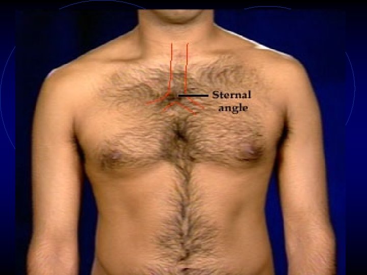
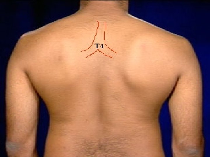
TRACHEA 1. Starts from cricoid cartilage to sternal angle anteriorly and T 4 spinous process posteriorly, where it divides into left and right main stem bronchi. This information is important in understanding D'spine sign seen in patients with large Mediastinal mass. 2. Trachea is slightly slanted to right. Bronchovesicular breathing heard in right infraclavicular region is due to this phenomenon. 3. Trachea has intra and extra thoracic components. This has important bearing in the understanding of physiology of variable obstruction.
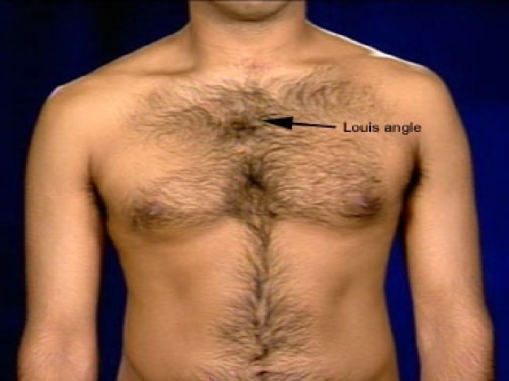
MANUBRIUM ANGLE • The angle between the body and Manubrium. Many important land marks occur at this level. It is called Louis Angle. • 2 nd rib articulates to Manubrium at this site. The ribs are counted anteriorly starting from this point. • Carina of trachea is at this level. • Mediastinum is divided into superior and inferior at this level.
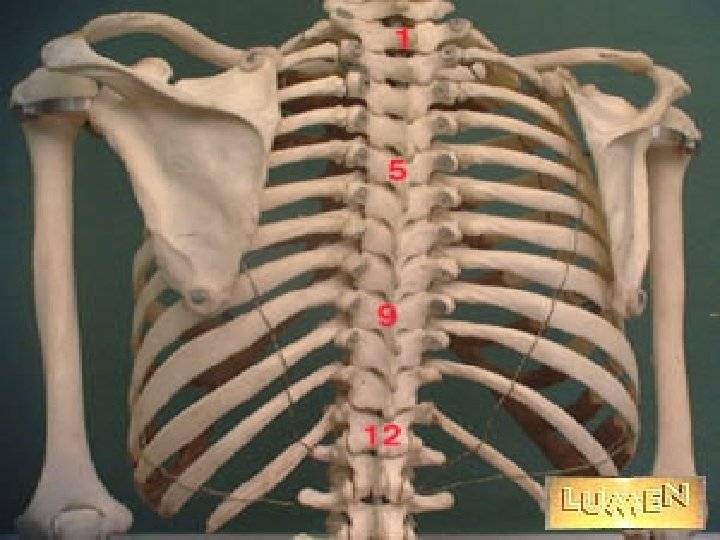
RIBS • Anteriorly ribs are counted down starting from 2 nd rib. There are 12 ribs and 11 interspaces. You can also count up from 12 th rib. Inferior angle of scapula sits on 7 th rib posteriorly.
SPACES • Anteriorly there are supra clavicular, • • infraclavicular, precardiac and Traube's space. Posteriorly we have interscapular, supra, and infra scapular spaces. Infraclavicular: Space below clavicle Supraclavicular: Space above clavicle Precardiac: Space in front of heart Traube's: Space overlying stomach Interscapular: Space between scapula Suprascapular: Space above scapula Infrascapular: Space below the scapula
LINES • Midsternal Line: A vertical line down the middle of • • sternum Parasternal Line: A vertical line along lateral edge of sternum Mid-Clavicular Line: A vertical line from middle of clavicle Anterior Axillary Line: A vertical line along anterior axillary fold Mid-Axillary Line: A vertical line at mid point between anterior and posterior axillary line. Posterior Axillary Line: Along post axillary fold Scapular Line: Inferior angle of scapula Vertebral line: Over spinous processes in the
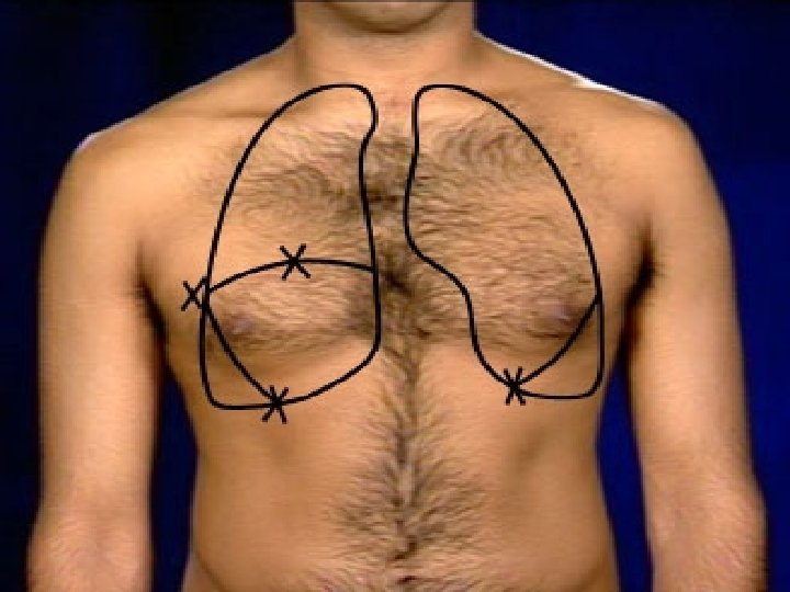
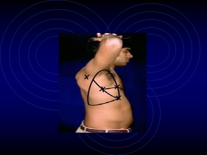
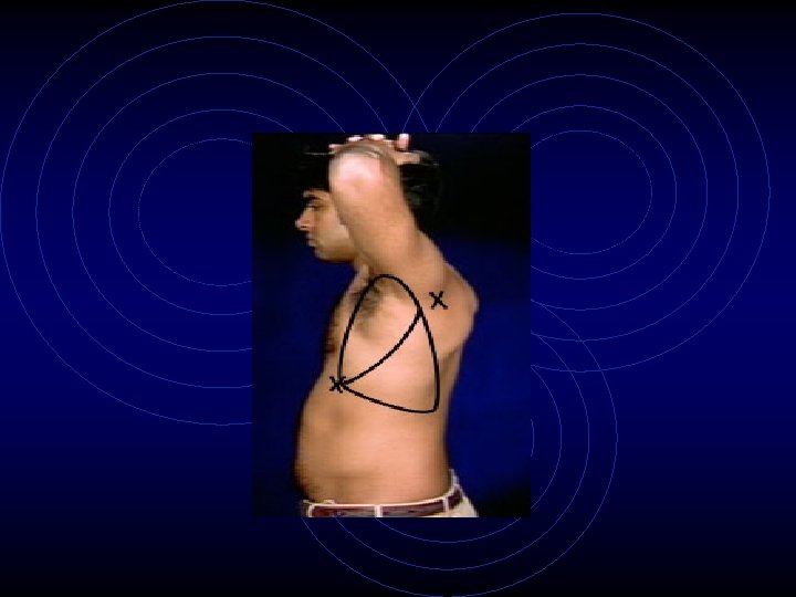
Right Lung: • With a marking pen start 3 centimeters above clavicle in midclavicular line, come down along right parasternal line , join to 6 th rib in midclavicular line, to 8 th rib in mid-axillary line, to 10 th rib posteriorly, to vertebral line posteriorly.
Left Lung: • At angle of Louis, follow the outer margin of heart to 6 th rib in mid-clavicular line. • Appreciate that apex of lung is just under the skin easily palpable in the supraclavicular space. • Pancoast tumor and TB occur at this site. Hence, the apex of lungs should be routinely examined.
Surface Anatomy of Lobes • Draw oblique fissure by drawing a line strait from 6 th rib in MCN. to 5 th rib in mid axillary line and along the medial margin of scapula (with the patients hands on head) to 3 rd spinous process • Transverse fissure can be drawn by drawing a line from 5 th rib in midaxillary line to 4 th rib anteriorly
Once the fissures are drawn over the outline of lungs, one can easily recognize the surface anatomy of lobes of lungs. One can then appreciate the importance of examining the patient all around the chest to cover the lobes. Most of lower lobe is in back, upper lobe is in front and all of middle lobe is in front. In the axilla all of the three lobes can be seen.
DIAPHRAGM
• Pleura: Once the diaphragm has been outlined you can appreciate that the pleural gutter is deep posteriorly. Fluid thus tends to accumulate posteriorly • Mediastinum is the space between lungs from inlet to outlet of thorax. Anteriorly it is between parasternal lines. Posteriorly it is at vertebral line. Mediastinum is narrow posteriorly and widens anteriorly. Inferiorly it extends to xiphisternum. Superiorly it starts at suprasternal notch. Since the inlet of thorax is slanted, only posterior Mediastinum extends to neck.
• Sternal angle separates superior from inferior Mediastinum. The inferior Mediastinum is divided into anterior, middle and posterior compartments. The space in front of heart is anterior Mediastinum and behind is posterior Mediastinum. Heart itself defines the middle Mediastinum. The posterior Mediastinum is divided into paravertebral and prevertebral space. Superior Mediastinum extends into the neck and is called cervico-Mediastinal space
It is important to know the structures in each compartment. In the differential of masses in the Mediastinum one uses this knowledge.
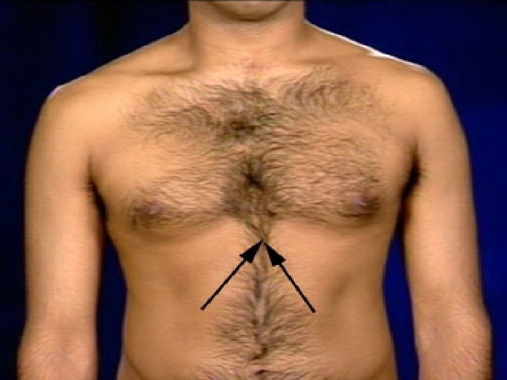
COSTAL ANGLE • Costal angle is formed by the 10 rib with Costal cartilage on either side and xiphisternum in the middle. The normal angle is. Both sides are symmetrical. Volume changes in each hemithorax will alter this relationship. Hyperinflated lungs will increase the Costal angle. Diaphragmatic paralysis also alters the symmetry of Costal angle.
• Spinous Process • The most prominent spinous process is 7 th cervical vertebra. You can count down the thoracic vertebra and the ribs using this landmark.
Respiratory Rate and Pattern of Breathing • The patient should not be aware that you are counting his respiratory rate. Count the respiratory rate while pretending to take the patient's pulse. • Note the rate, pattern and comfort of respiration.
Normal • Resting rate is between 10 -14 per minute, regular with no apparent discomfort. . • Chest wall and abdomen expand during inspiration and is symmetrical. • Abdominal component of expansion is dominant in men and thoracic component in women. • Periodic deep breathing (Sighs) less tha 5 per minute.
Abnormal Finding • Minor changes in rate and rhythm of respiration occur due to anxiety and while it may represent an abnormality, it may not be significant • Rate below 10/min: Bradypnea: (Narcotics, raised intracranial tension, myxedema) • Rate above 20/min: Tachypnea: (Interstitial, vascular and multitude of diseases, anxiety
Pattern • Periodic breathing. Cyclical increase and decrease • • in depth of respiration: Cheyne-stokes breathing: (CHF, Cerebrovascular insufficiency) Slow deep breathing: Kussmaul: (Ketoacidosis) Totally irregular with no pattern: Biot's breathing: (CNS injury) Periodic deep breathing: Sighs: (Anxiety state) Instead of simultaneous chest and abdominal expansion with inspiration abdomen retracts while chest expands: Abdominal paradox: (Diaphragmatic paralysis)
• On the side of unstable chest wall hemithorax retracts while the normal side expands with inspiration: Thoracic paradox: (Flail chest) • With lips pursed patient controls expiration slowly: Pursed lip breathing: (Obstructive lung disease) • No abdominal component : ( Acute abdomen) • No thoracic component: (Pleurisy, Chest wall pain, Ankylosing spondylitis)
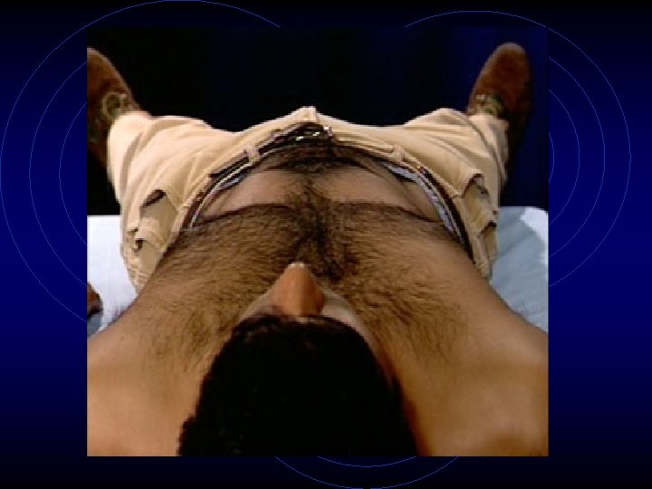
Discomfort • Labored breathing: (Heart and Lung diseases) • Unable to assume supine position because of worsening shortness of breath: Orthopnea: (CHF, Diaphragmatic paralysis, SVC syndrome, Anterior mediastinal mass) • Unable to erect position because of worsening shortness of breath, more comfortable in supine position : Platypnea: (Pulmonary spiders in cirrhotic)
Size of Thorax • The size of thorax is determined by the balance between elastic recoil of lungs and chest wall compliance. In normal, the FRC position is usually at 60% of the TLC. At this position muscle length tension curve is optimal for muscle contraction. If the elastic recoil of lung decreases the resting position of thorax will be larger, it maybe 80% of the TLC position. This position is very inefficient to generate force by muscles and leads to shortness of breath.
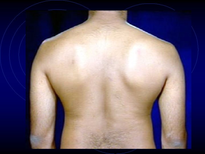
Symmetry of Hemithorax • Both sides are equal in size and asymmetry is abnormal. Unilateral lung or pleural disease alters negative pressure in pleura, affecting the resting size of hemithorax. e. g. In pneumothorax the negative pressure in pleura is lost and there is nothing to hold chest wall down. Hemithorax on that side will assume TLC position. In patients with atelectasis the negative pressure in pleura increases and the size of hemithorax will become smaller
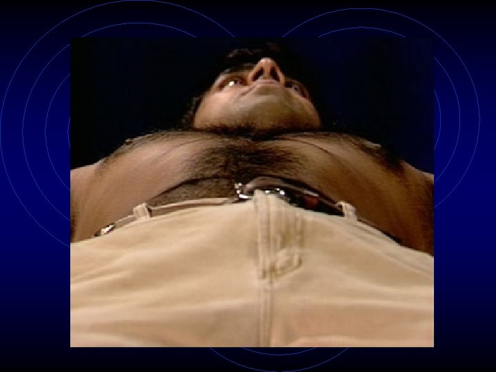
• It is best to assess symmetry of hemithorax with patient laying flat in bed without pillows. Stand either at head or foot end and look tangentially at the thorax level to assess asymmetry
Accessory Inspiratory Muscles •
Accessory Expiratory Muscles • Normal tidal expiration is passive and there is no muscle contraction. Expiratory muscle contraction is always accessory. When you force expiration, expiration muscles come into play. Abdominal muscles and intercostals are expiratory muscles. If a patient is contracting abdominal muscles for quiet respiration it is abnormal and he is attempting to force expiration.
Forced Expiration • Only peak flows can be increased by forced expiration. The flow rates cannot be increased for most of expiratory phase by forcing expiration. The increasing positive pressure in pleura compresses airway and further decreases airway size thus countering the increased force to expire.
• In patients with increased airway resistance patients attempt to increase airflow. They have two options either to adopt a rapid shallow breathing or to use pursed lip breathing to counter auto-peep and enhance emptying of lung. When airways are severely narrowed, air trapping occurs and patient may breath with a very high FRC and the only way he can breath is to adopt a rapid shallow breathing.
Negative Pleural Pressure Assessment • Only peak flows can be increased by forced expiration. The flow rates cannot be increased for most of expiratory phase by forcing expiration. The increasing positive pressure in pleura compresses airway and further decreases airway size thus countering the increased force to expire.
• In patients with increased airway resistance patients attempt to increase airflow. They have two options either to adopt a rapid shallow breathing or to use pursed lip breathing to counter auto-peep and enhance emptying of lung. When airways are severely narrowed, air trapping occurs and patient may breath with a very high FRC and the only way he can breath is to adopt a rapid shallow breathing.
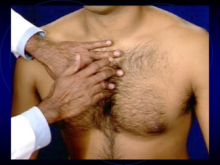
Method Of Exam 1. Position yourself in front of the patient and note the position of the thyroid cartilage. 2. Inspect for the symmetry of clavicular insertion of both sternomastoids. 3. Tracheal Position: Gently bend the head to relax the sternomastoids. By inserting your finger between the trachea and sternomastoid, assess and compare the space on either side
Trachea: Position To evaluate the position of the upper mediastinum • Normal: Trachea is slightly tilted to right. As a result, the clavicular insertion of right Sternomastoid is slightly more prominent and the space between trachea and sternomastoid is smaller compared to left.
Abnormal Finding • Tracheal deviation could be either due to Lung, pleural, Mediastinal or Chest wall disease. The mediastinum can be either pulled or pushed away from the lesion
• Lung Pull: ( Loss of lung volume) • Atelectasis • Fibrosis • Agenesis • Surgical resection Push: (Space occupying lesions) • Large mass lesions
Pleura 1. Push: • Pneumothorax • Pleural effusion 2. Pull: • Pleural fibrosis Mediastinal masses and thyroid tumors Kypho-scoliosis
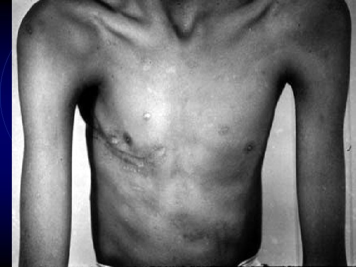
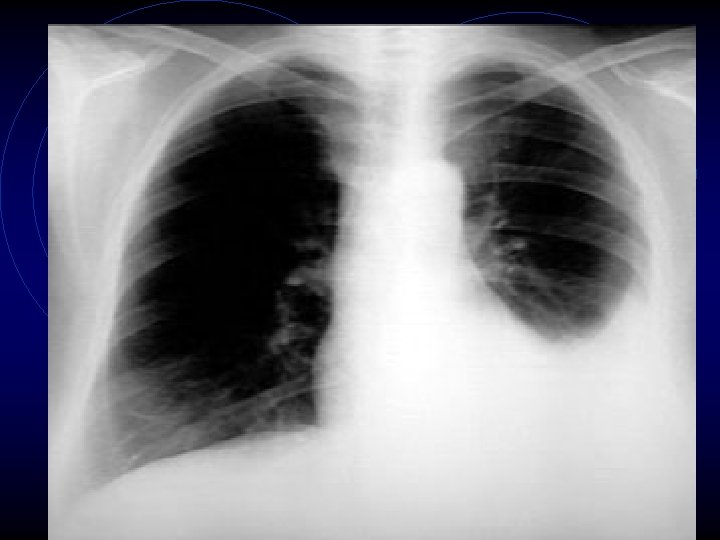
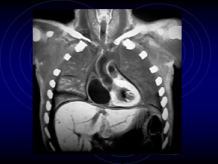
Atelectasis
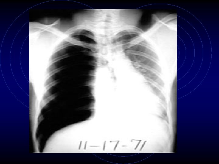
Resorptive atelectasis • When airways are obstructed there is no further ventilation to the lungs and beyond. In the early stages blood flow continues and gradually the oxygen and Nitrogen get absorbed, resulting in atelectasis. • The following in an example of right lung resorptive atelectasis. In this instance, atelectasis followed bronchial obstruction due to cancer.
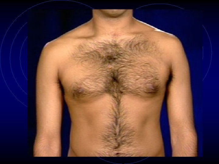
Chest: Observation Chest asymmetry • Kyphoscoliosis • Larger hemithorax : (Pneumothorax, Pleural effusion) • Smaller hemithorax: (Atelectasis, Pleural fibrosis, Agenesis of Lung)
Increased pleural negative pressure • Unilateral (airway obstruction) or bilateral (COPD, DIF, Asthma) • Intercostal and supraclavicular fossa retraction • Downward movement of trachea with quiet inspiration
Skin and soft tissue • Puncture sites and Scars (Thoracentesis, FNAB, Chest tube, Surgical scars)
Prominent collateral veins (SVC syndrome)
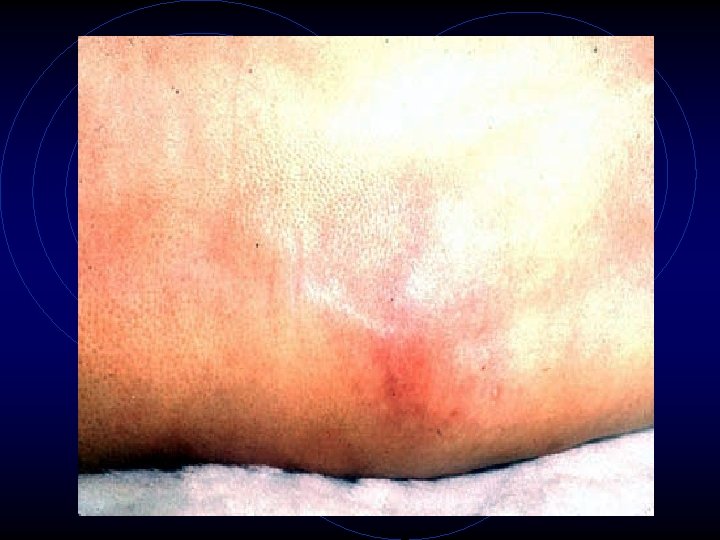
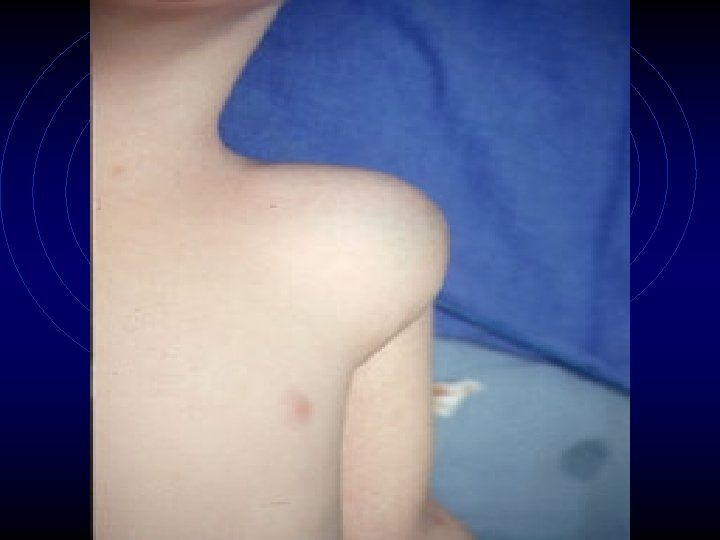
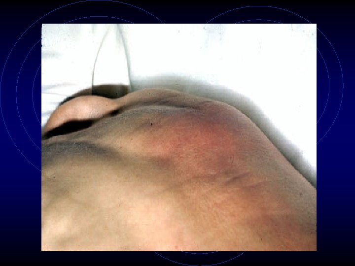
Swelling (Recent thoracentesis, Empyema, Mesothelioma, Empyema necessitatis, Cystic hygroma •
• Erythema (Empyema) • Warmth (Empyema) • Tenderness ( Empyema, Rib and chest wall lesions ) • Subcutaneous nodules (Metastasis)
Voice transmission • • • Patient to say "99" "1, 2, 3" or "E" Each time you lay your hands or listen All around the chest and compare Dorsal surface of your fingers or ulnar surface of your hand (tactile fremitus) Listen with diaphragm (vocal resonance) If increased have patient whisper Note the intensity Quality of pitch Compare
Tactile Fremitus •
Abnormal Finding • Decreased: (Pleural effusion, • • Pneumothorax, Atelectasis, Mass) Increased: (conditions giving bronchial breathing) Bronchophony: (Normal) Whispering pectoroliquy ( Normal ) Qualitative: Egophony
Lungs: Percussion • Percuss the lung fields, alternating, from top to bottom and comparing sides. • Percuss over the intercostal space and note the resonance and the feel of percussion. • Keep the middle finger firmly over the chest wall along intercostal space and tap chest over distal interphalangeal joint with middle finger of the opposite hand.
• The movement of tapping should come from the wrist. • Tap 2 -3 times in a row. • Do not leave the percussing finger on chest , otherwise you will dampen the sound. • Stand on one side and with your flat of hand, tap the chest from top to bottom and from side to compare. I use this method as a screening step to identify the area of abnormality
• The movement of tapping should come from the wrist. • Tap 2 -3 times in a row. • Do not leave the percussing finger on chest , otherwise you will dampen the sound. • Stand on one side and with your flat of hand, tap the chest from top to bottom and from side to compare. I use this method as a screening step to identify the area of abnormality
Movement of Diaphragm: • Identify the lower limit of resonance during deep inspiration and deep expiration. • This determines the range of movement of the diaphragm.
Normal • The lung is filled with air (99% of lung is air). • • Hence, percussion of it gives a resonance. This step helps identify areas of lung devoid of air. Appreciate the dullness of the left anterior chest due to heart and right lower chest due to liver. Note the hyper-resonance of the left lower anterior chest due to air filled stomach. Normally, the rest of the lung fields are resonant. Normal diaphragmatic excursion is 5 -6 cm.
Abnormal Finding Lungs fields Dullness: (Mass, Atelectasis, Consolidation, Pleural effusion) Hyper-resonance: (Emphysema, Asthma, Pneumothorax, Blebs)
• Decreased or increased resonance is abnormal. Increased resonances can be noted either due to lung distention as seen in asthma, emphysema, bullous disease or due to Pneumothorax. Decreased resonance is noted with pleural effusion and all other lung diseases. Experienced physicians are able to discriminate between dullness of pleural effusion from a consolidation or a mass lesion of lung. The dullness is flat and the finger is painful to percussion with pleural effusion. • Diaphragmatic motion
Diaphragmatic motion • Decreased diaphragmatic excursion: (Emphysema, paralysed diaphragm)
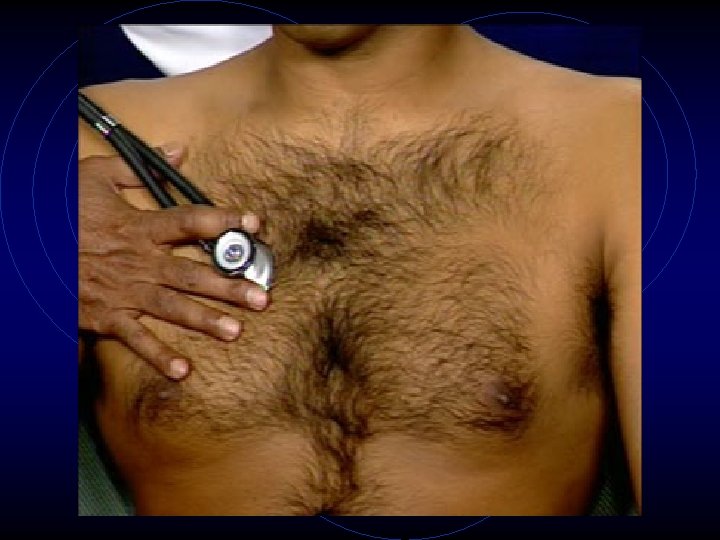
Auscultation of Lungs • While the patient breathes normally with mouth • • open, auscultate the lungs, making sure to auscultate the apices and middle and lower lung fields posteriorly, laterally and anteriorly. Alternate and compare sides. Use the diaphragm of the stethoscope. Listen to at least one complete respiratory cycle at each site. First listen with quiet respiration. If breath sounds are inaudible, then have him take deep breaths. First describe the breath sounds and then the adventitious sounds.
• Note the intensity of breath sounds and make a comparison with the opposite side. • Assess length of inspiration and expiration. Listen for the pause between inspiration, expiration and the quality of pitch of the sound • Also compare the intensity of breath sounds between upper and lower chest in upright position. Compare the intensity of breath sounds from dependent to top lung in the decubitus position. • Note the presence or absence of adventitious sounds
Normal • There are two normal breath sounds. Bronchial and vesicular. Breath sounds heard over the tracheobronchial tree are called bronchial breathing and breath sounds heard over the lung tissue are called vesicular breathing. The only place where tracheobronchial trees are close to chest wall without surrounding lung tissue are trachea, right sternoclavicular joints and posterior right interscapular space. These are the sites where bronchial breathing can be normally heard. In all other places there is lung tissue and vesicular breathing is heard
• The bronchial breath sounds over the trachea has a higher pitch, louder, inspiration and expiration are equal and there is a pause between inspiration and expiration.
• The vesicular breathing is heard over the thorax, lower pitched and softer than bronchial breathing. Expiration is shorter and there is no pause between inspiration and expiration. The intensity of breath sound is higher in bases in erect position and dependent lung in decubitus position
• The breath sounds are symmetrical and louder in intensity in bases compared to apices in erect position. No adventitious sounds are heard.
Abnormal Finding • Intensity of breath sounds, in general, is a good index of ventilation of the underlying lung. If the intensity increases there is more ventilation and vice versa. Breath sounds are markedly decreased in emphysema
• Symmetry: If there is asymmetry in intensity, the side where there is decreased intensity is abnormal.
• Bronchial breathing anywhere other than over the trachea, right clavicle or right interscapular space is abnormal. Presence of bronchial breathing would suggest
• • Consolidation Cavitation Complete alveolar atelectasis with patent airways Mass interposed between chest wall and large airways • Tension Pneumothorax • Massive pleural effusion with complete atelectasis of lung
• Experienced physicians could discriminate between consolidation and cavitation by noting the quality of bronchial breathing. In consolidation, the bronchial breathing is low pitched and sticky and is termed tubular type of bronchial breathing. In cavitary disease, it is high pitched and hollow and is called cavernous breathing. You can simulate this sound by blowing over an empty coke bottle. In tension pneumothorax bronchial breath sounds has a metallic quality and is called amphoric breathing
Adventitious sounds • • Wheeze Stridor (rhonchi) Crackles Pleural Rub 91
AUSCULTATION • Tracheal Breath Sounds: Loud, harsh, high pitched. • Bronchial Breath Sounds: Loud, high-pitched with air swishing past. • Bronchovesicular Sounds: Heard near branching of main bronchi, combination of bronchial and vesicular sounds. • Vesicular Sounds: Soft, low-pitched, airy, swishing, heard below the level of the bronchi
• PLEURAL FRICTION RUB: Grating sound heard during breathing that stops when the breath is held. Caused by friction of visceral and parietal pleura. • PULMONARY CONSOLIDATION: Occurs with late-stage lobar pneumonia. • BRONCHOPHONY: Increased transmission of sound to the lung periphery. Indicative of pulmonary consolidation
• WHISPERED PECTORILOQUY: Words being understood better when whispered. Also indicative of pulmonary consolidation. • EGOPHONY: "E" to "A" sound-changes. Indicative of pulmonary consolidation or pleural effusion.
• HAMMAN'S SIGN: Crunching, crackling sound over chest heard synchronous with the heart beat. Occurs with mediastinal emphysema -- air in the mediastinum. • CAUSES: Can follow thoracic surgery, trauma. • Boerhaave's Syndrome: Esophageal rupture causing air in mediastinum. Rare
LUNG DISEASES • Asthma • Atelectasis: Bronchial plug ------> decreased lung volume ------> higher lung density ------> lung mass is pulled toward chest wall by negative pressure • Tracheal deviation toward affected side • crackles, maybe • no breath sounds
• Bronchiectasis: Chronic bronchial dilation. • Caused by frequent pulmonary infections or pneumonia. • Large amounts of sputum will be expectorated when patient lies prone hanging toward floor. • Bronchitis: Acute (infectious) or chronic (smoker's) • Bronchiolitis: Common in infants and children
• • • Lung Cancer Cor Pulmonale Croup: Kids under 3 years old. Rapid, staccato coughs. Differential Diagnosis is between inflammatory Croup or Spasmodic Croup. Cystic Fibrosis Pleural Effusion: Dullness on percussion. Decreased fremitus. Reduced breath sounds. Emphysema Epiglottitis: In kiddies, don't inspect the pharynx without a chest tube nearby. Pneumonia