Physical and chemical properties of proteins Denaturation Physical


 ØProtomeric proteins:")
 Colloid (1 – 100 nm) Suspension")


: Destabilize hydrophobic")
.")


 Ø Organic solvents denature proteins by disrupting the side")
 Disrupt ionic bonds The reaction of")


 by")

 �Changes functional properties of proteins �Changes color")
 � 0. 1 M Na. OH")
 �Some amino acids become highly reactive �NH 3 groups in")
 Problem: Lysine is the limiting amino acid in cereal foods. Limiting")
 Isomerization (racemization) • L- to D-amino acids • We cannot")


 or Guanidinium")

![Two-state Unfolding of Protein �Keq=[N]/[U]= ( [θ]obs- [θ]D)/( [θ]N- [θ]D) = FN/(1 - FN)](https://slidetodoc.com/presentation_image_h2/14503ebb63b70f625c1f3e7bcb015fa3/image-46.jpg "Two-state Unfolding of Protein �Keq=[N]/[U]= ( [θ]obs- [θ]D)/( [θ]N- [θ]D) = FN/(1 - FN)")


 � DSC measures the heat required to raise the temperature of the solution")

 = -H/R � Van't Hoff")


 p. H: proteins are most stable in the")
 Disulfide bonds: It was observed that many extracellular")
 Not all residues make equal contributions to protein")


- Slides: 68
Physical and chemical properties of proteins. Denaturation.
Physical properties Ø Size Ø Colloidal solutions Ø Charge Ø UV absorption Ø Solubility
Protein molecular size Molecular weight: ØVary from 6000 to million Daltons (Da) ØProtomeric proteins: 50 000 to 100 000 Da ØOligomeric proteins: > 100 000 Da
Colloidal properties Ø Ø Solution (< 1 nm) Colloid (1 – 100 nm) Suspension (> 100 nm) Protein q q Particle size of 2~20 nm Protein solution has colloidal properties (high viscosity, high absorption capacity, light distraction, do not pass through a semipermeable membrane …
Protein charge The p. H at which the protein has zero netcharge is referred to as isoelectric point (p. I). Amphoteric properties.
UV absorption �Trp, Tyr, Phe, and His have aromatic groups of resonance double bonds. �Proteins have a strong absorption at 280 nm.
Solubility Ø Affected by the balance of hydrophobic and hydrophilic amino acids on its surface Ø Charged amino acids play the most important role in keeping the protein soluble Ø Solubility determined by repulsion forces among protein molecules and a hydration water layer 7
Influence of p. H on protein solubility Ø The proteins are least soluble at their isoelectric point (no net charge) Ø The protein become increasingly soluble as p. H is increased or decreased away from the p. I
Influence of salt on protein solubility Ø Positively and negatively charged small ions in solution can cluster around charged side groups Ø These ions can “screen” interacting side groups from each other Ø Charged side groups of proteins can collect a cloud of ions called a counterion atmosphere Ø Extent of the counterion atmosphere depends on the ionic strength of the charged ions in solution
Influence of salt concentration on protein solubility �At low salt concentrations protein solubility increases (salting-in): interactions between side groups at the p. I are screened which prevent aggregation. �At high salt concentrations protein solubility decreases (salting-out): salt ions bound to water molecules and disrupt hydrolytic layer of proteins. 10
Protein denaturation Proteins exist in two main states NATIVE STATE � Usually most stable � Usually most soluble � Polar groups usually on the outside � Hydrophobic groups on inside DENATURED STATE � Loss of native conformation �Altered secondary, tertiary or quaternary structure �Disruption of disulfide bonds (covalent) and non-covalent bonds (H-bond, ionic bond, hydrophobic interaction �Peptide bonds are not affected �The proteins can regain their 11 native state when the denaturing influence is removed. Renaturation.
Denaturing agents n Physical agents Thermal treatment �High temperature destabilizes the non-covalent interactions holding the protein together causing it to eventually unfold. Increase molecular energy and motion. �Hydrogen bonds are affected the most. �Freezing can also denature due to ice crystals and weakening of hydrophobic interactions. q %Denatured 100 0 0 T (C) 100
q Thermal treatment Ø At a slow increasing temperature, protein conformation remains intact in a relatively broad temperature range. Ø Abrupt loss of structure (and function) occurs in a narrow temperature range
q Thermal treatment: an example
Denaturing agents: Physical agents Hydrostatic pressure (5 000 to 10 000 atm): Destabilize hydrophobic interactions; Water molecules can penetrate hydrophobic protein core. q http: //www. researchgate. net/profile/Vadim_Mozhaev/publication/227836660_High_pressure_e ffects_on_protein_structure_and_function/links/0 f 31752 e 01 a 8 a 03 a 30000000. pdf UV radiation: similar to high temperature treatment effect: higher kinetic energy increases the vibration of molecules thus disrupting H-bonds. q
Denaturing agents: Physical agents q X-rays q Violent shaking (H-bond disruption).
Denaturing agents: Chemical agents Ø Ø Ø Acids and alkalis; Altered p. H Organic solvents (ether, alcohol) Salts of heavy metals (Pb, Hg) Chaotropic agents Detergents Reducing/oxidizing agents
Ø Acids and alkalis Disrupt ionic bonds Example: protein denaturation by gastric juices
Denaturing agents: Chemical agents n n p. H and denaturation Proteins are more stable against denaturation at their isoelectric point than at any other p. H. At extreme p. H values, strong intramolecular electrostatic repulsion caused by high net charge results in swelling and unfolding of the protein molecule. 100 %Denatured q 0 0 p. H 12
Ø Organic solvents (ether, alcohol) Ø Organic solvents denature proteins by disrupting the side chain intramolecular hydrogen bonding. Ø New hydrogen bonds are formed instead between the new organic solvent molecule and the protein side chains.
Ø Salts of heavy metals (Pb+2, Cd+2 Hg+2…) Disrupt ionic bonds The reaction of a heavy metal salt with a protein usually leads to an insoluble metal protein salt.
Ø Chaotropic agents A chaotropic agent is a molecule in water solution that can disrupt the hydrogen bonding network between water molecules. This has an effect on the stability of the native state of other molecules in the solution such as proteins. Compete for hydrogen bonds Examples: o urea o guanidinium chloride
Ø Detergents Disrupt hydrophobic interactions in protein molecules. Detergents are amphiphilic molecules which contain both hydrophobic and hydrophilic parts. Hydrophobic parts of the detergent associates with the hydrophobic parts of the protein. Hydrophilic parts of the detergent interact with water molecules Thus, hydrophobic parts of the protein do not need to interact with each other.
Ø Reducing agents Mercaptoethanol
Protein denaturation: consequences Change in physical, chemical and biological properties of proteins. Ø Increased viscosity Ø Altered functional properties Ø Loss of enzymatic activity
Ø Altered solubility � Denaturation of the protein can both increase or decrease solubility of proteins. � E. g. very high and low p. H denature but the protein is soluble since there is much repulsion Low p. H + + + � Very high or very low temperature on the other hand will lead to loss in solubility since exposed hydrophobic groups of the denatured protein lead to aggregation (may be desirable or undesirable in food products) Insoluble complex 26
Ø Increased digestibility Ø Denatured protein is more easily digested due to enhanced exposure of peptide bonds to enzymes. Ø Cooking causes protein denaturation and therefore, cooked food is more easily digested.
Chemical properties of proteins 1. Hydrolysis �Proteins can be hydrolyzed (the peptide bond) by acid or enzymes to give peptides and free amino acids (e. g. soy sauce, fish sauce etc. )
Chemical properties of proteins: consequences Ø Modifies protein functional properties E. g. increased solubility Ø Increases bioavailability of amino acids Excessive consumption of free amino acids is not good however
2. Maillard reaction (carbonyl - amine browning) �Changes functional properties of proteins �Changes color �Changes flavor �Decreases nutritional quality (amino acids less available)
3. Alkaline reactions �Soy processing (textured vegetable protein) � 0. 1 M Na. OH for 1 hr @ 60°C �Denatures proteins �Opens up its structure due to electrostatic repulsion �The peptide bond may also be hydrolyzed
Alkaline reactions (cont. ) �Some amino acids become highly reactive �NH 3 groups in lysine �SH groups and S-S bonds become very reactive (e. g. cysteine) Lysinoalanine formation (LAL) �Lysine becomes highly reactive at high p. H and reacts with dehydroalanine forming a cross-link �Lysine, an essential amino acid, becomes unavailable
Lysinoalanine formation (LAL) Problem: Lysine is the limiting amino acid in cereal foods. Limiting amino acid: Essential amino acid of least quantity. Lysinoalanine can lead to kidney toxicity in rats, and possibly humans.
Alkaline reactions (cont. ) Isomerization (racemization) • L- to D-amino acids • We cannot metabolize D-amino acids • Not a very serious problem in texturized vegetable protein production.
4. Oxidation �Lipid oxidation �Aldehyde, ketones react with lysine making it unavailable �Usually not a major problem �Methionine oxidation (no major concern) �Sulfoxide or sulfone �Met sulfoxide still active as an essential amino acid �Met sulfone – no or little amino acid activity http: //onlinelibrary. wiley. com/doi/10. 1111/15414337. 12127/epdf
Protein Stability �Protein stability is the net balance of forces, which determine whether a protein will be in its native folded conformation or a denatured state. �Protein stability normally refers to the physical (thermodynamic) stability, not the chemical stability.
Chemical Stability �Chemical stability involves loss of integrity due to bond cleavage. �deamination of asparagine and/or glutamine residues, �hydrolysis of the peptide bond of Asp residues at low p. H, �oxidation of Met at high temperature, �elimination of disulfide bonds �disulfide interchange at neutral p. H �Other processes include thiol-catalyzed disulfide interchange and oxidation of cysteine residues.
Protein Stability �The net stability of a protein is defined as the difference in free energy between the native and denatured state: �Both GN and GU contribute to G �The free energy may be readily calculated from the following relationships: K = [N]/[U] = FN/(1 - FN), FN = fraction folded DG = GN - GU = -RTln. K �Decreasing the energy of the folded state or increasing the energy of the unfolded state have
Protein Stability �Protein stability is important for many reasons: �Providing an understanding of the basic thermodynamics of the process of folding, �increased protein stability may be a multi-billion dollar value the in food and drug processing, and in biotechnology and protein drugs. �Two relatively recent innovations, which have had major impact in the study of thermodynamics of proteins were the development of very sensitive techniques, differential scanning calorimetry (especially by Privalov and Brandts) and sitedirected mutagenesis.
Stability of the Folded State �Measuring protein stability is measuring the energy difference between the U (unfolded) and F (folded) states. �The average stability of a monomeric small protein is about 5 - 10 kcal/mol, which is very small! DG = GN - GU = -RTln. K K=e-DG/RT = e-10 x 1000/(2 x 298) =2 x 10 7 �i. e. in aqueous solution, at room temperature, the ratio of folded : unfolded protein is 2 x 10 7 : 1!
Stability of the Folded State �K as the equilibrium constant, is the ratio of the forward (f) and the reverse (u) rate constant. K=kf/ku �If a typical protein refolds spontaneously with a rate constant of kf = 1 s-1, its rate of spontaneously unfolding under the same condition will be 10 -7 s-1. The half life is 0. 693/107 s = 80 days. �This suggests that the unfolding of proteins will only be transient. �We have to perturb the equilibrium to enable us to measure the unfolding of proteins using urea, p. H, etc.
Techniques for Measuring Stability �Any methods that can distinguish between U and F Absorbance (e. g. Trp, Tyr) Fluorescence (Trp)-difference in emission max & intensity. CD (far or near UV) - (2 o or 3 o) NMR DSC (calorimetry) Urea gradient gels - difference in the migrating rates between F and U. Catalytic activity Chromophoric or fluorophoric probes
Denaturing Proteins at Extreme p. Hs � High p. H and low p. H denature many, but not all proteins (many are quite stable at p. H 1!). � The basic idea is that the net charge on the protein due to the titration of all the ionizing groups leads to intramolecular charge-charge repulsion, which is sufficient to overcome the attractive forces (mostly hydrophobic and dispersive) resulting in at least partial unfolding of the protein. � The presence of specific counterion binding leads to formation of compact intermediate states such as the molten globule (substantial secondary structure, little or no tertiary structure, relatively compact size compared to the native state).
Denaturants � The effects of denaturants such as urea (usually 8 M) or Guanidinium Hydrochloride (usually 6 M Gu. HCl) are complex, and currently are best thought of as involving preferential solvation of the denatured (unfolded) state, involving predominantly hydrophobic related properties, and to a lesser extent H-bonding (both side-chains and backbone appear to be more soluble in the presence of the denaturants). � There is no a very good solvent because solvents that are good for the hydrophobic components are bad for the hydrophilic ones and vice versa. � As in the case of p. H-induced denaturation, not all proteins are unfolded by these denaturants. � Protein stability: SCN- < Cl- < Urea < SO 4 2 e. g. midpoints of unfolding transition for RNase: Gu. SCN = 0. 3 M, Gu. HCl = 0. 8 M, and urea nearly 3 M.
Denaturants
Two-state Unfolding of Protein �Keq=[N]/[U]= ( [θ]obs- [θ]D)/( [θ]N- [θ]D) = FN/(1 - FN) FN = fraction folded
Denaturants �It is common to extrapolate the data for the unfolding transition as a function of denaturant to 0 M to give the value in water (e. g. G(H 2 O)). DG D-N = DG H 20 D-N - m D-N [denaturant] DG H 20 D-N is about – 5 to – 10 kcal/mol �The extrapolation can have large errors.
Urea Unfolding of Barnase
m - value �m-value reflects the dependence of the free energy on denaturant concentration �Typically for urea m ~ 1 kcal/mol �For Gu. HCl m ~ 3 kcal/mol � The variation in slope (m) is believed to be due to change in the solvent accessible area of hydrophobic residues. The m-value is related to how cooperative the transition is, how much structure remains in the denatured state, perhaps how much denaturant binds to the unfolded state, etc. � It’s important to note that because of different values of m, two proteins that have Cm is such that one may appear more stable, but, in fact, the opposite is true in the stability (based on DG H 20 D-N).
Thermal Denaturation �The effects of temperature on protein structure have been, and are, controversial, since most proteins can show the phenomenon of cold denaturation, under appropriate conditions! �Disruption of hydrogen bonding and increasing hydrophobicity occurs with thermal denaturation.
(DSC) � DSC measures the heat required to raise the temperature of the solution of macromolecules relative to that required to the buffer alone (heat obtained by substracting two large numbers). � DSC can be used to directly measure the enthalpy and melting temperature of a thermally induced transition. At Tm (50% unfolded), DG = 0, DH = TDS
Thermal Denaturation �It is generally assumed that Cp is constant with respect to temperature. However, Privalov observed that Cp was positive for denaturation, i. e. the heat capacity Cp was greater for the unfolded state than the folded state. Cp = H/T = TS/T �It is probably the change in ordered water structure between the native and denatured states which accounts, at least in part, for the change in Cp.
Thermal Denaturation � The Van't Hoff eq: dln. K/d(1/T) = -H/R � Van't Hoff plots (ln. K vs. 1/T) of thermal denaturation of proteins are non-linear, indicating that H varies with temperature. � This implies that the heat capacity for the folded and unfolded proteins are different! DH/DT = Cp = (Cp. U - Cp. N) � Since H = Ho + Cp(T-To), S = So + Cp ln(T/To) and G(T) = Ho - T So + Cp [(T - To) - Tln(T/To )] where T 0 is any reference temperature (usually set = Tm). � The Gibbs Helmholz equation. G(T) = Hm(1 -T/Tm) - Cp[(Tm - T) + Tln(T/Tm)] � The temperature where S = 0, Ts = Tm exp(-Hm/[Tm. Cp])
Thermal Denaturation �There are two important forms of enthalpy as far as protein unfolding is concerned, �the Van't Hoff enthalpy, from the temperature dependence of the equilibrium constant, DHVH, �and the enthalpy measured calorimetrically (the area under the peak), DHcal. �If these are equal, it means there are no populated intermediates present at the Tm, i. e. the system is a two-state one. �For most proteins DHVH/Dhcal = 1. 05 ± 0. 03 for two-state.
Thermal Unfolding of Barnase
Thermophilic Proteins � Living organisms can be found in the most unexpected places, including deep sea vents at > 100 ºC and several hundred bars pressure, in hot springs, and most recently, deep in the bowels of the earth, living off H 2 formed by chemical decomposition of rocks! � The proteins found in thermophilic species are much more stable than their mesophilic counterparts (although this corresponds to only 3 - 8 kcal/mol of free energy). � However, the overall three-dimensional structures will be essentially the same for both thermophilic and mesophilic proteins. � It only takes stability of a couple of H-bonds, you can understand why there are no gross differences in structure between thermophilic and mesophilic proteins. � The upper limit of temperature growth for bacteria is about 110 º C. � Many of the species found in these extreme environments
Thermophilic vs Mesophilic Proteins � Thermophilic proteins have increased amounts of Arg, increased occurrence of Ala in helices, and Gly/Ala substitutions (which affect the entropy of the denatured state, and thus its free energy) and increased number of salt bridges. � Each of these alone makes only a small effect, but several such changes are enough. In general, it appears that there is no single determinant of increased thermal stability; each protein is a unique case, typically involving variations in hydrophobic interactions, H-bonds, electrostatic interactions, metalligand (e. g. Ca 2+) binding, and disulfide bonds. There is some suggestion that better packing may also play a role.
Stability-activity Trade-off? � Some enzymes from thermophiles that are very stable at normal temperatures have low activities at the lower temperatures. � There are is a compromise between the stability and activity in the structure of the active site of a protein. � There are several positions in the active site can be mutated to give more stable but less active protein. � Activity can then be increased further at an unacceptable expense to stability. � Active site of enzymes and binding sites of proteins are a general source of instability, because they contain groups that are exposed to solvent in order to bind substrates and ligands, and so are not paired with their normal types of partners.
Aldehyde Ferredoxin Oxidoreductase � The crystal structure of an unusual hyperthermophilic enzyme, aldehyde ferredoxin oxidoreductase, a tungsten-containing enzyme, has been solved. � The optimum temperature for this enzyme is > 95 C!! The amino acid composition is close to the average for all prokaryotic proteins except glutamine. It is 45 % helical, 14 % sheet. There are no disulfide bonds. � As observed with many othermophilic proteins there may be an increased number of salt bridges. � What may be significant is that the solvent accessible area is reduced, although the fraction of polar/hydrophobic is similar to other proteins.
Cold Denaturation �The free energy curve starts to drop at lower temperatures as predicted by thermodynamics of protein folding. �In the past few years, several proteins have been shown to exhibit cold denaturation under destabilizing conditions, in usually either low p. H or moderate denaturant concentration. �Fink, A. L. observed a cold Denaturation for a Staphylococcal Nuclease Mutant under neutral p. H and no-denaturant conditions.
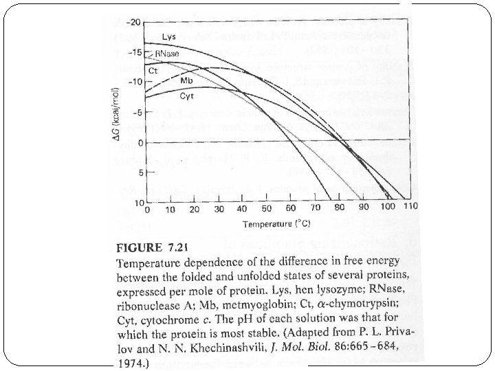
Factors Affecting Protein Stability � 1) p. H: proteins are most stable in the vicinity of their isoelectric point, p. I. In general, electrostatic interactions are believed to contribute to a small amount of the stability of the native state; however, there may be exceptions. � 2) Ligand binding: It has been known for a long time that binding ligands, e. g. inhibitors to enzymes, increases the stability of the protein. This also applies to ion binding --- many proteins bind anions in their functional sites.
Factors Affecting Protein Stability � 3) Disulfide bonds: It was observed that many extracellular proteins contained disulfide bonds; whereas intracellular proteins usually did not exhibit disulfide bonds. � In addition, for many proteins, if their disulfides are broken (i. e. reduced) and then carboxymethylated with iodoacetate, the resulting protein is denatured, i. e. unfolded, or mostly unfolded. � Disulfide bonds are believed to increase the stability of the native state by decreasing the conformational entropy of the unfolded state due to the conformational constraints imposed by cross-linking (i. e. decreasing the free energy of the unfolded state). Most protein have "loops" introduced by disulfides of about 15
Factors Affecting Protein Stability � 4) Not all residues make equal contributions to protein stability. In fact, it makes sense that interior ones, inaccessible to the solvent in the native state, should make a much greater contribution than those on the surface, which will also be solvent accessible in the unfolded state. �Proteins are very malleable, i. e. a mutation at a particular residue tends to be accommodated by changes in the position of adjacent residues, with little further propagation.
Denatured States �If the denatured state involves most residues in a fully extended peptide chain conformation, i. e. maximal solvent exposure, then substitutions involving solvent-exposed residues in the native state will have limited effect. �If, on the other hand, the denatured state have considerable residual structure, then it is also possible that mutations may affect the conformation and free energy of the unfolded state; in extreme cases, perhaps only the denatured state and not the native state!
wt m+ m-value m- � The m-value changes can be used to understand the nature of denatured state. � The effect of mutations to the protein stability can be estimated using the change of DG H 20 D-N � For some of the mutation, the m-value is changed. The different m-values related to the difference between the number of molecules of solvent bound in the native vs. denatured state. Since for the folded stated we have similar structure, the number of solvent molecules bound to the folded state is about the same, and the m-value difference reflects the different distribution of denatured state.
Different Unfolded States m+ mutant has a more exposed unfolded state than that of m - mutant. m+ mutant M- mutant smallest