Phylum Rotifera Phylum Rotifera 1850 described species most

Phylum Rotifera
")
Phylum Rotifera • 1850 described species • most live in freshwater (100’s / L) • Others live in salt water, soil, water film on moss, plankton • small (50 -500 μm) but complex • http: //www. youtube. com/watch? v=YF 8 OJt_pujc

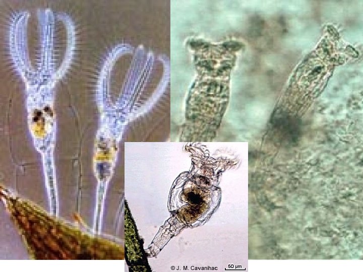
Corona Head Mastax Trophi Trunk Phylum Rotifera • 3 general regions: head, trunk, foot • pedal gland in toes glue rotifer to substrate • ciliary corona for feeding, locomotion Foot • complete digestive tract • mastax = muscular pharynx • trophus = grind food

Corona Head Mastax Trophi Trunk Foot Phylum Rotifera • protonephridia • smooth and striated muscle tissue • pseudocoelomate (good hydrostatic skeleton) • syncytial, cuticle-like epidermis (never molted) • parthenogenic

Fig. 1. Diversity of morphological adaptation in monogononts to different environments. Planktonic: 1 - Synchaeta, 2 - Polyarthra (fam. Synchaetidae); 3 - Asplanchna (fam. Asplanchnidae); 4 - Hexarthra (fam. Hexarthridae); 5 - Conochilus (fam. Conochilidae); 6, 7 - Keratella (fam. Brachionidae). Psammonic: 8 - Dicranophorus, 9 - Wierzejskiella (fam. Dicranophoridae). Phytophylic: 10 - Trichotria, 11 - Macrochaetus (fam. Trichotriidae). Periphytonic: 12 - Collotheca (fam. Collothecidae); 13 - Floscularia (fam. Flosculariidae). Parasitic: 14 - Claria (fam. Clariaidae); 15 - Balatro (fam. Dicranophoridae). Fig. 2. Unitypical morphological construction in bdelloids. 1 - Habrotrocha, 2 - Otostephanos, 3 - Ceratotrocha (fam. Habrotrochidae); 4 - Philodina, 5, 6 - Rotaria, 7, 8 - Dissotrocha, 9 - Macrotrachela, 10 - Mniobia (fam. Philodinidae); 10, 11 - Adineta (fam. Adinetidae); 12 - Philidinavus, 13 - Abrochta (fam. Philodinavidae).

A rotifer trophus

More trophi

Types of trophi for different types of foods Malleate: for gripping and grinding Forcipate: for gripping Ramate: Grinding only Incudate: seizing Uncinate: For pre-digested food Virgate: piercing and sucking

Acoelomate: e. g. platyhelminthes Coelomate: Mollusca to arthropoda, All deuterostoma Pseudocoelomate: e. g. rotifera, acanthocephala, nematoda
 Ova (2 n) mitosis Summer cycle (asexual reproduction) Ova")
Parthenogenesis Amictic females (2 n) Ova (2 n) mitosis Summer cycle (asexual reproduction) Ova (2 n) Amictic females (2 n) Spring (photoperiod, temp) Winter: Dormant zygote (2 n) Ova (1 n) diapause or cryptobiosis Sperm (1 n) Autumn cycle mitosis (sexual reproduction) Males (1 n) Mixis stimuli - photoperiod, temp - no food - overcrowded Mictic females (2 n) meiosis

P. Acanthocephala: Spiny headed worms
")
P. Acanthozoa: Spiny headed worms • 1100 spp, all parasites of vertebrates (esp fish) – Spiny head used for attachment – Lack gut • Larvae parasitize crustacea, insecta • Pseudocoelomate Intestine of eider duck with acanthocephalan parasites
.")
Acanthor Cystacanth Acanthella Acanthocephala·Fig. 2. Life cycle of common acanthocephalan species (cf. Table 1). A Macracanthorhynchus hirudinaceus; B. 1 The adults live in the intestine of their final hosts, being attached by their hooked proboscis. The penetration of the intestinal wall leads to inflamed protrusions (IP) appearing along the outer side. 2 After copulation the adult females excrete eggs for several months (patent period). These eggs are passed fully embryonated (i. e. they contain the hooked larva) with the feces of the host. 3 -6 Intermediate hosts ( Gammarus spp. or beetle larvae) become infected by ingesting infective eggs. Inside the intestine the acanthor is released from the egg (4; RA), enters the body cavity and is transformed into an larva (5). The latter matures within 60 -95 days (in M. hirudinaceus) and is described as an infective larva (). Infection of the final hosts occurs when they swallow infected intermediate hosts. The young worms reach sexual maturity within 60 -90 days in M. hirudinaceus (after 20 days in Polymorphus minutus) and start egg production (= end of prepatent period). AC, acanthor; BH, body hooks; IP, inflamed protrusion of IW; IW, intestinal wall; PH, proboscis hooks; RA, released acanthor

Acanthor Cystacanth Acanthella Acanthocephala·Fig. 3. Life cycle of two common acanthocephalan species parasitizing fish. A ; B. 1 Adults are attached to the intestinal wall of their final hosts, trouts (A) or chubs and other fish (B). 2 Fully embryonated eggs are passed with host's feces. 3 -6 Intermediate hosts (A Asellus aquaticus, B ostracod crustaceans) are infected by uptake of eggs. Inside their intestine the acanthor larva (4) is released from its enters the body cavity and becomes transformed into the acanthella larva (5). This stage differentiates to the infective larva without in about 30 -60 days (6) depending on outer conditions. Final hosts are infected by swallowing intermediate hosts. In A. anguillae a may also become involved. When bleaks and some other fish ingest intermediate hosts (Asellus aquaticus), the infective larva enters the fish viscera, but there is no further development, but quick depeneration. Infected paratenic hosts may be a second source of infection for the final host. Neoechinorhynchus rutili and A. anguillae reach sexual maturity in about 20 -30 or 40 -60 days respectively (prepatent period). Adults live only for about 2 -3 months (patent period). AC, acanthor; IP, inflamed protrusion of IW; IW, intestinal wall; LM, PH, proboscis hooks
- Slides: 17