Phylum Porifera Sponges Chapter 6 The Sponges Phylum

. • About")
. •")


 ostium 2) choanoderm 3)")


 incurent pore 2) incurrent canal")


 Porocytes (Water Flow Through Cell)")


 – Siliceous – Calcareous")


 six rayed spicules.")


- Slides: 52
Phylum Porifera: Sponges Chapter 6
The Sponges • Phylum Porifera (Latin porous, “pore”; ferre : “to bear”). • About 5, 500 living species most marine although there about 200 freshwater species.
Characteristics of Sponges • Metazoa: without true tissue. Cellular grade of complexity (Parazoa). • Adults asymmetrical or superficially radially symmetrical. • Unique flagellated cells the choanocytes that drive water through canals and chambers: the aquiferous system. • Adults sessile suspension feeders; larval stages are motile. • Reproduction sexual or asexual.
The Poriferan Bauplan • Two unique organizational attributes: – The aquiferous system – Highly totipotent nature of sponge cells
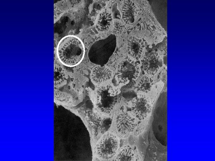
Overview of general structures • Sponges move water through their bodies using choanocytes • All cells are loosely arranged into a gelatinous matrix, the mesohyle • Water enters small holes called ostia • Water exits large holes called oscula • A skeleton helps maintain the structure of the sponge.
Body Structure and the Aquiferous System Dermal pores or ostia The mesohyl includes a non cellular coloidal mesoglea in which are embedded collagen fibers, spicules and various cells. Most of these cells are able to change from one type to another as required. 3) Water flows through the ostia all the chanels and eventually out through the osculum.
Types of Canal Systems • Most sponges can be separated based on their type of canal system. 1. Asconoid 2. Syconoid 3. Leuconoid
Asconoid sponges • Found in radially symmetrical calcarous sponges: rarely exceed 10 cm in height.
Asconoid sponges • Simple organization • Water moves through the ostia into the spongocoel • Choanoderm simple and continuous one cell thick.
Fig. 6. 3 Asconoid condition Choanoderm simple and continuous one cell thick.
Fig. 6. 3 Asconoid water flow 3 Water flow: 1) ostium 2) choanoderm 3) osculum 2 1
#1 #2 #3
Syconoid sponges • Syconoid condition: simple folding of the pinacoderm and choanoderm. • As complexity increases the mesohyl may thicken and appear to have two layers. • Water is brought in through the incurrent canals and then to radial canals (lined with choanocytes).
Fig. 6. 3 Syconoid condition Choanoderm restricted to chambers with an apopyle opening into the atrium.
Fig. 6. 3 Syconoid water flow Water flow: 1) incurent pore 2) incurrent canal 3) prosopyle 4) choanocyte chamber 5) apopyle 6) atrium 6) osculum
Leuconoid sponges • Additional folding of the choanoder and further thickening of the mesohyl. • Water is brought in through incurrent canals, and discharged through excurrent canals. • Most common type.
Fig. 6. 3 Leuconoid condition The atrium is reduced to a series of excurrent canals. Water flow: 1) dermal pore 2) incurrent canal 3) prosopyle 4) choanocyte chamber 5) apopyle 6) excurent canal 7) osculum
• The more complex a sponge condition the more particles it can filter from the water column.
Types of Cells I. III. IV. Pinacocytes (Epidermal Cell) Porocytes (Water Flow Through Cell) Choanocytes (Collar Cells) Archeocytes (Amoebocytes)
I. Pinacocytes • Cells of the external epithelium • Main functions: – Structure – Contraction
II. Porocytes • Cells which form pores • Function: to allow water flow
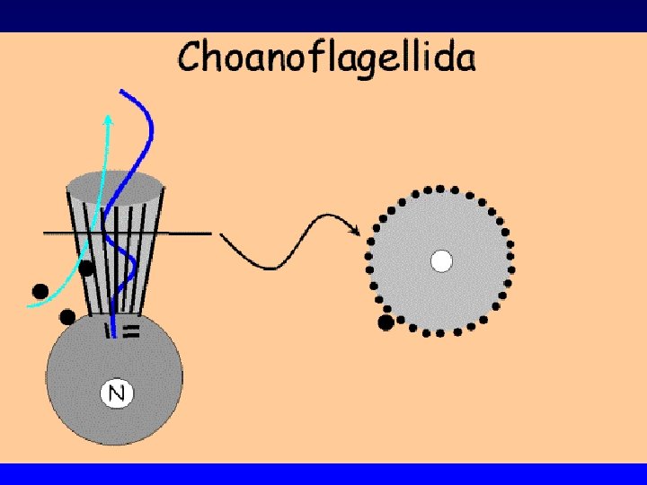
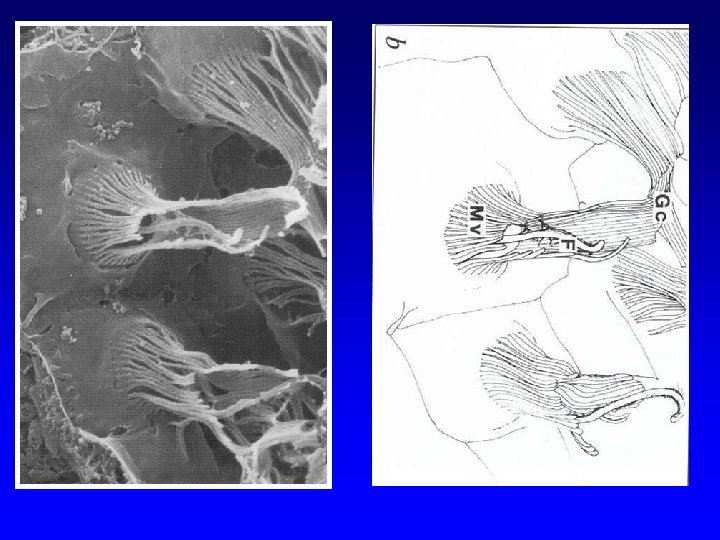
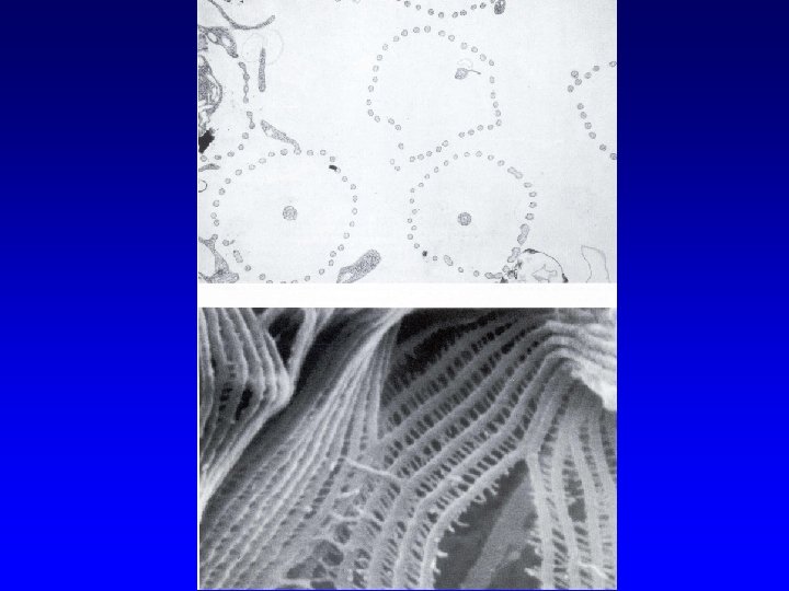
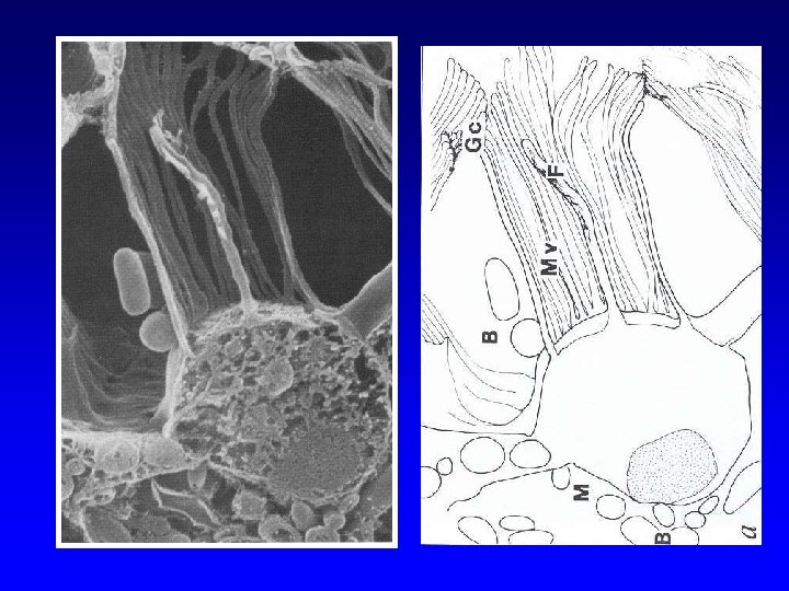
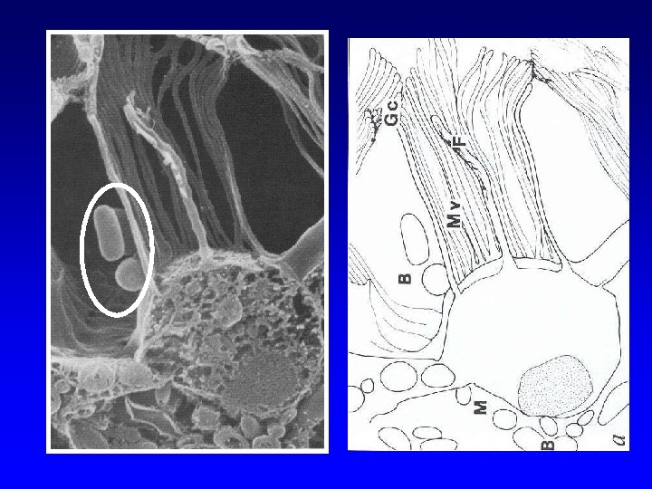
III. Chanocytes • Line the flagellated canals and chambers. • Main function: to create water flow.
Codosiga
Sponge feeding
Diameter of channels influences water flow velocity. Particles that are captured are in the 2 -5 µm range.
Predatory sponges Cladorhizid carnivorous sponge
The Harp Sponge, Chondrocladia lyra
IV. Archaeocytes • Amoeboid cells which can be non-sessile • Found in cellular matrix • Main function: – Digestion – Secrete structural components • Spongin • Spicules
Sponge feeding
Sclerocyte in the process of secreting a spicule
Keeping the mesohyle together • Spongin • Spines (spicules) – Siliceous – Calcareous
Spongin • Fibers of collagen
Spicules • Spines, when placed together form a very rigid skeleton
Megascleres And Microscleres m M
Main Groups of Sponges • P: Porifera – C: Calcarea – C: Hexactinellida – C: Desmospongiae
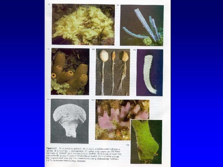
Calcarea • Calcareous sponges • Spicules composed of calcium carbonate • Small < 10 cm tall
Calcareous sponges
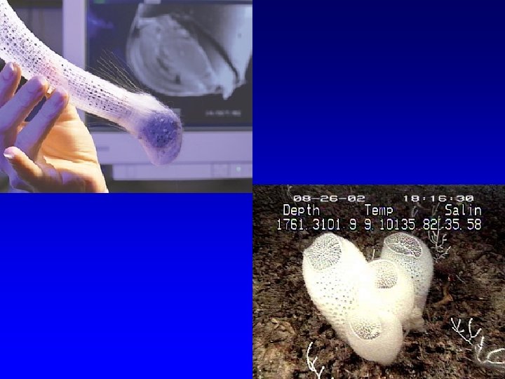
Hexactinellida • Glass sponges • Some spicules fused to form skeleton • Spicules made of glass (Siliceous spicules) six rayed • Deep water sponges
Siliceous spicules in Hexactinellida (Triaxon) six rayed spicules.
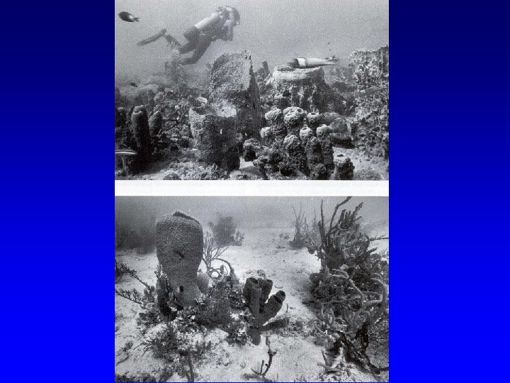
Desmospongiae • Common sponges • Skeleton is variable – Spicules – Spongin – both • Can be large
Siliceous Spiculse in Desmospongia Never six-rayed Microscleres Megascleres
Sclerospongiae Sclerosponges were first proposed as a class of sponges, in 1970 by Hartman and Goreau. Sclerosponges are sponges with a soft body that covers a hard, often massive limestone basal skeleton. Because of their long life span (500 -1, 000 years) it is thought that analysis of these sponges could extend data regarding ocean temperatures, salinity, and other variables farther into the past than has been previously possible. However, it was later found that the sclerosponges are not a monophyletic group (they are polyphyletic) and therefore this class is currently not recognized.
The Big Picture • Sponges are metazoans, but don’t have true tissues • They are an ancient group • Three main groups (taxonomic), which fall into three main structural groups • Four types of specialized cells *choanocyte*