
PHYLUM Platyhelminthes Dr Bindhusaran M D Hom Assistant
PHYLUM Platyhelminthes Dr. Bindhusaran M. D. (Hom. ) Assistant Professor Dept. of Pathology & Microbiology
Cestodes • Long segmented worms • Size few mm to several meters • Adult seen in intestine of man • Sexes are not separate • Alimentery canal is absent
TAENIA SOLIUM • pork tapeworm • Small intestine- upper jejeunum
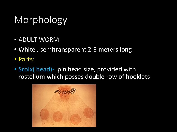
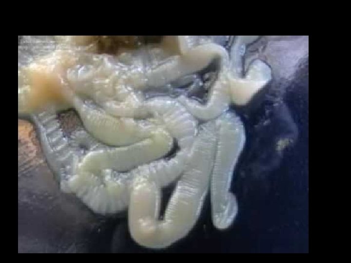
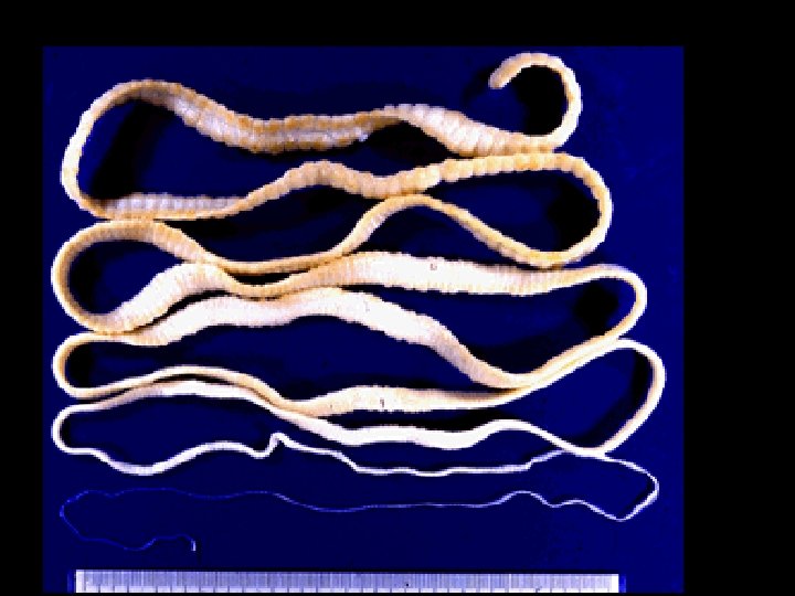
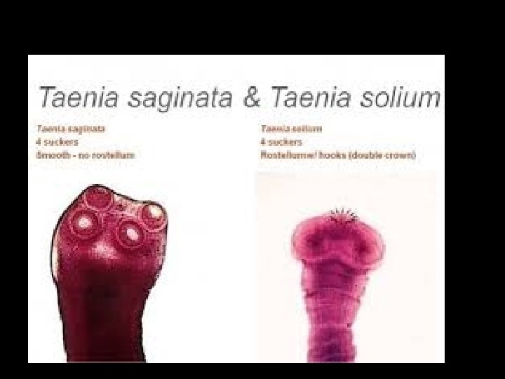
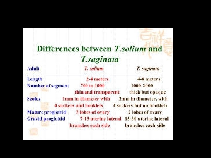
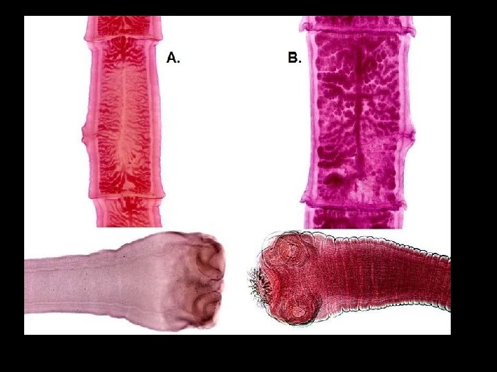
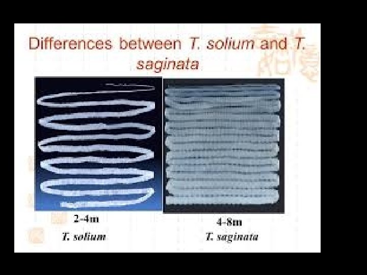
Morphology • ADULT WORM: • White , semitransparent 2 -3 meters long • Parts: • Scolx( head)- pin head size, provided with rostellum which posses double row of hooklets

morphology • Neck - short • Proglottides ( segments): 800 - 900 segments, • Immature, mature and gravid segments

segments • Gravid segment is 12 mm X 6 mm size • Common genital pore at margin which alternate iregularly between margins • Gravid uterus consist a median longitudinal stem and 5 – 10 lateral branches • Gravid segments expelled in chains • Worm has life span of 25 years
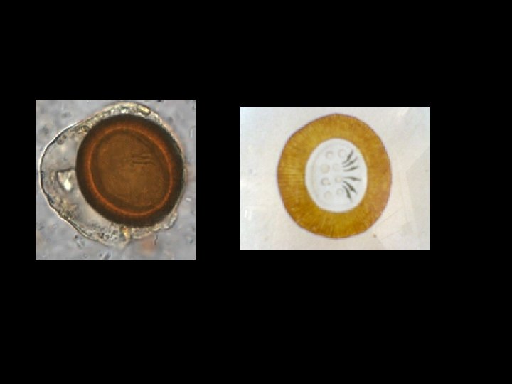
eggs • Spherical and brown in color • Thin outer shellv • Inner embryophore is brown , radially striated • Contain an oncosphere with 3 pairs of hooklets • Remain viable for 8 weeks • Infects human and pig

Life cycle • Two hosts • Man – definitive host • Pig – intermediate host
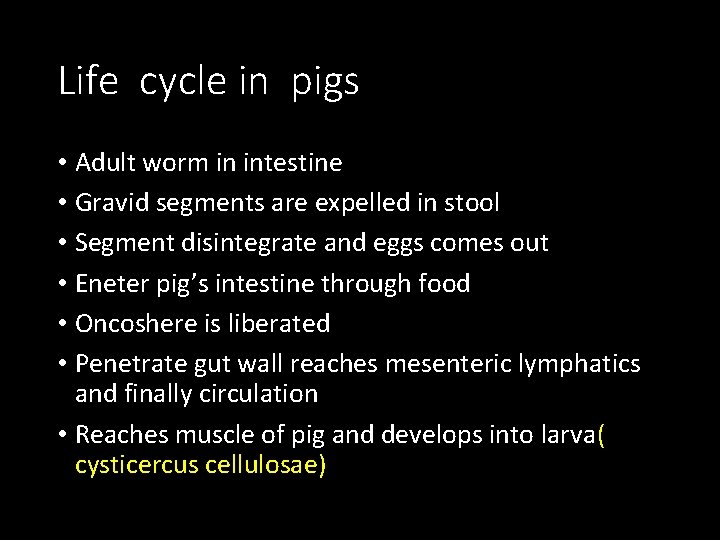
Life cycle in pigs • Adult worm in intestine • Gravid segments are expelled in stool • Segment disintegrate and eggs comes out • Eneter pig’s intestine through food • Oncoshere is liberated • Penetrate gut wall reaches mesenteric lymphatics and finally circulation • Reaches muscle of pig and develops into larva( cysticercus cellulosae)
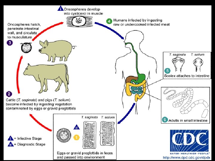
Human cycle • Infected eating infected pork with cysticercus • On coming to contact with bile exvaginates and scolex attaches to gutwall and grow into adult worm • Attain sexual maturity in 2 -3 months • Gravid segments are expelled
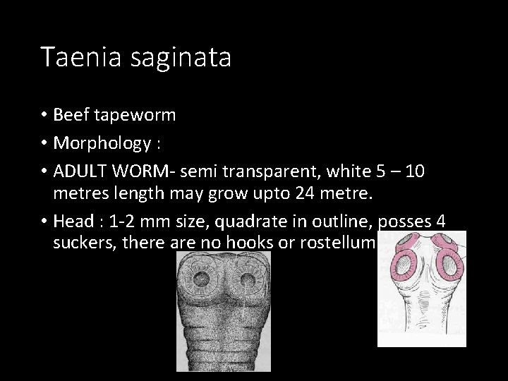
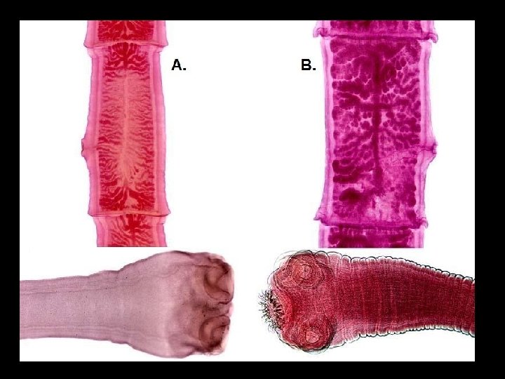
Taenia saginata • Beef tapeworm • Morphology : • ADULT WORM- semi transparent, white 5 – 10 metres length may grow upto 24 metre. • Head : 1 -2 mm size, quadrate in outline, posses 4 suckers, there are no hooks or rostellum
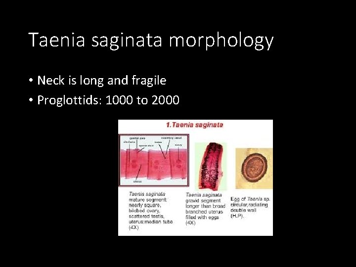
Taenia saginata morphology • Neck is long and fragile • Proglottids: 1000 to 2000
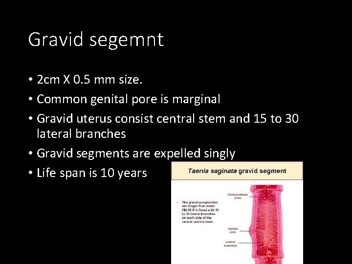
Gravid segemnt • 2 cm X 0. 5 mm size. • Common genital pore is marginal • Gravid uterus consist central stem and 15 to 30 lateral branches • Gravid segments are expelled singly • Life span is 10 years
eggs • Spherical and brown in color • Thin outer shellv • Inner embryophore is brown , radially striated • Contain an oncosphere with 3 pairs of hooklets • Remain viable for 8 weeks • Infective only to cattle
Life cycle in cattles • Adult worm in human intestine • Gravid segments are expelled in stool • In the soil Segment disintegrate and eggs comes out • Eneter cattles intestine through food while grazing • Oncoshere is liberated in the intestine • Penetrate gut wall reaches mesenteric lymphatics and finally circulation • Reaches muscle of cattle and develops into larva( cysticercus bovis)
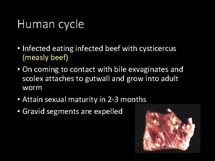
Human cycle • Infected eating infected beef with cysticercus (measly beef) • On coming to contact with bile exvaginates and scolex attaches to gutwall and grow into adult worm • Attain sexual maturity in 2 -3 months • Gravid segments are expelled
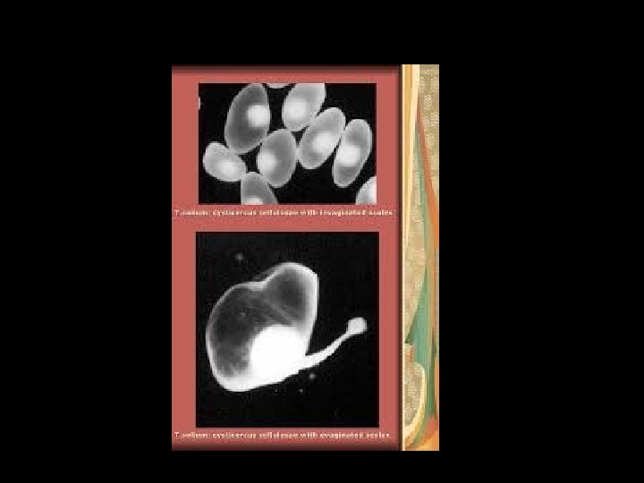
Larval development of T. saginata and T. solium • Cysticercus bovis: • This is the larval stage developing in the muscles of cow or buffalo • Measures 5 -10 mm breadth by 3 – 4 mm length • Conatin unarmed scolex • Can live for 8 month in the flesh of cattle • Can develop into adult worm when ingested by man
Cysticercus cellulosae • This is larval stage of T. solium developing in muscles of pig. • Mature cyst is opalescent ellipsoidal body and measures 8 to 10 mm width X 5 mm length • There is dense milky white spot at the side where the scolex with hooks remain invaginated( remain enclosed in a sheath) • Cyst contain fluid rich in salt and albuminous material • They can live for 8 months in pigs flesh and develops into worm when ingested by man • Cysticercus is found in man also
Pathogenesity • Adult worm usually do not produce symptoms • Occasionally vague abdominal discomfort, anaemia, chronic indigestion, diarrhea alternate constipation etc. • Larva Causes Cysticercosis
Cysticercosis • It is the development of larval stage of taenia solium the human body • by drinking contaminated water or by eating uncooked vegetables with taenia solium egg • Cysticercus cellulosae develops in human body • Clinical features depends upon the location of the cysticerci in the body
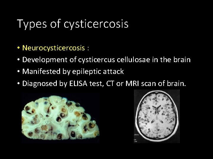
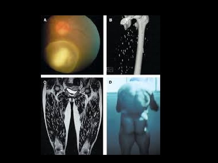
Types of cysticercosis • Neurocysticercosis : • Development of cysticercus cellulosae in the brain • Manifested by epileptic attack • Diagnosed by ELISA test, CT or MRI scan of brain.
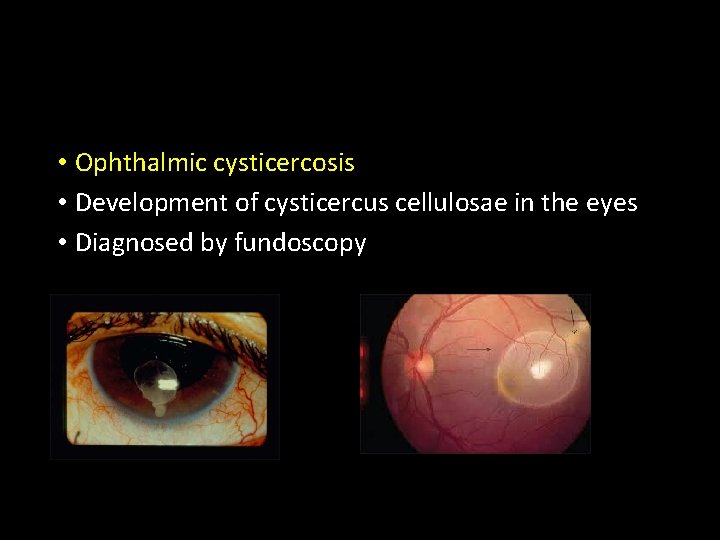
• Ophthalmic cysticercosis • Development of cysticercus cellulosae in the eyes • Diagnosed by fundoscopy
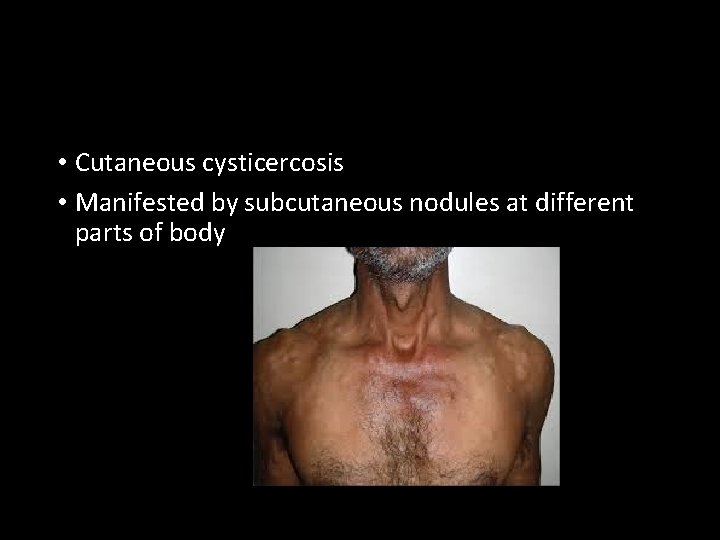
• Cutaneous cysticercosis • Manifested by subcutaneous nodules at different parts of body
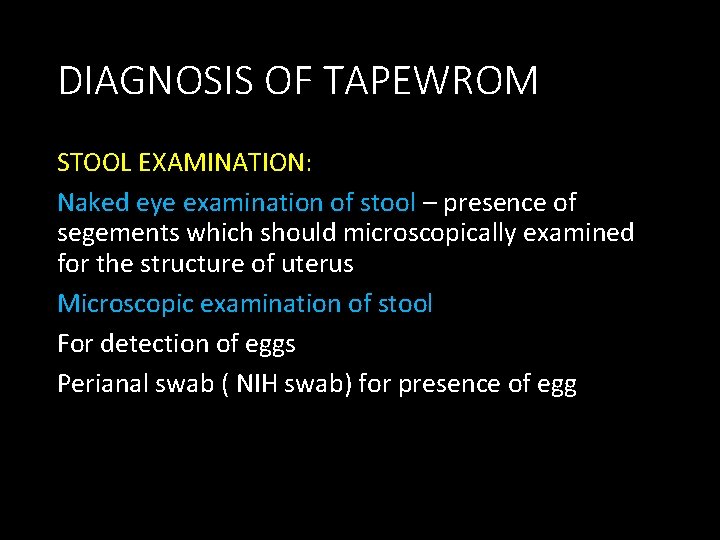
DIAGNOSIS OF TAPEWROM STOOL EXAMINATION: Naked eye examination of stool – presence of segements which should microscopically examined for the structure of uterus Microscopic examination of stool For detection of eggs Perianal swab ( NIH swab) for presence of egg
DIAGNOSIS OF CYSTICERCI • Biopsy of subcutaneous nodule contain cysticerci • X – ray examination of skull and soft tissues • CT scan of brain and affected organs • MRI of brain • Myelography to detect spinal cysticerci • Eosinophilia • Antigen can be detected in serum and CSF and ELISA
- Slides: 37