Phylum Nemathelminthes Round worms Class Nematoda General characters

 Class: Nematoda")
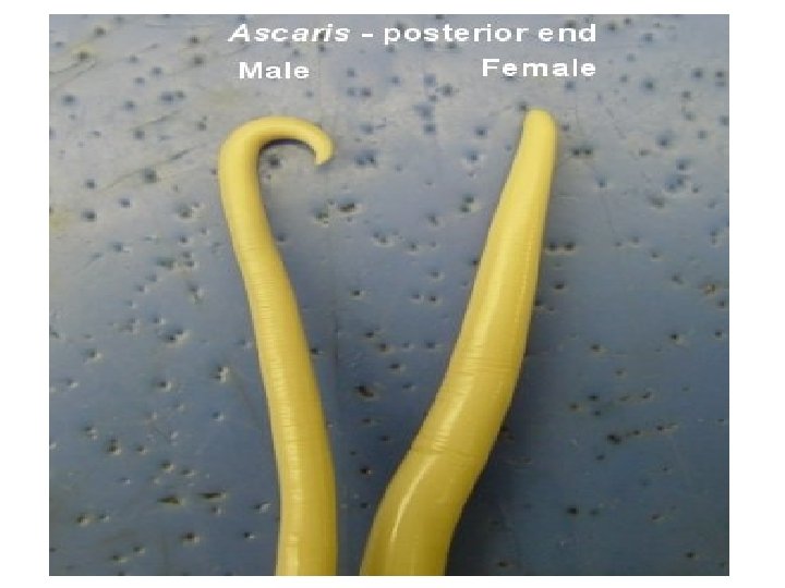
Separate sexes § Males smaller than females, and commonly has a curved")
The body wall is made up of 3 layers: a. Outer laminated non cellular")
 Digestive system: § A straight tube starting at the anterior head end, by")


 Reproductive organs: Male: curved posterior end. - In the form of single coiled")


 By ingestion of eggs: o Directly infective when passed in")


 Ascaris lumbricoides (The giant intestinal round worm) Distribution: § Cosmopolitan (more")
 Long cylindrical with tapering ends. 2) Pink or yellowish creamy in")


. Each")


: 2 mg/kg as a single oral dose Mebendazole (Antiver,")


 Geographical distribution: o Cosmopolitan, more common in children than")


 o the male dies after")


, Pyrantel pamoate (Combantrin) or Flubendazole (Fluvermol): as single oral dose")


 Whipworm Geographical distribution: • Cosmopolitan, more common in worm moist")


 or Flubendazole (Fluvermal). Control: – Treatment of infected")
- Slides: 84
Phylum: Nemathelminthes (Round worms) Class: Nematoda
General characters: 1)Separate sexes § Males smaller than females, and commonly has a curved posterior end. 2)Un-segmented, elongated cylindrical, round worms. 3)Body cavity containing body fluid, in which the digestive and genital systems float. 4)Size: varies from less than one mm to nearly one meter in length.
5)The body wall is made up of 3 layers: a. Outer laminated non cellular cuticle; which may inflate anteriorly forming cervical alae (Enterobius vermicularis) or posteriorly forming copulatory bursa (Ancytostoma). On the cuticle there may be papillae, expansions or spines. b. Thin syncytial layer or hypodermis that secretes the cuticle. c. Single muscular layer, divided into 4 quadrants by the hypodermal lines, its arrangement varies in different nematodes and helps in classification.
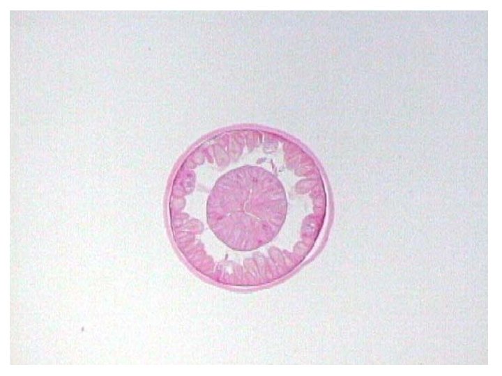
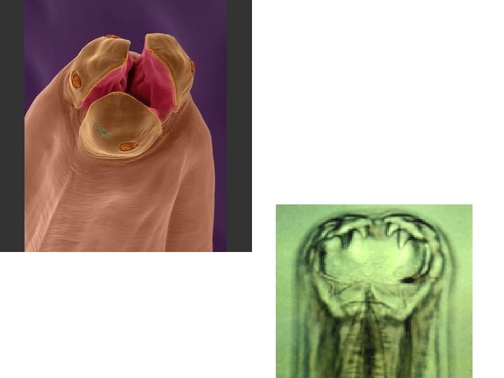
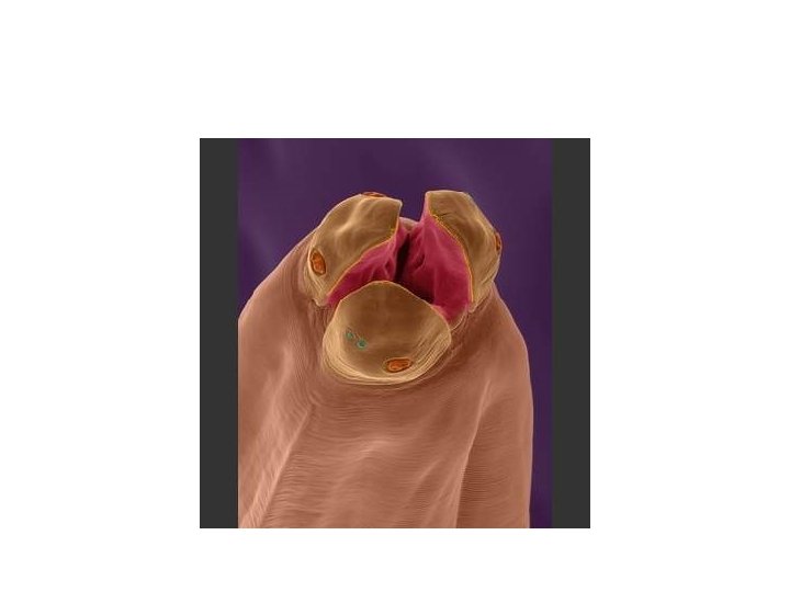
6) Digestive system: § A straight tube starting at the anterior head end, by the mouth which is provided with facilities for attachment e. g lips, teeth, and plates which ends by the anus posteriorly. § Mouth leads to oesophagus, with tri-radiate lumen (one dorsal and two subventral) , varies in shapes according to the species of the nematode parasite, and helps in identification.
Types of oesophagus: i-Cellular esophagus: a narrow of cells attached to the esophagus (Trichocephalus trichiuris , Trichinella spiralis & Capallaria). ii – Muscular: simple tube surrounded with a straited muscular wall: a) One segment: i- Club- shaped oesophagus (Ascaris). ii- Cylindrical esophagus (Filariae)
Club- shaped Cylindrical
b. Two segments: 1 - Double – bulbed oesophagus, with anterior club shaped portion and posterior spherical part (Oxyuris = Enterobius vermicularis) 2 - Rhabditiform oesophagus, with an anterior cylindrical portion and pyriform posterior portion and a narrow constriction in between (Strongyloides stercoralis).
Double – bulbed Rhabditiform
- After the esophagus is the intestine which proceeds until it opens in the anus on the ventral surface in female or it joins the genital duct which open in cloaca in male (subterminal except in Trichiuris and Trichinella).
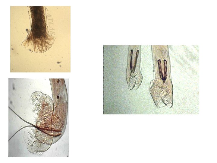
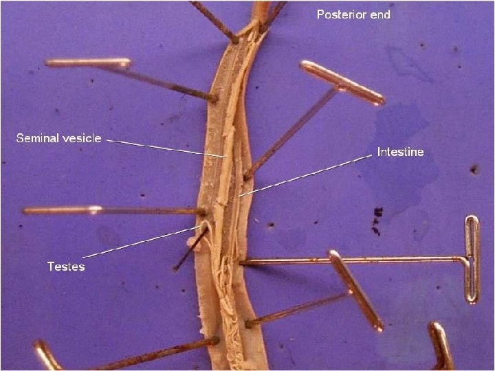
7) Reproductive organs: Male: curved posterior end. - In the form of single coiled convoluted tubule, Testes followed by vas deferens, seminal vesicle and finally opens by ejaculatory duct with the anus in the cloaca posteriorly. - The cuticle may show expansion (Copulatory bursa) with or without spicules that help in differentiation.
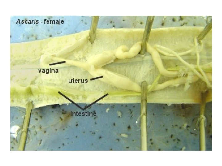
Female: - straight posterior end One or 2 genital systems joining to open by a single vagina in the vulva. - Each has a glandular ovary→ oviduct→ receptaculum seminis →uterus and vagina.
v Egg of parasitic nematode may hatch in the external environment and controlled by suitable factors as temperature, moisture and oxygen. Or it may hatch after ingested by the host and stimulated by carbon dioxide tension, salts, PH and temperature. In both types larva undergoes 4 molts until it becomes adult.
8 -Excretory system : consists of two longitudinal lateral canals running in the lateral lines. -the lateral canals join in terminal duct→ excretory pore in the region of the oesophagus. 9 - No circulatory system : the fluid in the body cavity contain heamoglobin, glucose, proteins, salts and vitamins that fulfill the function of blood. 10 - Nervous system: consists of nerve ring around the esophagus and 6 nerve trunks pass to the head and others to the posterior end. -Sensory organs are in the labial, cervical, anal & genital regions.
Transmission of nematodes 1) By ingestion of eggs: o Directly infective when passed in stool (Enterobius). o Infective after a period of maturation outside (Ascaris and Trichocephalus), by the larvated eggs. 2) Ingestion of larvae: o Larva itself: in Trichostrongylus colubriformes and Hookworms through food and drink. o Larva in muscles: Trichina capsule in muscles of pigs. o Larva in insect: Medina worm in cyclops. 3) Penetration of skin: o Larval penetration: Hook worm and Strongyloides. o Insect bite: Filarial worms.
Human nematodes are divided according to the habitat of adult worms into: Intestinal nematodes Small intestine: 1. Ascaris lumbricoides. 2. Hookworms (Ancylostoma duodenal and Necator americanus). 3. Strongyloides stercoralis (Dwarf thread worm). 4. Trichostrongylus colubriformis. 5. Capillaria philippinensis. 6. Trichinella spiralis. (considered as both intestinal and tissue nematode). •
Large intestine: 1. Enterobius vermicularis. 2. Trichiuris trichiura. Tissue nematodes 1. 2. 3. 4. Filariae. Dracunculus medinensis (Medina worm). Larva migrans (visceral and cutaneous). Trichinella spiralis (considered as both intestinal and tissue nematode).
Intestinal nematodes 1) Ascaris lumbricoides (The giant intestinal round worm) Distribution: § Cosmopolitan (more prevalent) in warm regions with poor sanitation. § Affect all ages but children are more commonly and heavily infected by their frequent exposure to contaminated soil.
Adult morphology: 1) Long cylindrical with tapering ends. 2) Pink or yellowish creamy in color. 3) Finely striated cuticle. 4) Mouth: at the anterior and, with 3 lips; one dorsal and two subventral with a small triangular buccal cavity. Each lip is provided with 2 sensory papillae and fine teeth. 5) Esophagus: muscular club- shaped followed by the intestine to open in anus in female and cloaca in male.
Female: o 20 -40 cm × 6 mm o with straight posterior end o two sets of genitalia o vulva at the junction of anterior and middle thirds of the body ventrally. Male: o 15 -20 cm × 3 mm o posterior end curved ventrally o one set of genital organs and 2 small equal spicules
Ascaris male posterior end
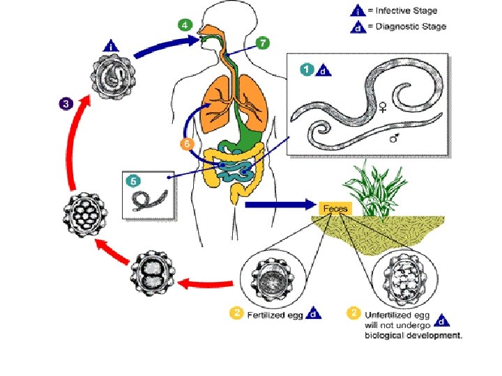
Life Cycle: Habitat: the adult worms live free in the small intestine (jejunum). Each female lays 200. 000 eggs per day regularly. Its life span is about one year. Definitive host: man. Infective stage: stage egg rhabditiform larva. containing a second stage Stages in the life cycle: egg in the soil larvated egg (with 2 nd stage rhabditiform larva) , ingested by man larva adult in the small intestine.
Size Shape Fertilized Unfertilized 60 × 45 µ 90 × 45 µ Oval with 2 coverings: 1. Outer thick regular albuminous mammillations. 2. Inner thick egg shell. Long and narrow Similar to 1. Less developed fertilized egg thin irregular except that the mammillated albuminous mammillations. layer is lost. 2. thin egg shell Color Brownish Content Immature ovum (one cell stage) Decorticated brownish Refractile granules
N. B. The unfertilized eggs are found not only in the absence of males but in about two fifths of all infections, since repeated copulations are necessary for the continuous production of fertile eggs.
Ascaris lumbricoides -- the human round worm
Ascaris • Fully embryonated eggs are swallowed and L 2 hatches in the stomach and penetrate stomach or duodenal mucosa • Larvae enter blood stream and leave through alveoli into lung • Larvae molt several times in the lungs L 3/L 4 move up and get swallowed • 2 -3 months after infection the adult worms start laying eggs (200, 000 daily) • Eggs are shed with the feces and embryonate within 2 -3 weeks
Eggs are voided with stool, mature in the soil after 2 weeks under suitable conditions (25ºc, humidity, shade and oxygen), develops 1 st stage Rhabditiform larva within the shell. After one more week, the larva molts into 2 nd stage rhabditiform larva within the egg (infective stage).
Methods of infection: v. Ingestion of mature eggs containing 2 nd stage rhabditiform larva, contaminating food (green raw vegetables), water or hands. Cockroaches and house flies may carry the larvated eggs to human food (mechanical transmission). v. Inhalation of mature eggs to the nasopharynx.
- Egg hatches in the small intestine, liberating the larva (freshly laid eggs with no larva inside are not capable of causing infection). - Rhabditiform larva comes out of the egg to the lumen of the small intestine, penetrate the mucosa to reach the circulation, carried to the right side of the heart pulmonary arteries lung. The larva breaks out of capillaries into lung alveoli and moults twice (2 nd and 3 rd moults).
- The larva creeps along the bronchioles bronchi trachea larynx pharynx, then swallowed to reach the small intestine where it moults once (4 th moult and becomes adult). - Eggs appear in feaces 2 -3 months after infection. - The life span of a female Ascaris in human body is about one year, then dies. So if a person shows infection with Ascaris for several years, it only means that this person is continuously re-infected.
N. B. In massive infection, some larvae may reach the general circulation to be filtered in various organs as abnormal foci in: lymph glands, spleen, kidneys, brain or spinal cord. When reaching the kidney they may find their way to urine and attract attention. In such ectopic sites, larvae are unable to grow to maturity and most of them are destroyed.
Pathogenesis and Clinical Manifestations Disease: Ascariais: The usual infection consisting of 5 – 10 worms, often goes unnoticed by the host and is discovered on a routine stool examination or by the discovery of an adult worm passed spontaneously in stool. The pathogenic effects of ascariasis are due to the following mechanisms: 1. Allergic reactions to the parasitic stages. 2. Effect of the adult on host nutrition. 3. Mechanical effect of the adult worm. 4. Wandering larvae and adults. 5. Microorganisms carried with larvae during migration.
v. Migrating larvae: 1. Lung: In light infection, there is slight damage with unnoticed pathological lesions. In heavy infection, the migrating larvae in the lungs result in lobular pneumonitis, there is cellular infiltration, serous exudates and some haemorrhage causing cough and bronchial irritation, (asthmatic attack), expectoration with blood stained sputum and aedema of lips, microscopically the larvae may be detected in the sputum, with many oesinophils. 2. General circulation: Occasionally some larvae reach the general circulation and distributed to various organs as lymph nodes, brain, spleen & kidneys (ectopic
v. Adult worm: in the intestine may cause abdominal discomfort with distension, colic, diarrhea or constipation, vomiting and dyspepsia due to production of anti-enzymes (anti-peptic and anti- tryptic substances that interfere with protein digestion). 1. Traumatic effects: a. In heavy infection intestinal obstruction. b. Obstruction of the bile ducts by the worms obstructive jaundice. c. Appendix appendicitis. d. Obstruction of ampulla of Vater acute hemorrhagic pancreatitis. e. Perforation of intestinal wall peritonitis.
f. Some worms may ascend via the stomach and oesophagus to the nasopharynx, enter the larynx causing suffocation especially in children. g. It may come out of mouth or nose or even go to Eustachian tube from the pharynx resulting in damage of the middle ear. 2. Toxic dead effects: metabolic by -products of living or worms may give rise to fever, allergic manifestations (urticaria and asthma) and nervous irritability (insomnia and even convulsions) 3. Nutritional impact: loss of appetite malnutrition and impairment of growth, with vitamin A and C deficiency.
Ascaris • Infection depends on fecal contamination of food, water or soil • Eggs are sensitive to sun light but otherwise extraordinarily resistant (ascarosides - special glycolipids secreted by the embryo) • Fertilized eggs are shorter and rounder than unfertilized
Ascaris • Occasional pulmonary symptoms • Intestinal phase mostly asymptomatic, but worms can lead to malnourishment in children • Dangerous complications are mostly observed in children under 10 • Volvulus, a mass of knotted worms obstructing the intestine • Penetration of the bile duct and liver by adult worms • Penetration of the intestinal wall, followed by peritonitis • Wandering and obstruction can be linked to certain medications
Diagnosis v. Clinical: symptom of intestinal ascariasis are indistinguishable from those of other intestinal helminthic infections. v. Laboratory 1. Detection of eggs in stool. (direct smear, after concentration, Stoll’s technique). 2. Detection of migrating larvae in sputum or better in gastric lavage contents. 3. Detection of adults passing out with or without stool or in vomitus. 4. Eosinophilia (7 – 12%). 5. Radiology: Barium meal shows cylindrical filling defect (string sign). 6. Biopsy.
Treatment Drugs Levamizol hydrochloride (Ketrax): 2 mg/kg as a single oral dose Mebendazole (Antiver, vermox) or Flubendazole (Fluvermal). Piperazine citrate, hydrate or adipate (parazine, vermizine or uvilon). Surgical For treatment of complications e. g intestinal obstruction, obstruction of appendix or bile ducts.
Control 1. 2. 3. 4. 5. 6. 7. 8. Mass treatment of infected persons. Sanitary disposal of excreta. Health education. Cleanliness (washing hands before meal). Proper washing of green raw vegetables. Pure water supply. Control of flies and other insects. Stool should not be used as a fertilizer unless being treated by chemicals or temperature of 50°C or higher to kill eggs.
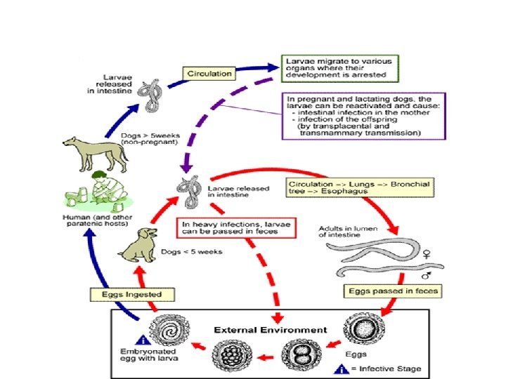
Toxocara canis & Toxocara cati These two species are ascarid parasites of dogs and cats. They are cosmopolitan. Both are similar in biology and morphology Adult Toxocara has three lips and a pair of cervical alae. Mature male: 4 -6 cm. Mature female: 6 -10 cm. Toxocara eggs: Dark brown with pitted shells Size: 85 × 75 Passes immature in dogs’ stool.
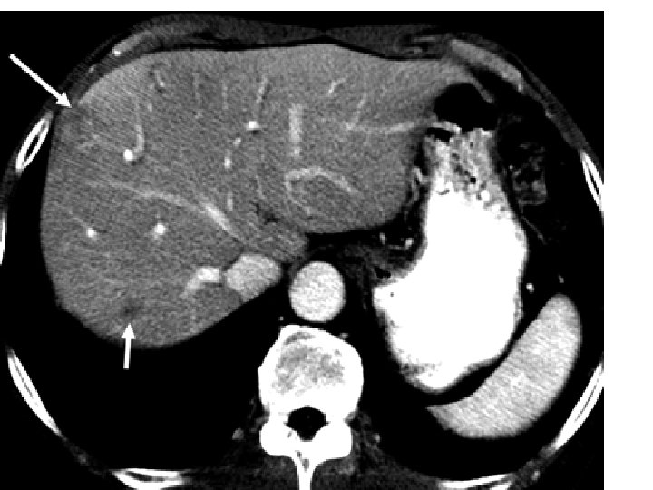
Life cycle: – Puppies are infected through mother’s milk soon after birth or prenatally. – They remain susceptible to reinfection until sexually mature, then acquire some resistance to reinfection. Mode of infection in man: • Accidental swallowing of larvated eggs of Toxocaca by man with contaminated food , drink or hands Visceral larva migrans. • The 2 nd stage rhabditiform larvae hatch in the small intestine, pierce the mucous membrane and are carried with blood to the liver, lungs, brain, eyes, heart and other tissues where they produce eosinophilic granulomatous lesions. Larvae remain for several weeks or months without any growth or development till they die.
Enterobius vermicularis (Oxyuris, pinworm, seatworm) Geographical distribution: o Cosmopolitan, more common in children than adults. o More common in temperate and cold climate than hot climate because of less frequent bathing and changing of under wears.
Morphology: o. Translucent cuticle, finely transversely striated. o. There are 2 wings like expansions (cervical alae) at the anterior end. The mouth with 3 small retractile lips, followed by small buccal cavity. o. The oesophagus: double- bulbed. Intestine opens by the anus ventrally, some distance from the posterior end. Female: 1 cm with a long thin sharply pointed tail occupying about 1/3 total length (hence the common name pin worm), 2 sets of genital organs and vulva at the junction of the anterior fourth with the rest of the body. Male: shorter than female 0. 5 cm, its posterior end curved ventrally, one set of genital organs that open with the anus in the cloaca and one spoon- shaped spicule.
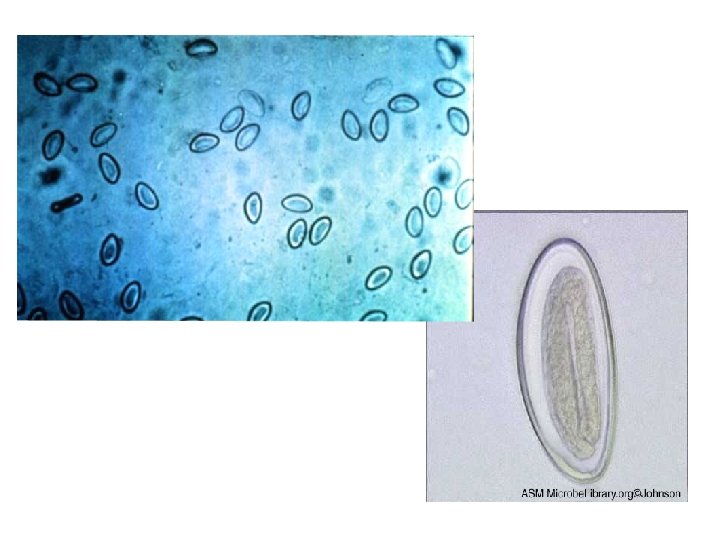
Eggs: • Size: 50 x 25µ • Shape: plano-convex • Shell: 2 layers and covered by a 3 rd outer thin albuminous sticky layer. • Colour : colourless. • Contents: fully developed larva.
female
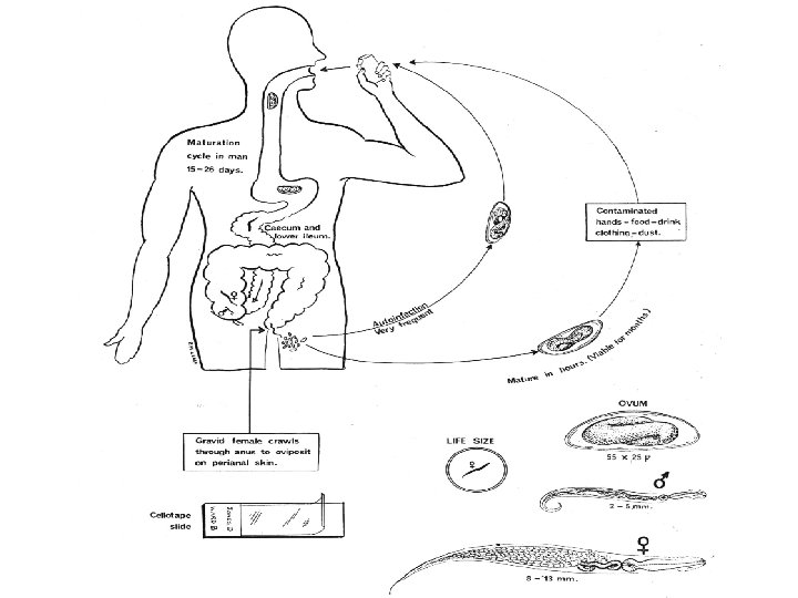
Life cycle: o Habitat: Adult worm live in the caecum, appendix and adjacent parts of small and large intestine. o Definitive host: Only man. o Infective stage: fully embryonated eggs containing fully developed larvae when laid, so eggs are immediately capable of starting infection when swallowed.
o The adults are short lived (40 -50 days) o the male dies after fertilization and expelled with feces o the gravid fertilized female migrate down the bowel (especially at night) when eggs are fully developed and come out of the anus to lay eggs around the anus and peri-anal region. o Each female deposits about 10. 000 eggs then dies after laying all its eggs. So continuous infection means repeated swallowing of eggs causing infection to go on. o It takes 2 -4 weeks to complete the life cycle (from time of swallowing eggs till eggs laid by the female).
Mode of infection: o Ingestion of eggs through contaminated food and drink. o Autoinfection: movements of female to peri-anal region at time of egg deposition causes intense irritation and itching eggs are carried under finger nails to the mouth after scratching of peri-anal skin (anus to mouth infection), this can be discovered in about one- third of infected children. o Retro-infection: eggs hatch on the peri-anal region and larvae migrate back through the anus to the rectum and caecum. o Air- borne infection: eggs can fall in the underwear and in the sheets and blankets. o Contact with patients (Direct hand to hand or indirect contact by handling contaminated articles as clothes, bed linens, toilet seats, door knobs )
Pathogenisis and clinical picture of Enterobiasis, Oxyuriasis: o. Perianal irritation, with nocturnal itching and enuresis, insomnia, irritability, restlessness, neurosis and inability to concentrate o. Pruritus ani due to: 1 - Nocturnal migration of the female worm on the perianal skin 2 - Skin sensitization due to worms ruptured during scratching 3 - Straitions on the cuticle. 4 - Sticky material on the eggs.
Pathogenisis and clinical picture – Vaginitis and salpingitis, with irritation, vaginal discharge, and even granuloma around eggs or worms. – Irritation of intestinal mucosa with minute ulcers, haemorrhage and 2 ry bacterial infection at site of attachment. – Obstructive appendicitis.
Diagnosis: o Clinical. o Laboratory: • Detection of adult worms in feces or on the peri-anal region. • Detection of eggs: a. In stool: seldom found, unless uterus of gravid female ruptures during migration to the peri-anal region. b. In urine of female patient. c. On peri-anal region by swab, which must be done early in the morning before defecation or bathing and should be repeated for several days before the patient is considered free.
Types of swabs: –Scotch adhesive tape swab. –Vaseline swab. –N. I. H. (National Institute of Health) swab.
Treatment: o Mebendazole (vermox), Pyrantel pamoate (Combantrin) or Flubendazole (Fluvermol): as single oral dose * a 2 nd dose must be given after 2 weeks to prevent re-infection. o Local: mercurial ointment is applied to the perianal skin especially at night relieve itching and kills females that come out to deposit eggs and prevent dispersal of eggs.
Control: – Mass treatment of the whole companions of the infected person. – Personal cleanliness. – Protection of food and drink from contamination by dust and hands of patients.
Adult Pinworm:
Life Cycle:
Females in perianal region:
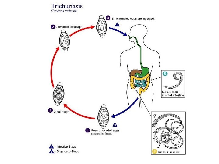
Trichuris trichiura (Trichocephalus trichiurus) Whipworm Geographical distribution: • Cosmopolitan, more common in worm moist regions. • More sensitive to the effect of desiccation and direct sun light than Ascaris.
Morphology : Adult body is demarcated into • an anterior thin part or whip-like (3/5) contains a cellular oesophagus, and • a posterior thick part bluntly rounded (2/5) contains the rest of organs. Male : 3 -4 cm. in length • coiled posterior end • single copulatory spicule inside a retractile sheath • terminal cloaca. Female : 4 -5 cm. in length • straight blunt posterior end • has one set of genitalia • the vulva opens at the junction of thin and thick parts. • Anal orifice is terminal
FEMALE
T. trichiurus egg • Barrel-shaped. • Yellowish-brown. • 50 X 25 µ. • Thick-shell. • Bipolar mucoid plugs. • Contains an immature embryo. • The egg requires 3 -5 weeks for the larva to develop inside and become infective.
Life Cycle • Habitat: large intestine mainly the caecum but is also found in the appendix and lower ileum. • Definitive host : man. • Reservoir host: some mammals. • Stages in the life cycle: Egg→larva → adult. • Infective stage: egg containing first stage larva. • Method of infection: Ingestion of larvated egg with contaminated food and drinks. • The female is oviparous. • Number of eggs deposited/female/day is about 2000 eggs. Ova pass with stool 2 months after infection. • Life span: 4 -6 years.
The adult parasite T. trichiurus deeply embedding its thin anterior portion into the sub-mucosa.
Pathogenicity and clinical manifestations Trichuriasis: o Light infection is asymptomatic. o Heavy chronic infection manifests with: • Frequent, small, blood-streaked diarrheal stools. • Abdominal pain and tenderness. • Dysentery. • Rectal prolapse. • Hypo- or hyperchromic anemia. ? ? • Appendicitis. • Protein loosing enteropathy. • Intestinal wall perforation and peritonitis. • Eosinophilia (30 -60 %) in acute heavy infection. of
Diagnosis: o Clinical: difficult. o Laboratory. • Stool examination for the characteristic egg (diagnostic stage). Ova should be quantified since light infections may not require treatment. • Proctoscopy: The worms can be seen attached to the inflamed and ulcerated rectal mucosa.
Treatment: – Mebendazole (Vermox or Antivir) or Flubendazole (Fluvermal). Control: – Treatment of infected patients. – Sanitary disposal of human stool. – Strict hygienic measures for hands, food and drink. – Control of house fly.