Phylum Ciliophora Group of protozoa characterized by head

Phylum - Ciliophora Group of protozoa characterized by head like structure called cilium which is important in : A – movement B - nutrition. Genus –Balantidium This parasite have two species : - Balantidium suis- infect the pigs Balantidium coli- infect the human. This disease is regarded as a zoonotic disease. Disease : Balantidiosis B. coli : - is the largest intestinal protozoa that effect human and can be seen macroscopically.

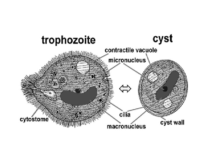
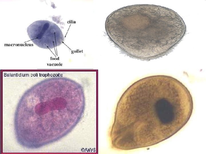
Morphology : - the parasite is observed in two stages only which are : - A – Trophozoite : 1 - Large size (40 µm to more than 70 µm). 2 - called the invasive stage. 3 - ovoidal in shape. 4 - covered by cilia & the anterior end are longer than posterior end. 5 - in the anterior end there is mouth called cytosome.

6 - the internal structures : A- have two nuclei , the macronucleus (the larger one) & micronucleus (the smaller one ) B- the macronucleus is kidney or bean in shape. C- the micronucleus is small spherical shape located in the concavity of macronucleus. D- have two secretory contractile vacuoles. E- numerous food vacuoles.

B- Cyst : 1 - typically spherical in shape. 2 - surrounded by thick wall (one or two layers ) by encystation in small intestine to protect the parasite from the host. 3 - contain the macronucleus and contractile vacuoles are visible in the cyst. 4 - the cilia are undetectable , only the roots may present.

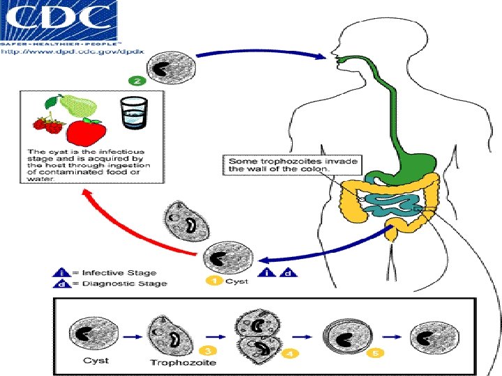
Multiplication : - occur by two methods: 1 - Asexual type by binary fission. 2 - Sexual type by conjugation. Life cycle : - direct life cycle or simple life cycle that the parasite does not need intermediate host. Mature cysts are passed with feces.
")
Transmission : - by ingestion of contaminated food or water (NOT in undercooked meat) containing the mature cysts. Excystation occurs in the small intestine, and the trophozoites colonize in the large intestine. Trophozoites undergo encystation to produce infective cysts. Cysts are the infective stage of this parasite.

Life Cycle – B coli Infective form: Cysts Mucosal & submucosal ulcers Ingestion: Faeco-oral No extra intestinal invasion Stomach: Resist acid Diarrhoea / Dysentery Small Intestine - excyst Large intestine – trophozoites Large intestine: encyst Passed out in feces

Pathogenesis • Balantidium coli produces proteolytic enzymes that break down and digest the intestinal epithelium. • Colon ulceration develops which allows for infiltration by lymphocytes and leukocytes. • Hemorrhaging and secondary bacterial infections will develop consequently • Perforation of the large intestine/colon and appendix occurs

Clinical Signs • Mild infections occur with diarrhea, • Abdominal pain • Alternating periods of constipation • Ulceration of the gut wall Diagnosis • Formed stools- Cysts • Diarrheic stools- Active Trophozoites

Diagnosis • Formed stools- Cysts • Diarrheic stools- Active Trophozoites Stool microscopy –in saline preparation , motile trophozoite -fresh diarrheic stool, Cyst-found occasionally in formed stool • When stool examination is negative-scrapings or biopsy specimens –sigmoidoscopy –useful in suspected cases

Treatment : Tetracycline -500 mg –four times -10 days Metronidazole/ Tinidazole -750 mg-three times-5 days

Prevention: Protection against contaminated food and water with feces containing cysts Personal hygiene, avoid food & water contamination, drink safe water

- Slides: 16