
Pheocromocytoma An Update in Genetic Profiling Diagnosis Treatment

- Slides: 51
Pheocromocytoma: An Update in Genetic Profiling, Diagnosis, Treatment Roula BOU KHALIL Endocrinology Division SGHUMC Assistant Professor, Balamand University
OUTLINE �Overview �Epidemiology �Updates in Genetics �Diagnosis (biochemical and imaging) �Treatment
OVERVIEW
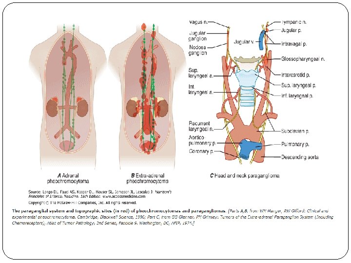
OVERVIEW �Pheochromocytoma is a tumor arising from adrenomedullary chromaffin cells that commonly produces one or more catecholamines: epinephrine, norepinephrine, and dopamine �Rarely, these tumors are biochemically silent. �Paraganglioma is a tumor derived from extra- adrenal chromaffin cells Ø sympathetic paravertebral ganglia of thorax, abdomen, and pelvis Ø parasympathetic ganglia located along the glossopharyngeal and vagal nerves in the neck and at the base of the skull, these do not produce catecholamines
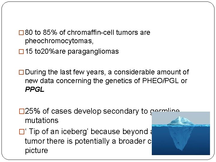
� 80 to 85% of chromaffin-cell tumors are pheochromocytomas, � 15 to 20%are paragangliomas � During the last few years, a considerable amount of new data concerning the genetics of PHEO/PGL or PPGL � 25% of cases develop secondary to germline mutations �‘ Tip of an iceberg’ because beyond a single tumor there is potentially a broader clinical picture
EPIDEMIOLOGY
� Prevalence of PPGL is not precisely known � Annual incidence of pheochromocytoma is approximately 0. 8 per 100, 000 person years � Prevalence of PPGL in patients with hypertension in general outpatient clinics varies between 0. 2 and 0. 6% � Autopsy studies demonstrate undiagnosed tumors in 0. 05– 0. 1% � In children with hypertension, prevalence of PPGL is approximately 1. 7% � Nearly 5% of patients with incidentally discovered adrenal masses on anatomical imaging prove to have a pheochromocytoma
� Equally in men and women �Mean age at diagnosis 4 th – 5 th decades � large number of patients have non classic symptoms such as abdominal pain, vomiting, dyspnea, heart failure, hypotension, or sudden death, suggesting that the majority of PHEOs are not diagnosed during life � Most PHEOs are sporadic with prevalence of malignancy 9% � About 10% of patients with PHEOs present with metastatic disease at the time of their initial work-up
�At least one-third of all patients with PPGLs have disease-causing germline mutations (inherited mutations present in all cells of the body) �The prevalence of PPGL in individuals carrying a germline mutation in PPGL susceptibility genes may be around 50%. �Patients with hereditary PPGLs typically present with multifocal disease and at a younger age than those with sporadic neoplasms
CLINICAL IMPORTANCE OF DIAGNOSIS � Cardiovascular morbidity and mortality due to excess catecholamines if left untreated � PPGL may enlarge mass effect � For familial disease, detection of a tumor in the proband may result in earlier diagnosis and treatment in other family members � Some PPGLs have malignant potential, defined as the presence of metastases in nonchromaffin tissue � Mutations in the gene encoding SDH subunit B (SDHB) can lead to metastatic disease in 40% or more
When to suspect pheochromocytoma? � Hyperadrenergic spells � Resistant hypertension � A familial syndrome that predisposes to catecholamine-secreting tumors (eg, MEN 2, NF 1, VHL) � A family history of PPGL � An incidentally discovered adrenal mass � Hypertension and new onset or atypical diabetes mellitus � Pressor response during anesthesia, surgery, or angiography � Onset of hypertension at a young age (eg, <20 years) � Idiopathic dilated cardiomyopathy � A history of gastric stromal tumor or pulmonary chondromas (Carney triad)
GENETICS
� Neurofibromatosis 1 (NF 1) � Von Hippel–Lindau (VHL) � Multiple endocrine neoplasia type 2 (MEN 2 A & MEN 2 B) � PPGL syndromes based on mutations of the genes for succinate dehydrogenase subunit D (SDHD), B (SDHB), and C (SDHC) � Others
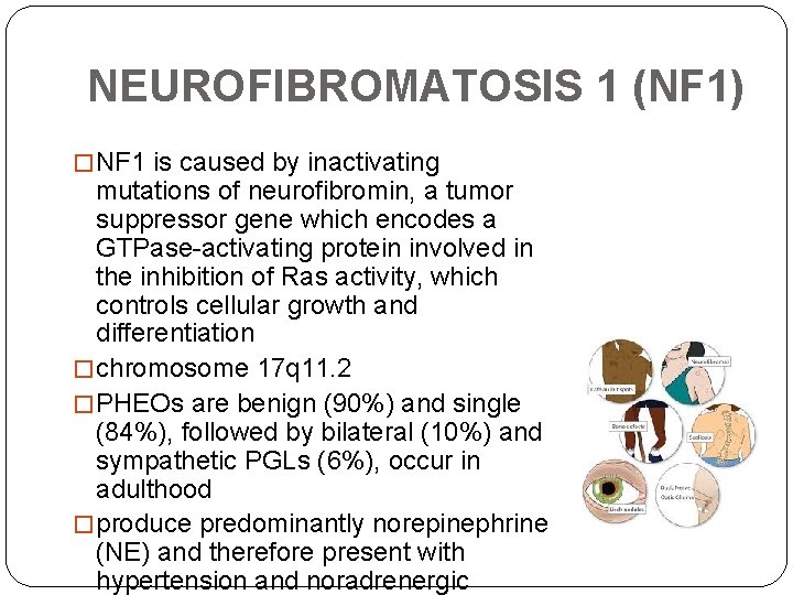
NEUROFIBROMATOSIS 1 (NF 1) � NF 1 is caused by inactivating mutations of neurofibromin, a tumor suppressor gene which encodes a GTPase-activating protein involved in the inhibition of Ras activity, which controls cellular growth and differentiation � chromosome 17 q 11. 2 � PHEOs are benign (90%) and single (84%), followed by bilateral (10%) and sympathetic PGLs (6%), occur in adulthood � produce predominantly norepinephrine (NE) and therefore present with hypertension and noradrenergic
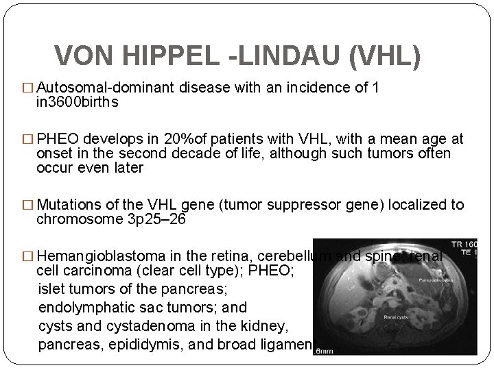
VON HIPPEL -LINDAU (VHL) � Autosomal-dominant disease with an incidence of 1 in 3600 births � PHEO develops in 20%of patients with VHL, with a mean age at onset in the second decade of life, although such tumors often occur even later � Mutations of the VHL gene (tumor suppressor gene) localized to chromosome 3 p 25– 26 � Hemangioblastoma in the retina, cerebellum and spine; renal cell carcinoma (clear cell type); PHEO; islet tumors of the pancreas; endolymphatic sac tumors; and cysts and cystadenoma in the kidney, pancreas, epididymis, and broad ligament
�PHEO may present as the first or only manifestation of VHL (VHL type 2 C) VHL carriers can present as apparently sporadic PHEO �Sympathetic PGL have been described also (10%) �Approximately half of PHEOs are bilateral and most produce NE (norepinephrine)
MULTIPLE ENDOCRINE NEOPLASIA 2 (MEN 2) � Autosomal-dominant syndrome caused by activating mutations in the RET proto-oncogene located on chromosome 10 q 11. 2 � MEN 2 A is characterized by medullary thyroid carcinoma (MTC), hyperparathyroidism, and PHEO � MEN 2 B is characterized by MTC, marfanoid habitus, mucosal ganglioneuromas, and PHEOs � PHEO occurs in approximately half of gene carriers and is almost always located within the adrenal glands, half of these are bilateral but asynchronous (up to 15 years apart)
�Codon 634 (MEN 2 A) or 918 (MEN 2 B) RET protooncogene mutations �Malignant PHEOs are uncommon <5% generally with large tumors �The pattern of catecholamine production in MEN 2 PHEO differs from that seen in other hereditary forms of PHEO. Epinephrine (E) +++ early clinical phenotype
PARAGANGLIOMAS (PGLs)
SYMPATHETIC PARAGANGLIOMAS �Derived from the sympathetic chain �Located in the chest, abdomen, or pelvis �Clinical picture due to either the secretion of catecholamines or the size of the tumors �The frequency of malignancy is much higher in sympathetic tumors with extraadrenal location
PARASYMPATHETIC PARAGANGLIOMAS �Usually found in the head and neck region �usually biochemically silent, and malignancy is seen in <10% of the cases �most frequent PGLs in the neck are carotid body tumors �most common below the neck are abdominal periaortic–pericaval tumors
�most frequent symptoms for the patients with head and neck tumors were palpable neck mass (55%) and cranial nerve palsies (16%), rarely hyperfunctioning �PGLs below the neck are more commonly hyperfunctioning �Retroperitoneal PGLs are more likely to be malignant with distant metastases or local invasion
SUCCINATE DEHYDROGENASE � The succinate dehydrogenase (SDH) is a mitochondrial enzyme complex with an important role in oxydative phosphorylation and intracellular oxygene sensing and signaling � Evidence that tumor genesis in PGL syndromes is linked to activation of hypoxia-related pathways � Constant signaling of hypoxia in the cell highly vascularized tumors � Disease-causing mutations in three genes (SDHB, SDHD, and SDHC)
PGL–SDHD � Autosomal-dominant syndrome � Familial and isolated head and neck parasympathetic PGLs and less frequently by sympathetic PGLs and PHEOs � Mutations in SDHD gene located on chromosome 11 q 21– 23 � generally benign, multifocal tumors � Maternal imprinting of SDHD, resulting in only paternal transmission of SDHD associated disease � PHEOs may be unilateral or bilateral and the mean age of diagnosis is 43 years
PGL–SDHB � Autosomal-dominant syndrome characterized by sympathetic extraadrenal PGLs and malignant disease � Inactivating mutations in the tumor suppressor SDHB gene located on chromosome 1 p 35– 36 � No maternal imprinting � Very strong association with a malignant intra- or extraadrenal phenotype � Malignant PHEOs are reported in 35 -40 % in these patients � Increased risk for renal cell carcinoma and papillary thyroid cancer
PGL–SDHC �Mutations in SDHC gene located on chromosome 1 q 21 �Autosomal dominant �No maternal imprinting �Benign and seldom multifocal head and neck parasympathetic PGL Ø In the last few years, four new genes (SDHA, SDHAF 2, MAX, and TMEM 127 ) have been found to be associated with predisposition to these tumours
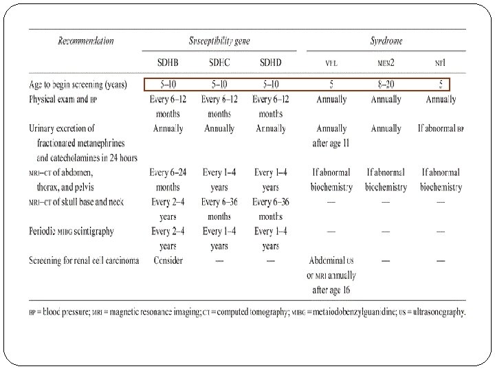
GENETIC TESTING � Germline mutations are responsible for about 25% of cases instead of 10% thought to be hereditary previously � 7. 5– 27% of tumors without an obvious syndrome or family history result from otherwise unsuspected germline mutations � Familial PPGLs inherited as autosomal-dominant (50%) chance of passing on the mutation to each child � Family history can be found; however, SDHD and SDHB mutations have age related penetrance reaching 100% by age 70 years � Sudden death should also be recorded
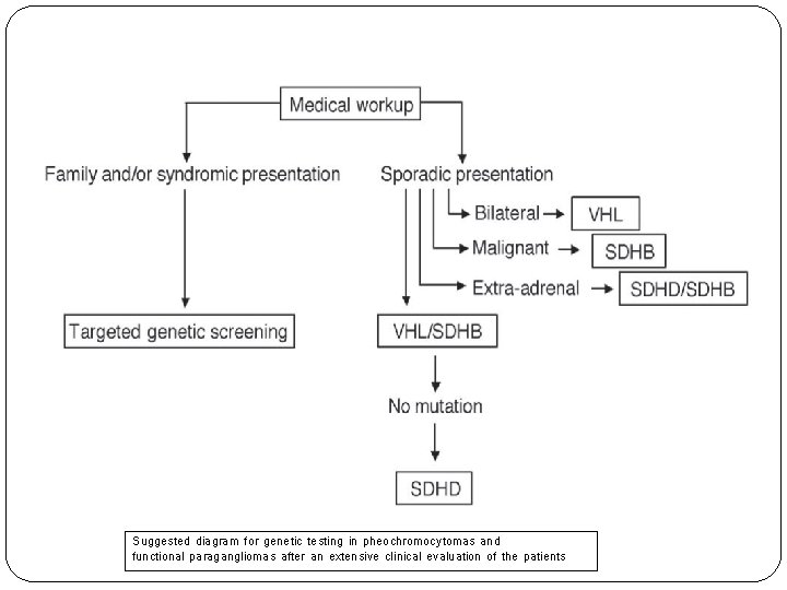
GENETIC TESTING � Younger age as hereditary PPGL occur at younger age than sporadic tumors � Genetic testing is more necessary in young adults, especially for VHL disease and SDHB � Extraadrenal, multifocal disease SDHB/SDHD gene mutations � Endocrine Society guidelines (2015) recommend that all patients with PPGLs should be engaged in shared decision making for genetic testing (but not necessarily done in each patient)
Suggested diagram for genetic testing in pheochromocytomas and functional paragangliomas after an extensive clinical evaluation of the patients
CLINICAL PRESENTATION
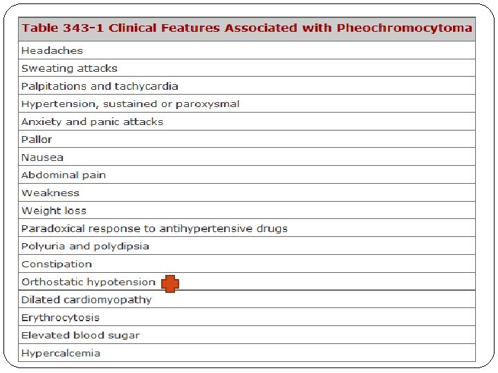
� TYPICAL SYMPTOMS Ø Sudden rise of BP with concurrent episodes of headache (80%), diaphoresis (70%), and palpitations (60%) pallor Ø Episodes usually last minutes or hours Ø Paroxysms may not recur for months or may recur many times daily � Other symptoms may include anxiety (50%), a sense of dread, tremor, or paresthesias � Cardiovascular symptoms (arrythmia, HF, cardiomyopathy) � Neurologic manifestations (stroke, confusion, seizures) � About 8% of patients may be completely asymptomatic, usually those with familial forms of the disease or with large, cystic tumors
HYPERTENSION �Hypertension is paroxysmal in 48% of patients, persistent in 29%, and 13% have normal BP �NE-secreting tumors are usually associated with sustained hypertension. �Tumors that secrete large amounts of E together with NE are associated with episodic hypertension. �Pure E-producing tumors can produce hypotension rather than hypertension
DIAGNOSIS
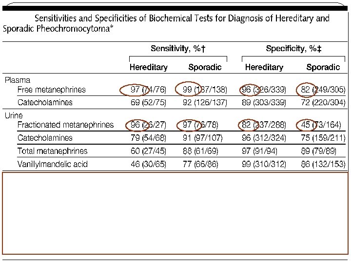
BIOCHEMICAL DIAGNOSIS � Plasma free or urinary fractionated metanephrines as initial testing � Despite the convenience of a spot urine sample, there is no evidence to suggest that this should replace the standardized 24 -hour urine collection method � When measuring the 24 -hour urinary excretion of fractionated metanephrines, urinary creatinine should be measured to verify completeness of the urine collection � Use liquid chromatography with mass spectrometric or electrochemical detection methods rather than other laboratory methods
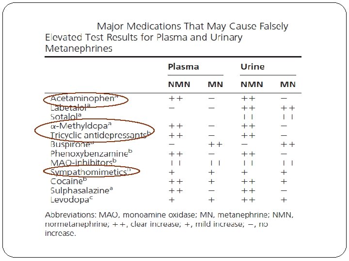
� For measurements of plasma metanephrines, draw blood with the patient in the supine position � Higher concentrations of plasma metanephrines in upright positions of blood sampling than in supine positions � Patients should be fully recumbent for at least 30 minutes before sampling � Solitary increases in either normetanephrine or metanephrine elevated 3 -fold or more above upper cutoffs are also rare as false positives � Elevations of both normetanephrine and metanephrine are rare as false- positives � For plasma free metanephrines, dietary considerations are only relevant when measurements include the dopamine metabolite 3 methoxytyramine (overbight fast)
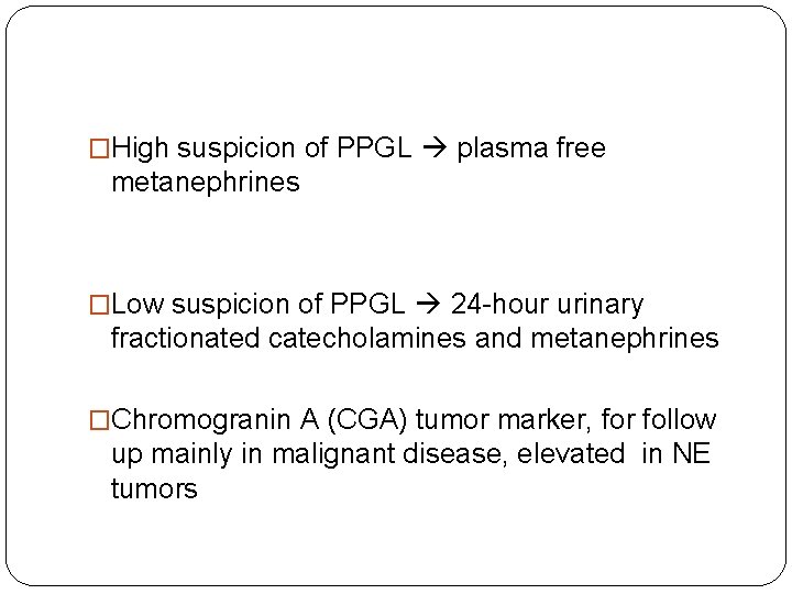
�High suspicion of PPGL plasma free metanephrines �Low suspicion of PPGL 24 -hour urinary fractionated catecholamines and metanephrines �Chromogranin A (CGA) tumor marker, for follow up mainly in malignant disease, elevated in NE tumors
IMAGING STUDIES � Imaging studies to locate PPGLs should be initiated once there is clear biochemical evidence of a PPGL � CT rather than MRI as the first-choice imaging modality �high HU density on noncontrast CT, marked enhancement with IV contrast on CT with delayed contrast washout [<50% at 10 mins], cystic & hemorrhagic changes, bilaterally, or larger size [>4 cm]) � A high signal intensity (bright) T 2 -weighted MRI image may be of value for the detection of PPGLs; however, a recent study showed that in pheochromocytomas this finding is relatively uncommon
�MRI recommended in patients with metastatic PPGLs, for detection of skull base and neck paragangliomas, in patients with an allergy to CT contrast, and in patients in whom radiation exposure should be limited (children, pregnant women, patients with known germline mutations, and those with recent excessive radiation exposure) � 123 I-metaiodobenzylguanidine (MIBG) scintigraphy in patients with metastatic PPGLs, in some patients with an increased risk for metastatic disease due to large size of the primary tumor, or recurrent disease
FDG PET � 18 F-FDG PET/CT is the preferred imaging modality over 123 I-MIBG scintigraphy in patients with known metastatic PPGLs �Sensitivity of 18 F-FDG PET was shown to be between 74 and 100%, with the highest performance for metastatic, particularly SDHBrelated PPGLs �Other imaging — 111 -In-pentetreotide scintigraphy (Octreoscan), DOPA PET
Perioperative Medical Management
� All patients with a hormonally functional PPGL should undergo preoperative blockade to prevent perioperative cardiovascular complications � Adrenergic receptor blockers as the first choice � Calcium channel blockers are the most often used add-on drug class to further improve blood pressure control � Preoperative coadministration of -adrenergic receptor blockers is indicated to control tachycardia only after administration of -adrenergic receptor blockers � Methyl-paratyrosine (metyrosine) inhibits catecholamine synthesis and may be used in combination with adrenergic receptor blockers for a short period before surgery
�Phenoxybenzamine is the preferred drug for preoperative preparation to control blood pressure and arrhythmia in most centers in the United States. It is an irreversible, long-acting, nonspecific alpha-adrenergic blocking agent, 20 and 100 mg daily, orthostasis, nasal stuffiness and fatigue �With their more favorable side-effect profiles, selective alpha 1 -adrenergic blocking agents (eg, prazosin, terazosin, or doxazosin) are utilized in many centers or are preferred
� Retrospective studies report that –adrenergic receptor blockers should be started at least 7 days preoperatively (better 10 – 14 days) � high-sodium diet a few days after the start of-adrenergic receptor blockade � Continuous administration of saline (1– 2 L) is also helpful if started the evening before surgery � Optimal target blood pressure? A target blood pressure of less than 130/80 mm. Hgwhile seated and greater than 90 mm Hg systolic while standing seems reasonable � Note that complete prevention of intraoperative hypertension and tachycardia cannot be achieved by any doses and combinations of antihypertensive and other drugs � Treatment options for hypertensive crises include intravenous sodium nitroprusside, phentolamine, or nicardipine
�Laparoscopic adrenalectomy �Open adrenalectomy (large tumors > 8 cm, malignant disease) �Partial adrenalectomy with cortical sparing in familial pheochromocytoma (High incidence of bilateral disease) to prevent permanent glucocorticoid deficiency.
�Major potential postoperative complications are hypertension, hypotension, and rebound hypoglycemia �Blood pressure, heart rate and plasma glucose levels should be closely monitored for 24– 48 hours �Measure plasma or urine levels of metanephrines on follow-up to diagnose persistent disease (2 -4 weeks after surgery) �Lifelong annual biochemical testing to assess for