Phase 1 CVR wrap up ANATOMY AND EMBRYOLOGY

Phase 1 CVR wrap up ANATOMY AND EMBRYOLOGY OF THE HEART Nneoma Uzo 24. 11. 2016 The Peer Teaching Society is not liable for false or misleading information…

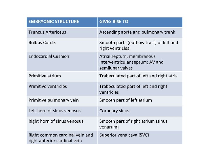
EMBRYOLOGY The peer teaching society is not liable for false or misleading information

Aortic Arch development

FETAL CIRCULATION

Fetal post-natal derivatives • • Allantois --> Urachus Ductus arteriosus Ductus venosum Foramen ovale Notochord Umbilical arteries Umbilical vein Median unbilical ligament Ligamentum arteriosum Ligamentum venosum Fossa ovalis Nucleus pulposus Medial umbilical ligament Ligamentum teres hepatis

ANATOMY

THE HEART
: • Fibrous Pericardium • Parietal")
The Pericardium Consists of three layers (outer to inner): • Fibrous Pericardium • Parietal layer of serous pericardium • Visceral layer of serous pericardium NOTE: The pericardial cavity lies between parietal and visceral layers.

Pericardial Sinuses • Oblique pericardial sinus: posterior surface of the heart • Transverse pericardial sinus: found superiorly on the heart.
: Right ventricle • Posterior (or base): Left atrium")
Heart Surfaces • Anterior (or sternocostal): Right ventricle • Posterior (or base): Left atrium • Inferior (or diaphragmatic): Left and right ventricles • Right pulmonary: Right atrium • Left pulmonary: Left ventricle

Heart Borders • Right border: Right atrium • Left border: Left ventricle (and some of the left atrium) • Inferior border: Left ventricle and right ventricle • Superior border: Right and left atrium and the great vessels

Chambers of the Heart • Right Atrium: - Crista terminalis - Fossa ovalis - The SAN - Musculi Pectinati - Tricuspid valve - Coronary Sinus: opens into right atrium between IVC and right atrioventricular orifice

Chambers of the Heart • Left Atrium - Smooth interior - Trabeculated auricular appendage - Mitral valve: 2 cusps NOTE: Fossa ovalis is not visible on this side

Chambers of the Heart • Right ventricle - Trabeculae carnae Chordae Tendineae Papillary muscle Pulmonary valves (outflow of blood)

Chambers of the Heart • Left ventricle - Trabeculae carneae - 2 Papillary muscles attached to the cusps of the mitral valve
 - Mitral (left) • Semilunar valves")
The Valves • Atrioventricular valves - Tricuspid (right) - Mitral (left) • Semilunar valves - Aortic - Pulmonary NOTE: Valves prevent the backflow of blood.

Great vessels of the heart • Aorta: Brachiocephalic trunk, left common carotid artery and left subclavian artery (T 12) • Pulmonary arteries: left and right pulmonary arteries • Pulmonary veins: 4 pulmonary veins • Superior Vena Cava: formed by the merging of the brachiocephalic veins • Inferior Vena Cava: initially formed by the common iliac veins (T 8)

CORONARY ARTERY ANATOMY
: Posterior Descending artery, right (acute) marginal artery")
• The right coronary artery (RCA): Posterior Descending artery, right (acute) marginal artery • The Left main coronary artery (LCA): Left circumflex coronary artery, left anterior descending artery, left (obtuse) marginal artery
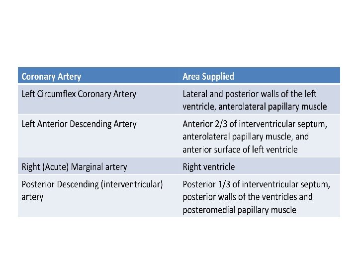

NOTE: SA and AV nodes are usually supplied by RCA
 = PDA arises from RCA • Left-dominant circulation (30%)")
REMEMBER… • Right-dominant circulation (90%) = PDA arises from RCA • Left-dominant circulation (30%) = PDA arises from LCX • Codominant circulation (20%) = PDA arises from both LCX and RCA

QUESTIONS? The Peer Teaching Society is not liable for false or misleading information…

- Slides: 25