
PHARYNXIII Presented by Dr Sushma Tomar Associate Professor
PHARYNX-III Presented by : - Dr. Sushma Tomar Associate Professor Department of
• Muscles of Pharynx. Lesson Plan • Constrictor muscles. • Applied aspects. • Gaps in pharyngeal wall. • Longitudinal muscles. • Nerve supply of pharynx. • Pharyngeal Plexus of nerves. • Arterial supply of pharynx. • Venous and Lymphatic drainage of pharynx.
Muscles of Pharynx

Constrictor Muscles v Constrictor muscles form the bulk of muscle layer. Origin • From the margins of posterior openings of nasal, oral, and laryngeal cavities. Insertion • Into the median fibrous raphe on the posterior aspect of pharynx. v Inferior constrictor overlaps the middle constrictor. v Middle constrictor overlaps the superior constrictor. Superior Constrictor Middle Constrictor Inferior Constrictor

Origin- Superior Constrictor • Pterygoid hamulus. • Pterygomandibular raphe. • Medial surface of mandible at upper end of mylohyoid line. • Side of posterior part of tongue. Insertion- Pterygoid Hamulus • Pharyngeal tubercle. • Pharyngeal raphe. Nerve supply- • Pharyngeal branch of Vagus nerve ( carrying fibers of cranial root of Pterygomandibular accessory nerve). Raphe Action • Helps in deglutition.

Origin- Middle Constrictor • Lower part of Stylohyoid ligament. • Lesser cornu of hyoid bone. • Upper border of greater cornu of hyoid bone. Insertion- Stylohyo id Ligamen t • Pharyngeal raphe. Nerve supply- Lesse r cornu • Pharyngeal branch of Vagus nerve ( carrying fibers of cranial root of Great accessory nerve). Action • Helps in deglutition. er cornu
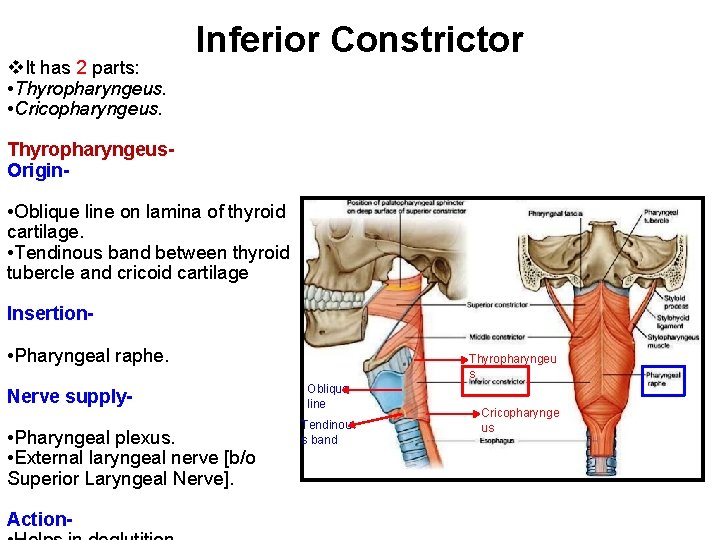
v. It has 2 parts: • Thyropharyngeus. • Cricopharyngeus. Inferior Constrictor Thyropharyngeus. Origin- • Oblique line on lamina of thyroid cartilage. • Tendinous band between thyroid tubercle and cricoid cartilage Insertion- • Pharyngeal raphe. Nerve supply- • Pharyngeal plexus. • External laryngeal nerve [b/o Superior Laryngeal Nerve]. Action- Thyropharyngeu s Oblique line Tendinou s band Cricopharynge us

Inferior Constrictor contd… Cricopharyngeus. Origin- • Cricoid cartilage. Insertion- • Pharyngeal raphe. Nerve supply- • Recurrent laryngeal nerve [b/o Vagus nerve]. Action • Helps in deglutition. Cricoid Cartila ge Cricopharyng eus
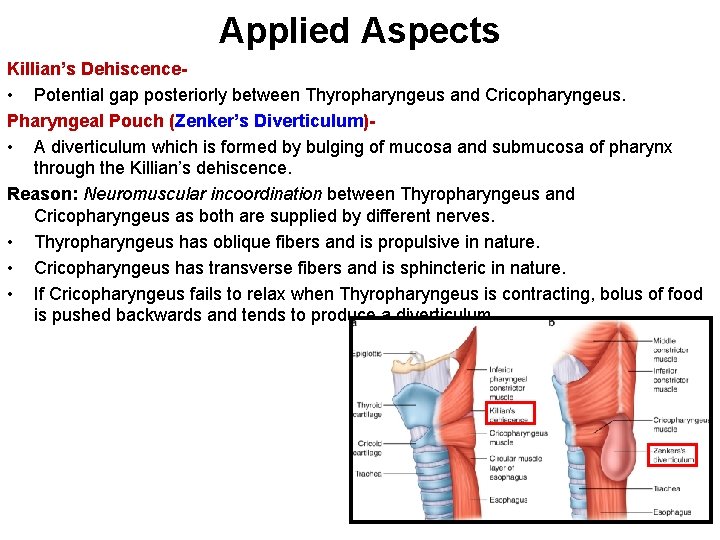
Applied Aspects Killian’s Dehiscence • Potential gap posteriorly between Thyropharyngeus and Cricopharyngeus. Pharyngeal Pouch (Zenker’s Diverticulum) • A diverticulum which is formed by bulging of mucosa and submucosa of pharynx through the Killian’s dehiscence. Reason: Neuromuscular incoordination between Thyropharyngeus and Cricopharyngeus as both are supplied by different nerves. • Thyropharyngeus has oblique fibers and is propulsive in nature. • Cricopharyngeus has transverse fibers and is sphincteric in nature. • If Cricopharyngeus fails to relax when Thyropharyngeus is contracting, bolus of food is pushed backwards and tends to produce a diverticulum.
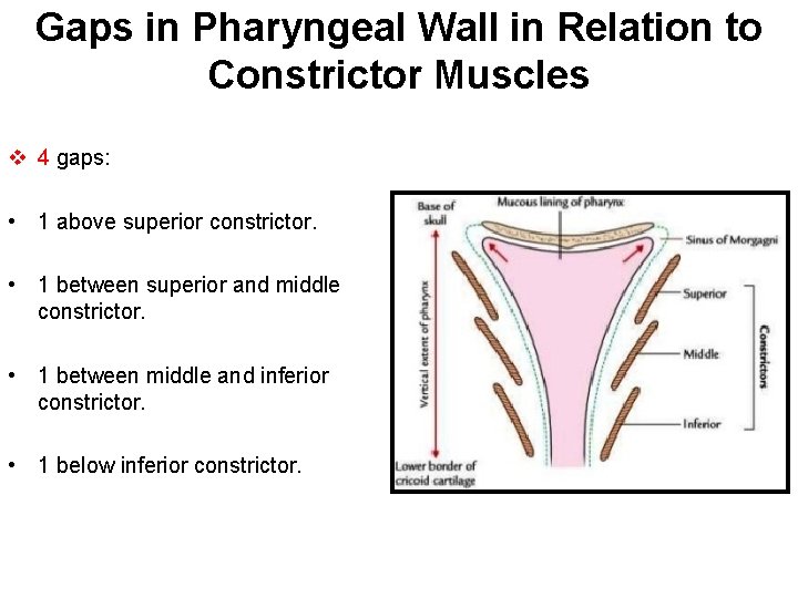
Gaps in Pharyngeal Wall in Relation to Constrictor Muscles v 4 gaps: • 1 above superior constrictor. • 1 between superior and middle constrictor. • 1 between middle and inferior constrictor. • 1 below inferior constrictor.
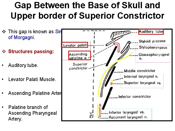
Gap Between the Base of Skull and Upper border of Superior Constrictor v This gap is known as Sinus of Morgagni. v Structures passing: • Auditory tube. • Levator Palati Muscle. • Ascending Palatine Artery. • Palatine branch of Ascending Pharyngeal Artery.

Gap Between the Superior Constrictor and Upper border of Middle Constrictor v Structures passing: • Stylopharyngeus Muscle. • Glossopharyngeal Nerve.
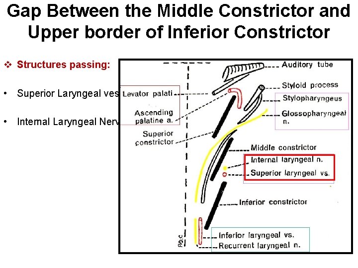
Gap Between the Middle Constrictor and Upper border of Inferior Constrictor v Structures passing: • Superior Laryngeal vessels. • Internal Laryngeal Nerve.
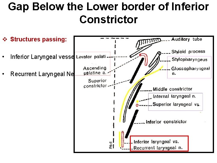
Gap Below the Lower border of Inferior Constrictor v Structures passing: • Inferior Laryngeal vessels. • Recurrent Laryngeal Nerve.
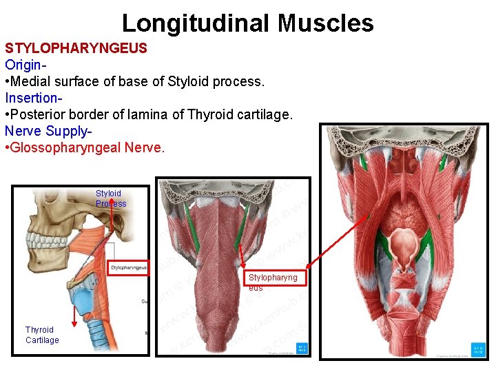
Longitudinal Muscles STYLOPHARYNGEUS Origin • Medial surface of base of Styloid process. Insertion • Posterior border of lamina of Thyroid cartilage. Nerve Supply • Glossopharyngeal Nerve. Styloid Process Stylopharyng eus Thyroid Cartilage
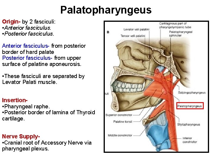
Palatopharyngeus Origin- by 2 fasciculi: • Anterior fasciculus. • Posterior fasciculus. Anterior fasciculus- from posterior border of hard palate Posterior fasciculus- from upper surface of palatine aponeurosis. • These fasciculi are separated by Levator Palati muscle. Insertion • Pharyngeal raphe. • Posterior border of lamina of Thyroid cartilage. Nerve Supply • Cranial root of Accessory Nerve via pharyngeal plexus.
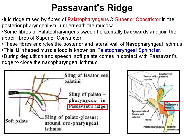
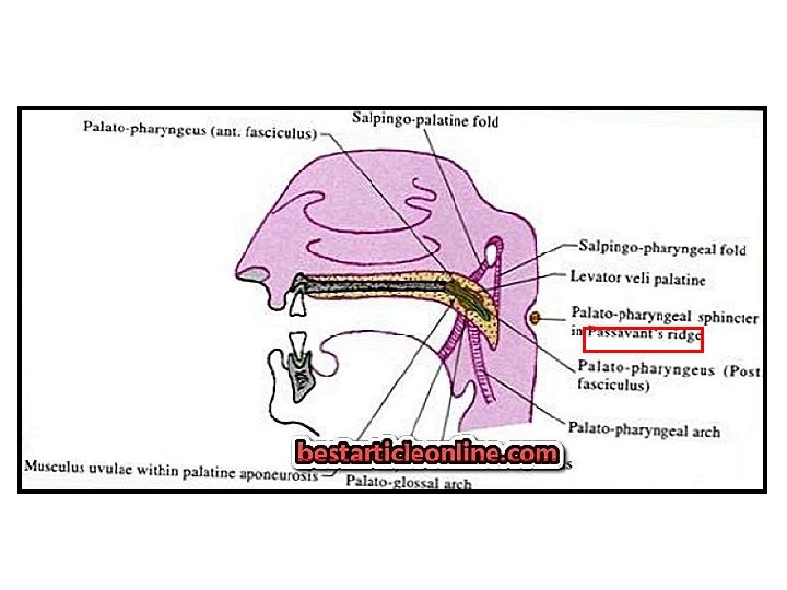
Passavant’s Ridge • It is ridge raised by fibres of Palatopharyngeus & Superior Constrictor in the posterior pharyngeal wall underneath the mucosa. • Some fibres of Palatopharyngeus sweep horizontally backwards and join the upper fibres of Superior Constrictor. • These fibres encircles the posterior and lateral wall of Nasopharyngeal Isthmus. • This ‘U’ shaped muscle loop is known as Palatopharyngeal Sphincter. • During deglutition and speech, soft palate comes in contact with Passavant’s ridge to close the nasopharyngeal isthmus.
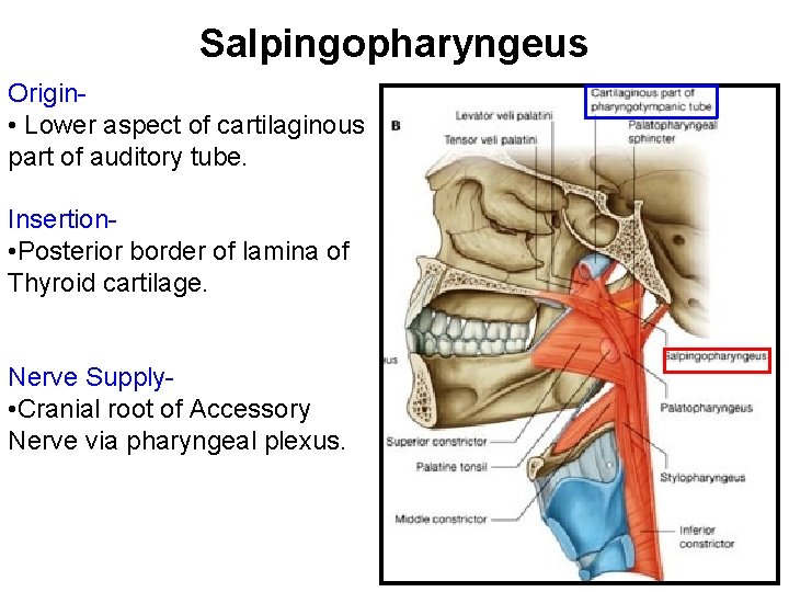
Salpingopharyngeus Origin • Lower aspect of cartilaginous part of auditory tube. Insertion • Posterior border of lamina of Thyroid cartilage. Nerve Supply • Cranial root of Accessory Nerve via pharyngeal plexus.
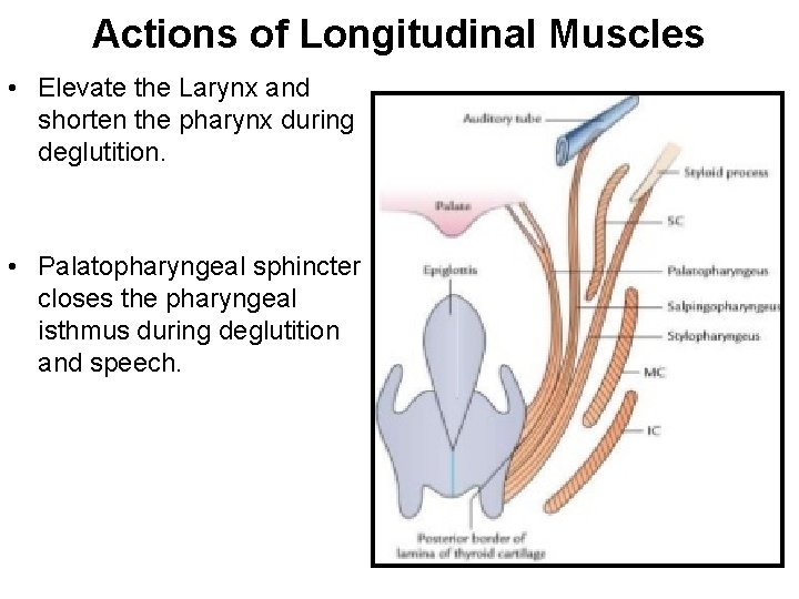
Actions of Longitudinal Muscles • Elevate the Larynx and shorten the pharynx during deglutition. • Palatopharyngeal sphincter closes the pharyngeal isthmus during deglutition and speech.
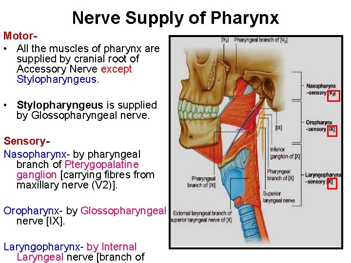
Nerve Supply of Pharynx Motor • All the muscles of pharynx are supplied by cranial root of Accessory Nerve except Stylopharyngeus. • Stylopharyngeus is supplied by Glossopharyngeal nerve. Sensory. Nasopharynx- by pharyngeal branch of Pterygopalatine ganglion [carrying fibres from maxillary nerve (V 2)]. Oropharynx- by Glossopharyngeal nerve [IX]. Laryngopharynx- by Internal Laryngeal nerve [branch of
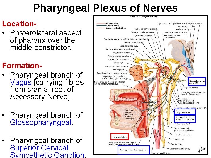
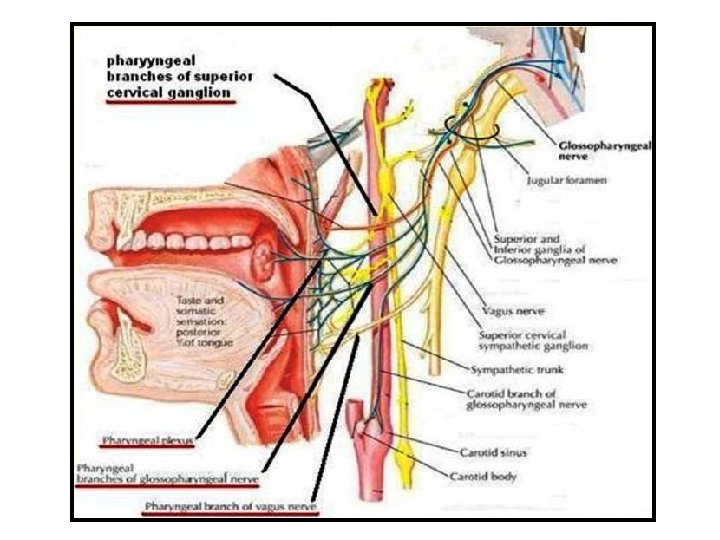
Pharyngeal Plexus of Nerves Location • Posterolateral aspect of pharynx over the middle constrictor. Formation • Pharyngeal branch of Vagus [carrying fibres from cranial root of Accessory Nerve]. • Pharyngeal branch of Glossopharyngeal. • Pharyngeal branch of Superior Cervical Sympathetic Ganglion.
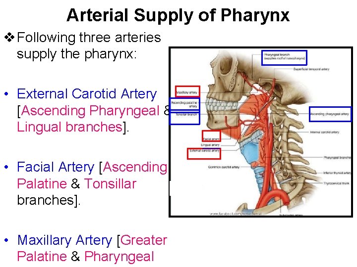
Arterial Supply of Pharynx v Following three arteries supply the pharynx: • External Carotid Artery [Ascending Pharyngeal & Lingual branches]. • Facial Artery [Ascending Palatine & Tonsillar branches]. • Maxillary Artery [Greater Palatine & Pharyngeal
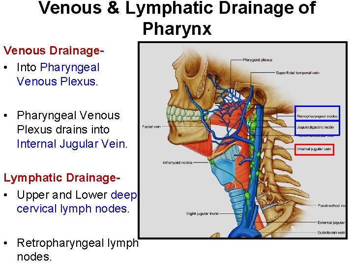
Venous & Lymphatic Drainage of Pharynx Venous Drainage • Into Pharyngeal Venous Plexus. • Pharyngeal Venous Plexus drains into Internal Jugular Vein. Lymphatic Drainage • Upper and Lower deep cervical lymph nodes. • Retropharyngeal lymph nodes.

- Slides: 26