Pharynx peripharyngeal spaces Mark Kozsurek M D Ph


 ascending palatine")


- Slides: 32
Pharynx, peripharyngeal spaces Mark Kozsurek, M. D. , Ph. D. mark@kozsurek. hu ED II. , 18/02/2019
What is the pharynx? n n Pharynx is a musculofascial half-cylinder lacking the anterior wall. According to the anterior relations, pharynx can be devided into three parts: nasopharynx, oropharynx and laryngopharynx. It is the common pathway for the air and the food. Extends from the base of skul to the level of the 5 th-6 th vertebrae where is replaced by the esophagus.
Skeletal framework Anterior and superior margin of the pharynx is attached to bones, cartilages and ligaments. The two sides merge posteriorly in the midline constituting the pharyngeal raphe. Line of attachment of the pharynx to the base of the skull.
The superior line of attachment includes the posterior margin of the medial plate of the pterygoid process, the pterygoid hamulus and the pharyngomandibular raphe. The middle line of attachment follows the inferior portion of the stylohyoid ligament, the lesser horn and then the greater horn of the hyoid bone. The inferior line of attachment is represented by the oblique line of the thyroid cartilage, a tendinous thickening of the fascia of the cricothyroid muscle and the cricoid cartilage.
Pharyngeal wall is mainly formed by skeletal muscles and fasciae. The latter ones are especially important where the muscular wall is interrupted. 1. Tunica mucosa epithelium lamina propria 2. Tunica fibrosa = pharyngobasillar fascia: a thick layer right under the mucosa that covers the inner surface of the muscles. 3. Tunica muscularis: circular layer: constrictors longitudinal layer: levators 4. Tunica adventitia = buccopharyngeal fascia: much thinner and is the posterior continuation of the fascia covering the outer surface of the buccinator muscle. Histologically might be considered as the tunica adventitia of the pharynx.
Superior constrictor: pterygopharyngeal part buccopharyngeal part mylopharyngeal part glossopharyngeal part Middle constrictor: ceratopharyngeal part chondropharyngeal part Note the gaps among the constrictors!!! Inferior constrictor: thyropharyngeal part cricopharyngeal part
Levators: Salpingopharyngeus Palatopharyngeus Stylopharyngeus
Where the muscles are missing. . . 1. At the Mylohyoid-Middle constrictor-Superior constrictor triangle: • Hyoglossus and Styloglossus muscles (Hyoglossus isolates lateral and medial sulci of the tongue) • lingual and hypoglossal nerves and deep lingual vein, submandibular duct – lateral sulcus • lingual artery and dorsal lingual vein, glossopharyngeal nerve – medial sulcus
2. At the thyrohyoid membrane: internal branch of superior laryngeal nerve (ex vagus n. ), superior laryngeal artery (ex sup. thyroid a. ex ext. carotid a. ), superior laryngeal vein (to sup. thyroid v. to internal jugular v. ) 3. At the inferior edge of the Inferior constrictor: recurrent laryngeal nerve (ex vagus n. ), inferior laryngeal artery (ex inf. thyroid a. , ex thyrocervical trunk ex subclavian a. ), inferior laryngeal vein (to inf. thyroid v. to left brachiocephalic v. )
Pharyngeal compartments: nasopharynx, oropharynx and laryngopharynx Identify the following structures: choana, pharyngobasillar fascia, pharyngeal fornix, pharyngeal tonsil, salpingopharyngeal fold, pharyngeal recess, salpingopalatine fold, torus tubarius, tubal tonsil, torus levatorius!
Following the removal of the mucous membrane pharyngeal muscles as well as the cartilagineous part of the tympanopharyngeal (or auditory) tube become visible.
The following structures should be identified: root of the tongue, palatoglossal and palatopharyngeal folds, tonsillary fossa, palatine tonsil, oropharynx, pharyngeal recess.
Please find: piriform recess, aryepiglottic fold, cuneiform tubercle, corniculate tubercle, interarythenoid notch, fold of the superior laryngeal nerve, greater horn of the hyoid bone, superior horn of the thyroid cartilage.
Tonsils
hemicapsule: paltine tonsil
respiratory epithelium: pharyngeal tonsil
mucous glands: lingual tonsil
Blood supply, innervation, lymphatic drainage
SUPERIORLY: • • • ascending pharyngeal a. (ex ext. carotid a. ) ascending palatine a. (ex facial a. ) small branches of the lingual and maxillary a. INFERIORLY: • pharyngeal branches of the inferior thyroid artery (ex thyrocervical trunk ex subclavian a. )
The pharyngeal veinous plexus communicates with the pterygoid plexus in the infratemporal fossa that drains toward the facial and retromandibular veins and into the external and internal jugular veins. Lymphatic vessels from the pharynx drain into the retropharyngeal and paratracheal nodes which are subgroups of deep cervical nodes (situated mainly around the internal jugular vein).
Three CNs are enough for the complete innervation of the pharynx.
Pharyngeal plexus: pharyngeal branches of the glossopharyngeal and vagus nerves and postganglionic sympathetic fibres from the superior cervical ganglion. Ambiguus nucleus (BM), dorsal vagus nucleus (GVM), lateral nucleus of ala cinerea (GVS)
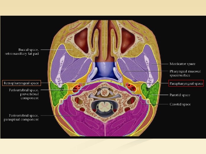
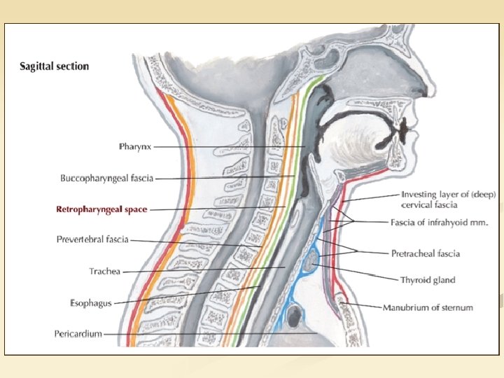
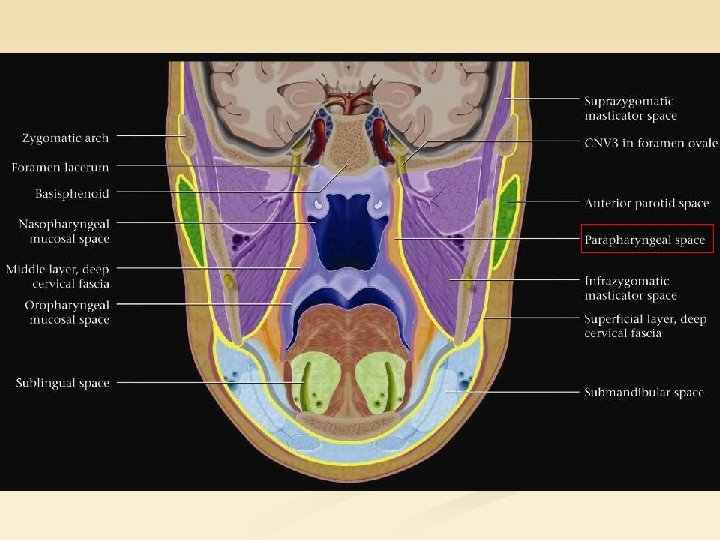
Peripharyngeal spaces
In a broader sense retropharyngeal space is found behind the posterior wall of the pharynx and anterior to the prevertebral muscles. In more precise anatomical descriptions retropharyngeal space is defined as an area behind the buccopharyngeal fascia (adventitia of the pharynx) and in front of the alar fascia. As the alar fascia fuses with the pharyngeal raphe, infections can not easily spread to the contralateral side. The danger space is between the alar fascia and the prevertebral fascia and has no septum in the midline.
Clinical considerations
Adenoid hypertrophy and otitis media
Peritonsillar and retropharyngeal abscess
Thank you for your attention!