Pharynx Larynx Pharynx Subdivisions and borders Pharyngeal muscles

Pharynx & Larynx

• Pharynx – Subdivisions and borders – Pharyngeal muscles – Blood supply and innervation • Larynx – Laryngeal cartilages Paired vs. unpaired Basic structure Membranes and ligaments – Vocal cords and folds – Muscles and innervation – Blood supply
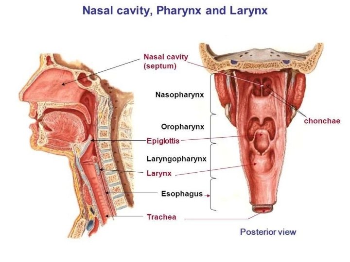

• Is a wide muscular tube • 12 cm in length • Located posterior to the nasal and oral cavities • Extends inferiorly, posterior to the larynx • Extends from the cranial base to the inferior border of the cricoid cartilage (anteriorly) and inferior border of C 6 (posteriorly) [Extends from the base of the skull to the level of the C 6 vertebra] where it is continuous with the oesophagus • Widest opposite the hyoid bone and narrowest at the junction where it joins the oesophagus

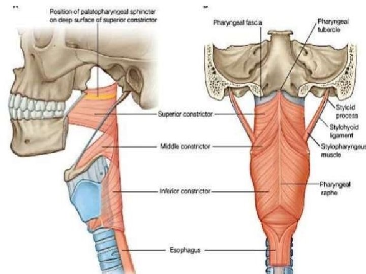
Pharyngeal wall The wall of the Pharynx consists of five layers: 1. Mucous membrane 2. Submucosa 3. Pharyngobasilar fascia 4. Pharyngeal muscles (3 constrictors) a. Stylopharyngeus b. Salpingopharyngeus C- Palatopharyngeus 5. Buccopharyngeal fascia

• Nasopharynx • Oropharynx • Laryngopharynx

• Respiratory function • Roof and Posterior wall: Continuous surface that lies inferior to the body of the sphenoid bone and the basilar part of the occipital bone • Pharyngeal tonsils: Found in the mucous membrane of the roof and the posterior wall of the nasopharynx

• Digestive function • Helps in the process of deglutition • Borders Superiorly: Soft Palate Inferiorly: Base of the Tongue Laterally: Palatoglossal and Palatopharyngeal arches and palatine tonsils

• Palatine tonsils – Collections of lymphoid tissue on either side of the Oropharynx between the arches • Tonsillar bed – Superior constrictor of the pharynx and the pharyngobasilar fascia form the tonsillar bed

ARTERIAL SUPPLY OF THE TONSIL 1 Tonsillar A. From facial A. 2 Lingual A. 3 Ascending palatine A. 4 - Maxillary artery. Acute follicular tonsillitis

• • • Extends from the superior border of the epiglottis and the pharyngoepiglottic folds to the inferior border of the cricoid cartilage Borders Posteriorly: related to the bodies of the C 4 - C 6 vertebrae. Posterior and lateral walls: Middle and Inferior constrictor muscles

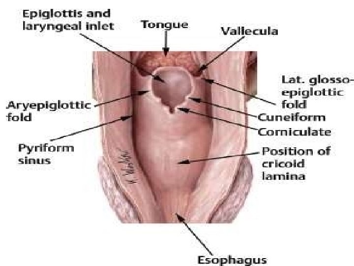
• Palatopharyngeus and Stylopharyngeus muscles form the walls • Piriform recess, small depression of the laryngopharyngeal cavity on either side of the laryngeal inlet • Separated from the laryngeal inlet by the aryepiglottic fold

• External circular layer • Internal Longitudinal layer 2 layers of voluntary muscle:

External circular layer • Constrictor muscles • Primarily responsible for constricting the pharynx during swallowing Both types are innervated by the vagus nerve, except for the stylopharyngeus, which is innervated by the glossopharyngea l nerve.

• Internal Longitudinal layer • Elevate/shorten and widen the pharynx during swallowing and speaking • Palatopharyngeus • Stylopharyngeus • Salpingopharyngeus Palatopharyngeus and salpingopharyngeus are innervated by the pharyngeal branch of CNX and the pharyngeal plexus • Stylopharyngeus is innervated by CN IX

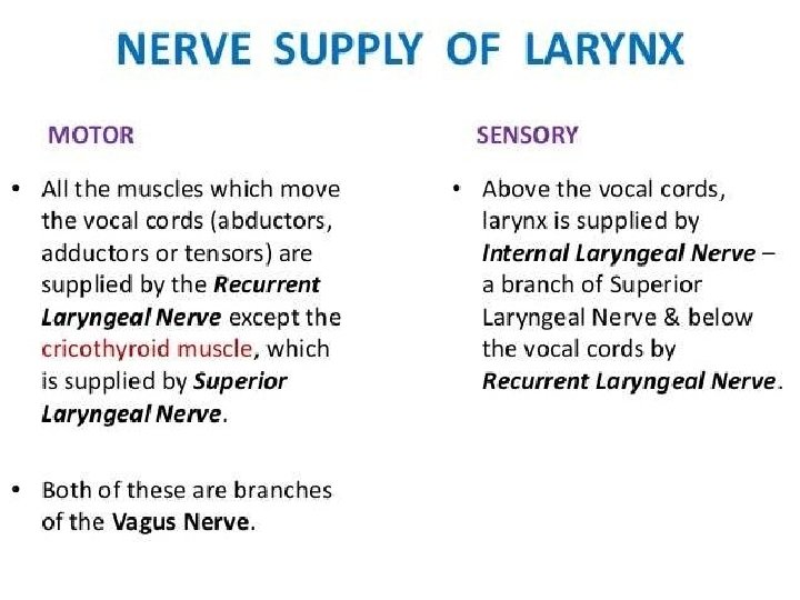
Sensory: Each of the three sections of the pharynx have a different innervation: The nasopharynx is innervated by the maxillary nerve (CN V 2). The oropharynx by the glossopharyngeal nerve (CN IX). The laryngopharynx by the vagus nerve (CN X). Motor: All the muscles of the pharynx are innervated by the vagus nerve (CN X), except for the stylopharyngeus, which is innervated by the glossopharyngeal nerve (CN IX).

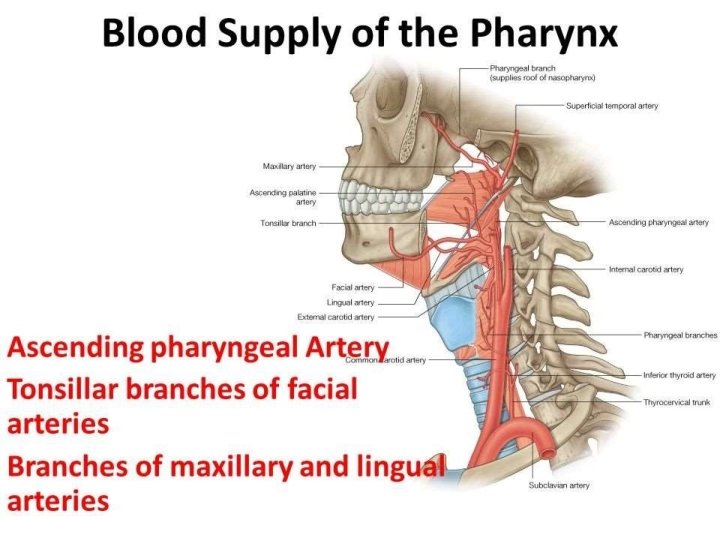
Blood Supply of the pharynx: Arterial supply is via branches of the external carotid artery: • Ascending pharyngeal artery • Ascending palatine artery • Tonsillar branches of the facial artery • Branches of the maxillary and lingual arteries • Pharyngeal branches of the inferior thyroid artery Venous drainage is achieved by the pharyngeal venous plexus, which drains into the internal jugular vein.
 Inferior pharyngeal constrictor is found in the laryngopharynx and")
Clinical Relevance: Pharyngeal Diverticulum (Pouch) Inferior pharyngeal constrictor is found in the laryngopharynx and has two components. The superior component (cartilage and the inferior component (cricopharyngeus) has horizontal fibres that attach to the cricoid cartilage This area between the two is a weak area in the muscosa. Normally during swallowing, the thryopharyngeus contracts as the cricopharyngeus relaxes, allowing the bolus of food to be propelled into the oesophagus and preventing the intrapharyngeal pressure form rising. If this coordinated relaxation of the cricopharyngeus does not occur, the intrapharyngeal pressure tends to rise and pharyngeal mucosa forms a midline diverticulum in the area between the thyropharyngeus and cricopharyngeus. It is possible for food to accumulate here, leading to dysphagia.

Barium swallow X-ray

Nasopharyngeal tonsil • Adenoids is the hypertrophied mass of lymphoid tissue situated at the junction of roof & post. wall of nasopharynx. • The mass of lymphoid tissue is termed as Adenoids only when it is hypertrophied. • It usually undergoes atrophy by puberty (13 - 14 yrs)

Waldeyer’s Ring • Waldeyer's tonsillar ring includes 1. Adenoid tonsil 2. Two tubal tonsils 3. Two palatine tonsils 4. Lingual tonsil.

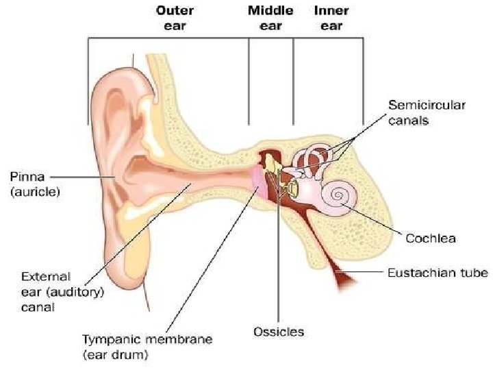
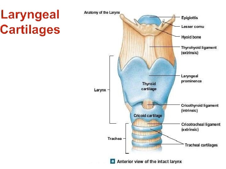
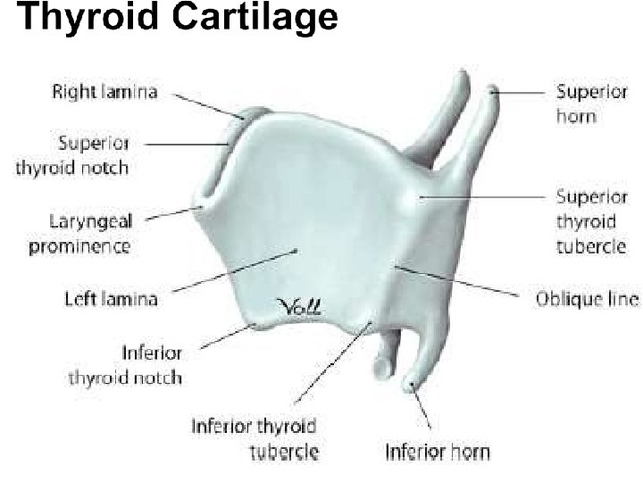
LARYNX • Is the upper , expanded part of the windpipe which is modified for phonation • Supported by a number of cartilages
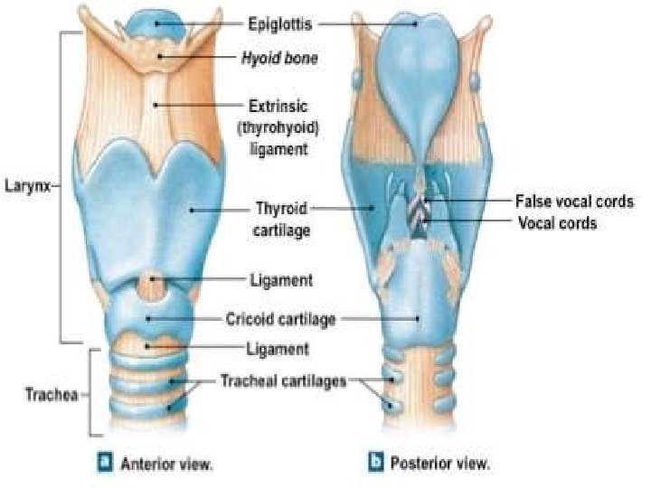

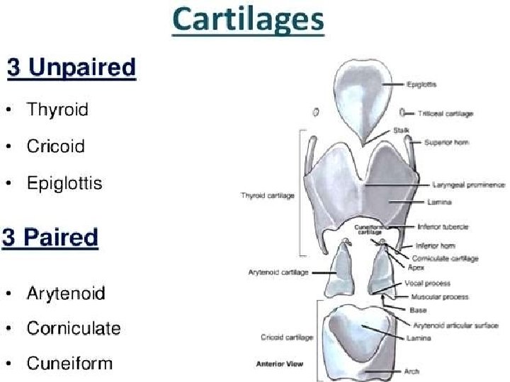
• Complex organ of voice production • Composed of 9 cartilages connected by membranes and ligaments • Contains the vocal folds • Located anteriorly in the neck • Vertebral levels C 3 -C 6 • Connects the inferior part of the Oropharynx to the trachea • Lies anterior to the Laryngopharnx • Secondary function is to guard the air passages

• Main divisions of the Laryngeal Cavity 1 Laryngeal Vestibule 2 Laryngeal cavity 3 - Infraglottic cavity
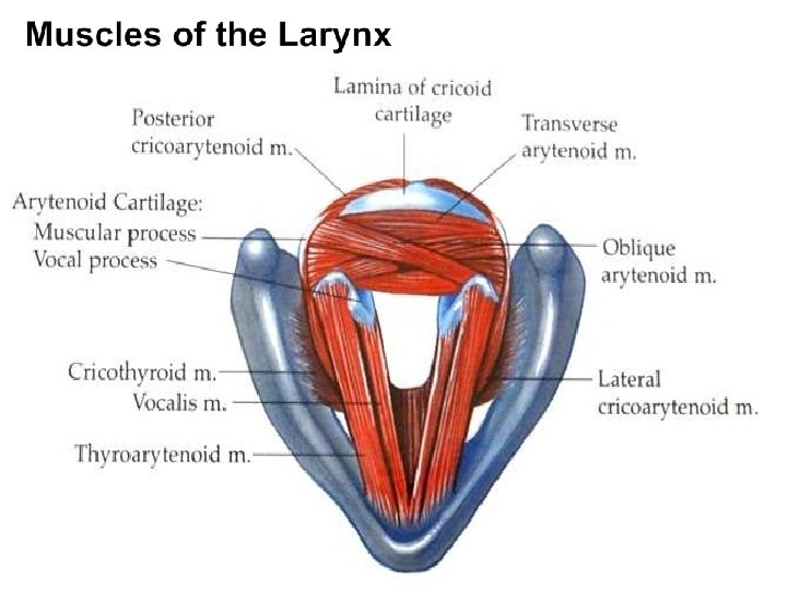


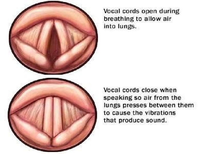


Muscle Origin Insertion Innervation Action Cricothyroid Antero-lateral part of cricoid cartilage Inferior margin and inferior horn of thyroid cartilage External laryngeal nerve Thyro-arytenoid Lower ½ of posterior aspect of angle of thyroid laminae and cricothyroid ligament Anterolateral arytenoid surface Posterior Cricoarytenoid Posterior surface of lamina of cricoid cartilage Stretches and tenses vocal ligament Relaxes valocal ligament Abducts vocal folds Vocal process of arytenoid cartilage Inferior laryngeal nerve (terminal part of recurrent laryngeal nerve from vagus) Adducts vocal folds Lateral cricoarytenoid Arch of arytenoid cartilage Transverse & Oblique arytenoids One arytenoid cartilage Contra-lateral arytenoid cartilage Adducts arytenoid cartilages Vocalis Lateral surface of vocal process of arytenoid cartilages Ipsilateral vocal ligament Relaxes posterior vocal ligament while maintaining tension of anterior part

• Superior Laryngeal artery Supplies the internal surface of the larynx • Cricothyroid artery Supplies the cricothyroid muscle • Inferior Laryngeal artery Supplies the mucous membrane and the muscles of the inferior part of the larynx
- Slides: 42