
Pharmacology of Antiarrhytmic Drug M Saifur Rohman dr
- Slides: 82
Pharmacology of Antiarrhytmic Drug M. Saifur Rohman, dr. Sp. JP. Ph. D. FICA Department of Cardiology and Vascular Medicine Faculty of Medicine, Brawijaya University
Mechanism of Arrhythmia • 1. 2. 3. • 1. 2. Abnormal heart pulse formation Sinus pulse Ectopic pulse Triggered activity Abnormal heart pulse conduction Reentry Conduct block
Classification of Arrhythmia • Abnormal heart pulse formation 1. 2. 3. 4. Sinus arrhythmia Atrial arrhythmia Atrioventricular junctional arrhythmia Ventricular arrhythmia • Abnormal heart pulse conduction 1. 2. 3. 4. Sinus-atrial block Intra-atrial block Atrio-ventricular block Intra-ventricular block • Abnormal heart pulse formation and conduction
Anti-tachycardia agents • 1. 2. 3. 4. 5. Modified Vaugham Williams classification I class: Natrium channel blocker II class: ß-receptor blocker III class: Potassium channel blocker IV class: Calcium channel blocker Others: Adenosine, Digital
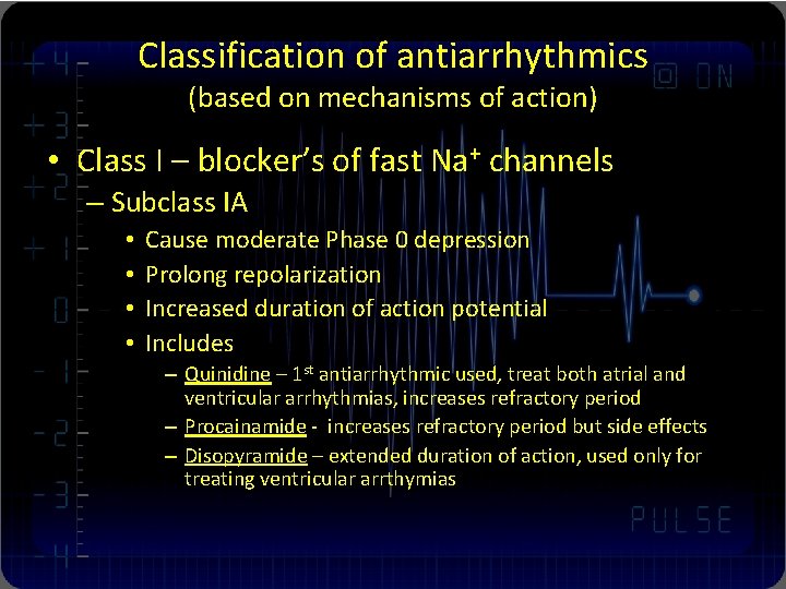
Classification of antiarrhythmics (based on mechanisms of action) • Class I – blocker’s of fast Na+ channels – Subclass IA • • Cause moderate Phase 0 depression Prolong repolarization Increased duration of action potential Includes – Quinidine – 1 st antiarrhythmic used, treat both atrial and ventricular arrhythmias, increases refractory period – Procainamide - increases refractory period but side effects – Disopyramide – extended duration of action, used only for treating ventricular arrthymias
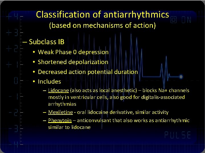
Classification of antiarrhythmics (based on mechanisms of action) – Subclass IB • • Weak Phase 0 depression Shortened depolarization Decreased action potential duration Includes – Lidocane (also acts as local anesthetic) – blocks Na+ channels mostly in ventricular cells, also good for digitalis-associated arrhythmias – Mexiletine - oral lidocaine derivative, similar activity – Phenytoin – anticonvulsant that also works as antiarrhythmic similar to lidocane
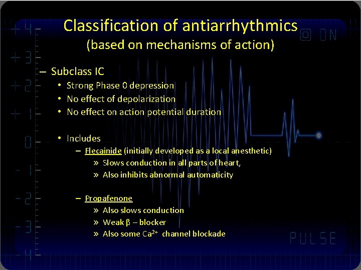
Classification of antiarrhythmics (based on mechanisms of action) – Subclass IC • Strong Phase 0 depression • No effect of depolarization • No effect on action potential duration • Includes – Flecainide (initially developed as a local anesthetic) » Slows conduction in all parts of heart, » Also inhibits abnormal automaticity – Propafenone » Also slows conduction » Weak β – blocker » Also some Ca 2+ channel blockade
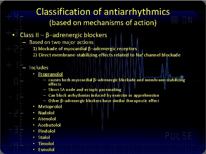
Classification of antiarrhythmics (based on mechanisms of action) • Class II – β–adrenergic blockers – Based on two major actions 1) blockade of myocardial β–adrenergic receptors 2) Direct membrane-stabilizing effects related to Na+ channel blockade – Includes • Propranolol – causes both myocardial β–adrenergic blockade and membrane-stabilizing effects – Slows SA node and ectopic pacemaking – Can block arrhythmias induced by exercise or apprehension – Other β–adrenergic blockers have similar therapeutic effect • • Metoprolol Nadolol Atenolol Acebutolol Pindolol Stalol Timolol Esmolol
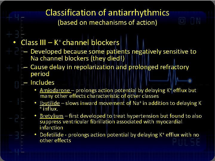
Classification of antiarrhythmics (based on mechanisms of action) • Class III – K+ channel blockers – Developed because some patients negatively sensitive to Na channel blockers (they died!) – Cause delay in repolarization and prolonged refractory period – Includes • Amiodarone – prolongs action potential by delaying K+ efflux but many other effects characteristic of other classes • Ibutilide – slows inward movement of Na+ in addition to delaying K + influx. • Bretylium – first developed to treat hypertension but found to also suppress ventricular fibrillation associated with myocardial infarction • Dofetilide - prolongs action potential by delaying K+ efflux with no other effects
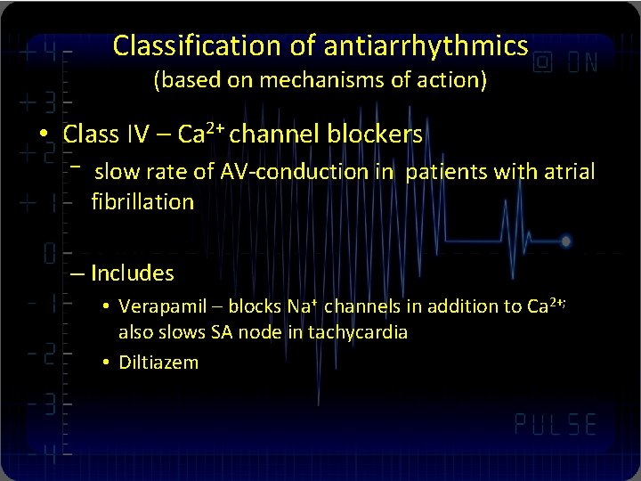
Classification of antiarrhythmics (based on mechanisms of action) • Class IV – Ca 2+ channel blockers – slow rate of AV-conduction in patients with atrial fibrillation – Includes • Verapamil – blocks Na+ channels in addition to Ca 2+; also slows SA node in tachycardia • Diltiazem
Anti-bradycardia agents 1. ß-adrenic receptor activator 2. M-cholinergic receptor blocker 3. Non-specific activator
Clinical usage Anti-tachycardia agents: • 1. 2. 3. Ia class: Less use in clinic Guinidine Procainamide Disopyramide: Side effect: like Mcholinergic receptor blocker
Anti-tachycardia agents: • Ib class: Perfect to ventricular tachyarrhythmia 1. Lidocaine 2. Mexiletine
Anti-tachycardia agents: • Ic class: Can be used in ventricular and/or supra-ventricular tachycardia and extrasystole. 1. Moricizine 2. Propafenone
Anti-tachycardia agents: • II class: ß-receptor blocker 1. Propranolol: Non-selective 2. Metoprolol: Selective ß 1 -receptor blocker, Perfect to hypertension and coronary artery disease patients associated with tachyarrhythmia.
Anti-tachycardia agents: • III class: Potassium channel blocker, extendspectrum anti-arrhythmia agent. • Amiodarone: Perfect to coronary artery disease and heart failure patients • Sotalol: Has ß-blocker effect • Bretylium
Anti-tachycardia agents: IV class: be used in supraventricular tachycardia 1. Verapamil 2. Diltiazem • Others: Adenosine: be used in supraventricular tachycardia •
Anti-bradycardia agents • • Isoprenaline Epinephrine Atropine Aminophylline
Proarrhythmia effect of antiarrhythmia agents • Ia, Ic class: Prolong QT interval, will cause VT or VF in coronary artery disease and heart failure patients • III class: Like Ia, Ic class agents • II, IV class: Bradycardia
Non-drug therapy • Cardioversion: For tachycardia especially hemodynamic unstable patient • Radiofrequency catheter ablation (RFCA): For those tachycardia patients (SVT, AF, AFL) • Artificial cardiac pacing: For bradycardia, heart failure and malignant ventricular arrhythmia patients.
Sinus Arrhythmia
Sinus tachycardia • Sinus rate > 100 beats/min (100 -180) • Causes: 1. Some physical condition: exercise, anxiety, exciting, alcohol, coffee 2. Some disease: fever, hyperthyroidism, anemia, myocarditis 3. Some drugs: Atropine, Isoprenaline • Needn’t therapy
Sinus Bradycardia Sinus rate < 60 beats/min Normal variant in many normal and older people Causes: Trained athletes, during sleep, drugs (ßblocker) , Hypothyriodism, CAD or SSS • Symptoms: 1. Most patients have no symptoms. 2. Severe bradycardia may cause dizziness, fatigue, palpitation, even syncope. • Needn’t specific therapy, If the patient has severe symptoms, planted an pacemaker may be needed. • • •
Sinus Arrest or Sinus Standstill • Sinus arrest or standstill is recognized by a pause in the sinus rhythm. • Causes: myocardial ischemia, hypoxia, hyperkalemia, higher intracranial pressure, sinus node degeneration and some drugs (digitalis, ß-blocks). • Symptoms: dizziness, amaurosis, syncope • Therapy is same to SSS
Sinoatrial exit block (SAB) • SAB: Sinus pulse was blocked so it couldn’t active the atrium. • Causes: CAD, Myopathy, Myocarditis, digitalis toxicity, et al. • Symptoms: dizziness, fatigue, syncope • Therapy is same to SSS
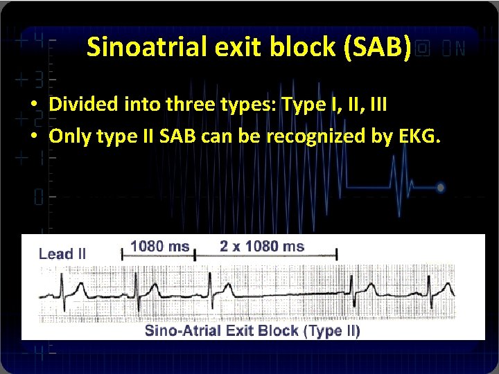
Sinoatrial exit block (SAB) • Divided into three types: Type I, III • Only type II SAB can be recognized by EKG.
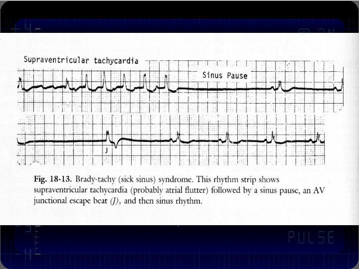
Sick Sinus Syndrome (SSS) • SSS: The function of sinus node was degenerated. SSS encompasses both disordered SA node automaticity and SA conduction. • Causes: CAD, SAN degeneration, myopathy, connective tissue disease, metabolic disease, tumor, trauma and congenital disease. • With marked sinus bradycardia, sinus arrest, sinus exit block or junctional escape rhythms • Bradycardia-tachycardia syndrome
Sick Sinus Syndrome (SSS) • EKG Recognition: 1. 2. 3. 4. 5. 6. Sinus bradycardia, ≤ 40 bpm; Sinus arrest > 3 s Type II SAB Nonsinus tachyarrhythmia ( SVT, AF or Af). SNRT > 1530 ms, SNRTc > 525 ms Instinct heart rate < 80 bmp
Sick Sinus Syndrome (SSS) • Therapy: 1. Treat the etiology 2. Treat with drugs: anti-bradycardia agents, the effect of drug therapy is not good. 3. Artificial cardiac pacing.
Atrial arrhythmia
Premature contractions • The term “premature contractions” are used to describe non sinus beats. • Common arrhythmia • The morbidity rate is 3 -5%
Atrial premature contractions (APCs) • APCs arising from somewhere in either the left or the right atrium. • Causes: rheumatic heart disease, CAD, hypertension, hyperthyroidism, hypokalemia • Symptoms: many patients have no symptom, some have palpitation, chest incomfortable. • Therapy: Needn’t therapy in the patients without heart disease. Can be treated with ß-blocker, propafenone, moricizine or verapamil.
Atrial tachycardia • Classify by automatic atrial tachycardia (AAT); intra-atrial reentrant atrial tachycardia (IART); chaotic atrial tachycardia (CAT). • Etiology: atrial enlargement, MI; chronic obstructive pulmonary disease; drinking; metabolic disturbance; digitalis toxicity; electrolytic disturbance.
Atrial tachycardia • May occur transient; intermittent; or persistent. • Symptoms: palpitation; chest uncomfortable, tachycardia may induce myopathy. • Auscultation: the first heart sound is variable
Intra-atrial reentry tachycardia (IART) • 1. 2. 3. 4. 5. ECG characters: Atrial rate is around 130 -150 bpm; P’ wave is different from sinus P wave; P’-R interval ≥ 0. 12” Often appear type I or type II, 2: 1 AV block; EP study: atrial program pacing can induce and terminate tachycardia
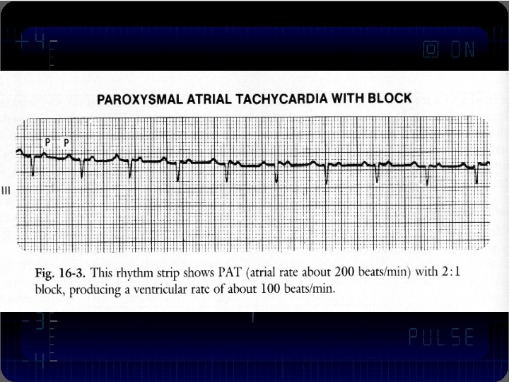
Automatic atrial tachycardia (AAT) • 1. 2. 3. 4. 5. 6. ECG characters: Atrial rate is around 100 -200 bpm; Warmup phenomena P’ wave is different from sinus P wave; P’-R interval≥ 0. 12” Often appear type I or type II, 2: 1 AV block; EP study: Atrial program pacing can’t induce or terminate the tachycardia
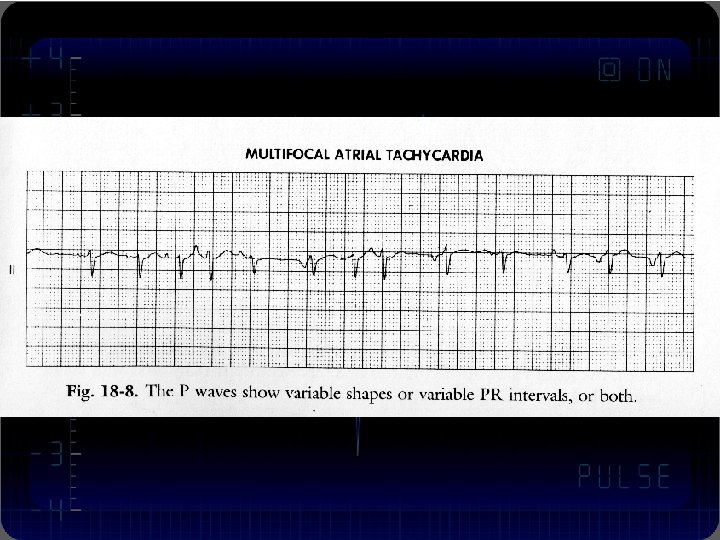
Chaotic atrial tachycardia (CAT) • • 1. 2. 3. 4. 5. Also termed “Multifocal atrial tachycardia”. Always occurs in COPD or CHF, Have a high in-hospital mortality ( 25 -56%). Death is caused by the severity of the underlying disease. ECG characters: Atrial rate is around 100 -130 bpm; The morphologies P’ wave are more than 3 types. P’-P’, P’-R and R-R interval are different. Will progress to af in half the cases EP study: Atrial program pacing can’t induce or terminate the tachycardia
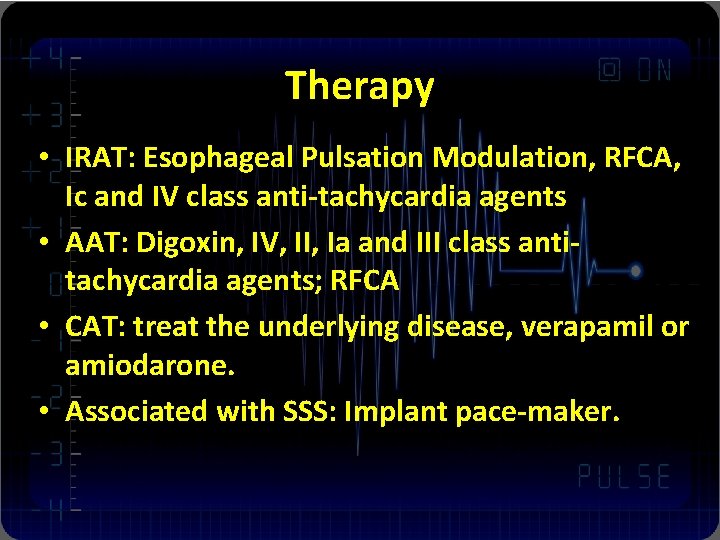
Therapy • IRAT: Esophageal Pulsation Modulation, RFCA, Ic and IV class anti-tachycardia agents • AAT: Digoxin, IV, II, Ia and III class antitachycardia agents; RFCA • CAT: treat the underlying disease, verapamil or amiodarone. • Associated with SSS: Implant pace-maker.
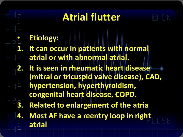
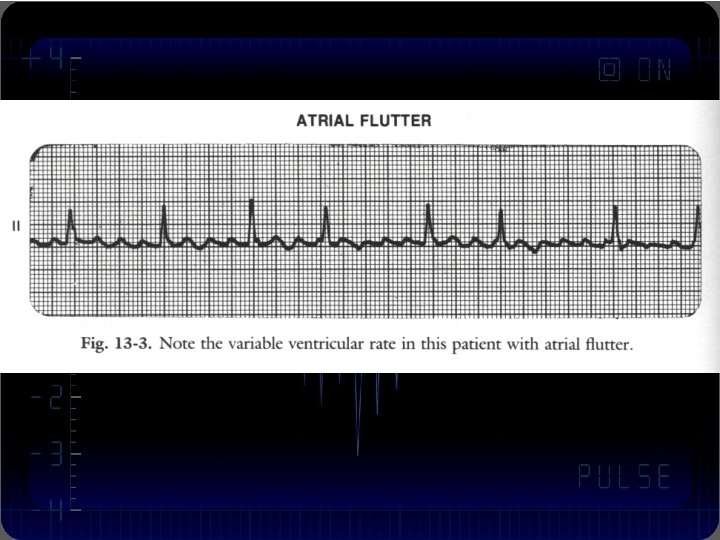
Atrial flutter • Etiology: 1. It can occur in patients with normal atrial or with abnormal atrial. 2. It is seen in rheumatic heart disease (mitral or tricuspid valve disease), CAD, hypertension, hyperthyroidism, congenital heart disease, COPD. 3. Related to enlargement of the atria 4. Most AF have a reentry loop in right atrial
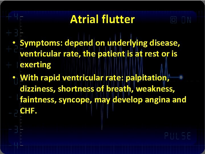
Atrial flutter • Symptoms: depend on underlying disease, ventricular rate, the patient is at rest or is exerting • With rapid ventricular rate: palpitation, dizziness, shortness of breath, weakness, faintness, syncope, may develop angina and CHF.
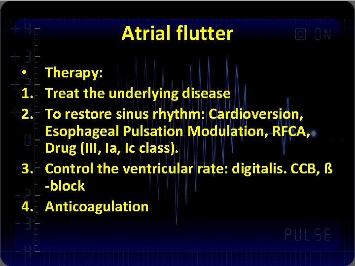
Atrial flutter • Therapy: 1. Treat the underlying disease 2. To restore sinus rhythm: Cardioversion, Esophageal Pulsation Modulation, RFCA, Drug (III, Ia, Ic class). 3. Control the ventricular rate: digitalis. CCB, ß -block 4. Anticoagulation
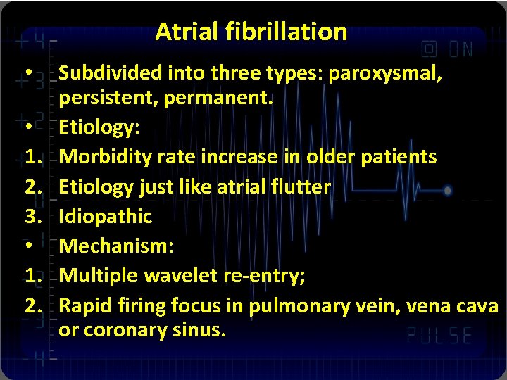
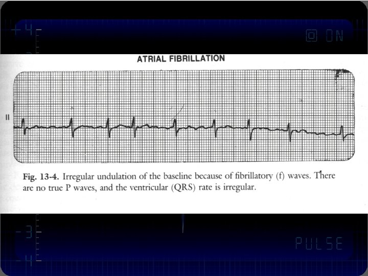
Atrial fibrillation • • 1. 2. 3. • 1. 2. Subdivided into three types: paroxysmal, persistent, permanent. Etiology: Morbidity rate increase in older patients Etiology just like atrial flutter Idiopathic Mechanism: Multiple wavelet re-entry; Rapid firing focus in pulmonary vein, vena cava or coronary sinus.
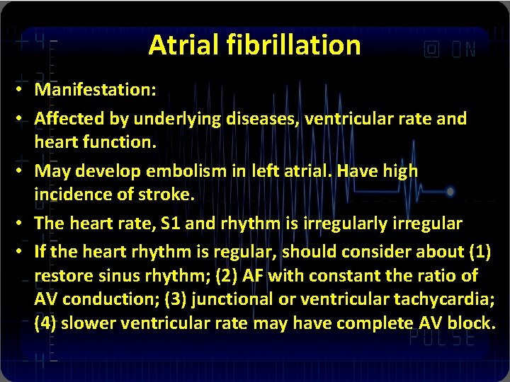
Atrial fibrillation • Manifestation: • Affected by underlying diseases, ventricular rate and heart function. • May develop embolism in left atrial. Have high incidence of stroke. • The heart rate, S 1 and rhythm is irregularly irregular • If the heart rhythm is regular, should consider about (1) restore sinus rhythm; (2) AF with constant the ratio of AV conduction; (3) junctional or ventricular tachycardia; (4) slower ventricular rate may have complete AV block.
Atrial fibrillation • Therapy: 1. Treat the underlying disease 2. Restore sinus rhythm: Drug, Cardioversion, RFCA, Maze surgery 3. Rate control: digitalis. CCB, ß-block 4. Antithrombotic therapy: Aspirine, Warfarin
Atrioventricular Junctional arrhythmia
Atrioventricular junctional premature contractions • Etiology and manifestation is like APCs • Therapy the underlying disease • Needn’t anti-arrhythmia therapy.
Nonparoxysmal AV junctional tachycardia • Mechanism: relate to hyper-automaticity or trigger activity of AV junctional tissue • Etiology: digitalis toxicity; inferior MI; myocarditis; acute rheumatic fever and postoperation of valve disease • ECG: the heart rate ranges 70 -150 bpm or more, regular, normal QRS complex, may occur AV dissociation and wenckebach AV block
Nonparoxysmal AV junctional tachycardia • Therapy: • Treat underlying disease; stopping digoxin, administer potassium, lidocaine, phenytoin or propranolol. • Not for DC shock • It can disappear spontaneously. If had good tolerance, not require therapy.
Paroxysmal tachycardia • Most PSVT (paroxysmal supraventricular tachycardia) is due to reentrant mechanism. • The incidence of PSVT is higher in AVNRT (atrioventricular node reentry tachycardia) and AVRT (atioventricular reentry tachycardia), the most common is AVNRT (90%) • Occur in any age individuals, usually no structure heart disease.
Paroxysmal tachycardia • Manifestation: • Occur and terminal abruptly. • Palpitation, dizziness, syncope, angina, heart failure and shock. • The sever degree of the symptom is related to ventricular rate, persistent duration and underlying disease
Paroxysmal tachycardia • ECG characteristic of AVNRT 1. Heart rate is 150 -250 bpm, regular 2. QRS complex is often normal, wide QRS complex is with aberrant conduction 3. Negative P wave in II III a. VF, buried into or following by the QRS complex. 4. AVN jump phenomena
Paroxysmal tachycardia • ECG characteristic of AVRT 1. Heart rate is 150 -250 bpm, regular 2. In orthodromic AVRT, the QRS complex is often normal, wide QRS complex is with antidromic AVRT 3. Retrograde P’ wave, R-P’>110 ms.
Paroxysmal tachycardia • Therapy: • AVNRT & orthodromic AVRT 1. Increase vagal tone: carotid sinus massage, Valsalva maneuver. if no successful, 2. Drug: verapamil, adrenosine, propafenone 3. DC shock • Antidromic AVRT: 1. Should not use verapamil, digitalis, and stimulate the vagal nerve. 2. Drug: propafenone, sotalol, amiodarone • RFCA
Pre-excitation syndrome (W-P-W syndrome) There are several type of accessory pathway 1. Kent: adjacent atrial and ventricular 2. James: adjacent atrial and his bundle 3. Mahaim: adjacent lower part of the AVN and ventricular • Usually no structure heart disease, occur in any age individual •
WPW syndrome • Manifestation: • Palpitation, syncope, dizziness • Arrhythmia: 80% tachycardia is AVRT, 15 -30% is AFi, 5% is AF, • May induce ventricular fibrillation
WPW syndrome • Therapy: 1. Pharmacologic therapy: orthodrome AVRT or associated AF, AFi, may use Ic and III class agents. 2. Antidromic AVRT can’t use digoxin and verapamil. 3. DC shock: WPW with SVT, AF or Afi produce agina, syncope and hypotension 4. RFCA
Ventricular arrhythmia
Ventricular Premature Contractions (VPCs) • Etiology: 1. Occur in normal person 2. Myocarditis, CAD, valve heart disease, hyperthyroidism, Drug toxicity (digoxin, quinidine and anti-anxiety drug) 3. electrolyte disturbance, anxiety, drinking, coffee
VPCs • 1. 2. 3. 4. Manifestation: palpitation dizziness syncope loss of the second heart sound
PVCs Therapy: treat underlying disease, antiarrhythmia No structure heart disease: Asymptom: no therapy Symptom caused by PVCs: antianxiety agents, ßblocker and mexiletine to relief the symptom. • With structure heart disease (CAD, HBP): 1. Treat the underlying diseas 2. ß-blocker, amiodarone 3. Class I especially class Ic agents should be avoided because of proarrhytmia and lack of benefit of prophylaxis • • 1. 2.
Ventricular tachycardia • Etiology: often in organic heart disease CAD, MI, DCM, HF, long QT syndrome Brugada syndrome • Sustained VT (>30 s), Nonsustained VT • Monomorphic VT, Polymorphic VT
Ventricular tachycardia • • 1. 2. 3. 4. 5. 6. Torsades de points (Tdp): A special type of polymorphic VT, Etiology: congenital (Long QT), electrolyte disturbance, antiarrhythmia drug proarrhythmia (IA or IC), antianxiety drug, brain disease, bradycardia
Ventricular tachycardia • Accelerated idioventricular rhythm: 1. Related to increase automatic tone 2. Etiology: Often occur in organic heart disease, especially AMI reperfusion periods, heart operation, myocarditis, digitalis toxicity
VT • Manifestation: 1. Nonsustained VT with no symptom 2. Sustained VT : with symptom and unstable hemodynamic, patient may feel palpitation, short of breathness, presyncope, angina, hypotension and shock.
VT • ECG characteristics: 1. Monomorphic VT: 100 -250 bpm, occur and terminate abruptly, regular 2. Accelerated idioventricular rhythm: a runs of 3 -10 ventricular beats, rate of 60 -110 bpm, tachycardia is a capable of warm up and close down, often seen AV dissociation, fusion or capture beats 3. Tdp: rotation of the QRS axis around the baseline, the rate from 160 -280 bpm, QT interval prolonged > 0. 5 s, marked U wave
Treatment of VT 1. Treat underlying disease 2. Cardioversion: Hemodynamic unstable VT (hypotension, shock, angina, CHF) or hemodynamic stable but drug was no effect 3. Pharmacological therapy: ß-blockers, lidocain or amiodarone 4. RFCA, ICD or surgical therapy
Therapy of Special type VT Accelerated idioventricular rhythm: usually no symptom, needn’t therapy. Atropine increased sinus rhythm Tdp: Treat underlying disease, Magnesium iv, atropine or isoprenaline, ßblock or pacemaker for long QT patient 3. temporary pacemaker • • 1. 2.
Ventricular flutter and fibrillation • Often occur in severe organic heart disease: AMI, ischemia heart disease • Proarrhythmia (especially produce long QT and Tdp), electrolyte disturbance • Anaesthesia, lightning strike, electric shock, heart operation • It’s a fatal arrhythmia
Ventricular flutter and fibrillation Manifestation: Unconsciousness, twitch, no blood pressure and pulse, going to die • Therapy: 1. Cardio-Pulmonary Resuscitate (CPR) 2. ICD •
Cardiac conduction block Block position: Sinoatrial; intra-atrial; atrioventricular; intra -ventricular • Block degree 1. Type I: prolong the conductive time 2. Type II: partial block 3. Type III: complete block •
Atrioventricular Block • AV block is a delay or failure in transmission of the cardiac impulse from atrium to ventricle. • Etiology: Atherosclerotic heart disease; myocarditis; rheumatic fever; cardiomyopathy; drug toxicity; electrolyte disturbance, collagen disease, lev’s disease.
AV Block AV block is divided into three categories: 1. First-degree AV block 2. Second-degree AV block: further subdivided into type I and type II 3. Third-degree AV block: complete block l
AV Block Manifestations: First-degree AV block: almost no symptoms; Second degree AV block: palpitation, fatigue Third degree AV block: Dizziness, agina, heart failure, lightheadedness, and syncope may cause by slow heart rate, Adams-Stokes Syndrome may occurs in sever case. • First heart sound varies in intensity, will appear booming first sound • •
AV Block • Treatment: 1. I or II degree AV block needn’t antibradycardia agent therapy 2. II degree II type and III degree AV block need antibradycardia agent therapy 3. Implant Pace Maker
Intraventricular Block • 1. 2. 3. 4. Intraventricular conduction system: Right bundle branch Left anterior fascicular Left posterior fascicular
Intraventricular Block • Etiology: • Myocarditis, valve disease, cardiomyopathy, CAD, hypertension, pulmonary heart disease, drug toxicity, Lenegre disease, Lev’s disease et al. • Manifestation: • Single fascicular or bifascicular block is asymptom; tri-fascicular block may have dizziness; palpitation, syncope and Adamsstokes syndrome
Intraventricular Block • 1. 2. 3. Therapy: Treat underlying disease If the patient is asymptom; no treat, bifascicular block and incomplete trifascicular block may progress to complete block, may need implant pace maker if the patient with syncope