Permanent Maxillary Premolars First Premolars General Characteristics Arch


 – Distinct mesial concavity – Cervical cross-section:")
 – Most common form – Short root")
 – – Resembles Type II except: Longer")


 • No deep mesial")


- Slides: 55
Permanent Maxillary Premolars First Premolars
General Characteristics: • Arch position: 4 th tooth from midline – Between the canine and 2 nd premolar • Universal #5 and # 12
General Form • Proximal geometric form is trapezoidal • Occlusally, resembles 6 -sided figure • Facially, similar to canine (5 -sided)
Form & Functions: • Two fairly equal cusps: facial and lingual • Presence of an occlusal table • Assists in tearing and piercing • Essentially a “grinding” tooth
Development Timeline: • Initial calcification: 1 1/2 to 1 3/4 years • Enamel completed: 5 - 6 years • Eruption: 10 - 11 years • Root completed: 12 - 13 years
Facial View • Closely resembles canine • Prominent buccal ridge with depressions on either side • Facial HOC at cervical third
Facial view, mesial outline: • Mesial HOC at junction of occlusal-middle thirds • Outline cervical to contact area slightly concave
Facial view, distal outline: • Similar to mesial outline • Less cervical concavity • HOC slightly more cervical than mesial
Facial view, occlusal outline: • Similar to canine except: • Cusp tip not as prominent • Cusp tip located just distal to midline of root* – Longer MB cusp ridge – Shorter DB cusp ridge *(unique)
Lingual View • Lingual narrower M-D than facial • Lingual cusp slightly shorter than facial (shortest of maxillary premolar cusps) • Lingual cusp offset to mesial • Lingual HOC at middle third
Mesial View • Trapezoidal geometric form • F and L cusps centered over root trunk • Mesial marginal groove visible
Mesial view. . . • Mesial concavity from root trunk to cervical portion of crown* • Mesial contact area facial to F-L midline
Distal View • Similar to mesial view except: • Shorter O-C than mesial – Distal marginal ridge more cervical – More of occlusal surface visible • No distinct distal concavity • Distal contact area slightly more cervical
Occlusal View • Outline is hexagon • Wider F-L than M-D • Outline tapers towards lingual • Lingual cusp offset to mesial • Mesial marginal ridge shorter than distal marginal ridge
Occlusal view. . . • Mesial outline indented by mesial marginal groove • M and D pits wellseparated • Less secondary grooves than 2 nd maxillary premolar
Occlusal view. . . • DB cusp ridge and distal marginal ridge angle is acute • MB cusp ridge and mesial marginal ridge angle is 90 o
Root Form • Single root (Type I) – Distinct mesial concavity – Cervical cross-section: kidney shape
Root form: • Bifurcated root (Type II) – Most common form – Short root trunk – Two roots: F and L, almost equal lengths
Root form: • Laminated root (Type III) – – Resembles Type II except: Longer root trunk F and L roots joined by lamination Mid-root cross-section: hour-glass shape
Root variations: • Trifurcation possible – 2 buccal roots and 1 lingual • Pulp canals: separate or connected • 2 pulp horns
How To Tell Right From Left: • • • Buccal cusp tipped distally* MB cusp ridge longer than DB* Mesial concavity* Lingual cusp tipped mesially* Mesial marginal groove*
Maxillary Second Premolar
General Characteristics: • Arch position: 5 th tooth from midline • Universal #4 and #13
Differences between 1 st and 2 nd: • Crown dimensions of 2 nd smaller than 1 st • 1 st more angular, 2 nd more rounded* • F and L cusps more equal in height with 2 nd*
Differences… • No mesial concavity or mesial marginal groove with 2 nd* • 2 nd normally single rooted • M and D pits closer together in 2 nd* • More supplemental grooves in 2 nd*
Developmental Timeline: • • Initial calcification: 2 - 2 1/4 years Enamel completed: 6 - 7 years Eruption: 11 - 12 years Root completed: 12 - 14 years
Facial View • Similar to 1 st except: – Buccal cusp of 2 nd not as long or pointed – Cusp tip mesial to root midline – MB cusp slope shorter than DB* – M and D contact areas slightly more cervical than 1 st
Lingual View • Similar to 1 st – Lingual cusp longer (equal to facial cusp) • Lingual cusp slightly offset to mesial
Mesial View • Similar to 1 st except: – – F and L cusps more equal height* No mesial concavity* Usually no mesial marginal groove Mesial contact area more cervical
Distal View • Similar to 1 st except: – F and L cusps more equal height – Distal contact area more cervical
Occlusal View • • • Line angles more rounded than 1 st* Central groove shorter* M and D pits closer together* More supplemental anatomy* M and D halves appear more symmetrical
Root Form • Usually single rooted (bifurcation rare but possible) • No deep mesial depression* • Root cross-section is ovoid
How To Tell Right From Left: • Lingual cusp tipped mesially* • MB cusp ridge shorter than DB • Root apex deviation usually towards distal
How To Tell 1 st from 2 nd: • 1 st usually bifurcated roots* • Occlusal outline of 1 st more angular, more rounded with 2 nd* • 1 st has mesial concavity*
1 st from 2 nd. . . • Central groove of 1 st longer • M and D pits of 1 st further apart* • MB cusp ridge of 1 st longer than DB (opposite with 2 nd) • Less supplemental grooves in 1 st
Tooth ID Test
Differences between 1 st and 2 nd: • Crown dimensions of 2 nd smaller than 1 st • 1 st more angular, 2 nd more rounded* • F and L cusps more equal in height with 2 nd*
Differences… • Buccal cusp tipped distally* • MB cusp ridge longer than DB* • Mesial concavity*
Differences… • No mesial concavity or mesial marginal groove with 2 nd* • 2 nd normally single rooted • M and D pits closer together in 2 nd* • More supplemental grooves in 2 nd*
Item 1 #9 Facial Mesial Lingual Occlusal Distal
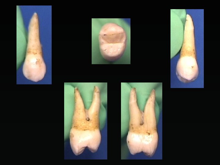
Item 2 #5 Facial Mesial Lingual Occlusal Distal
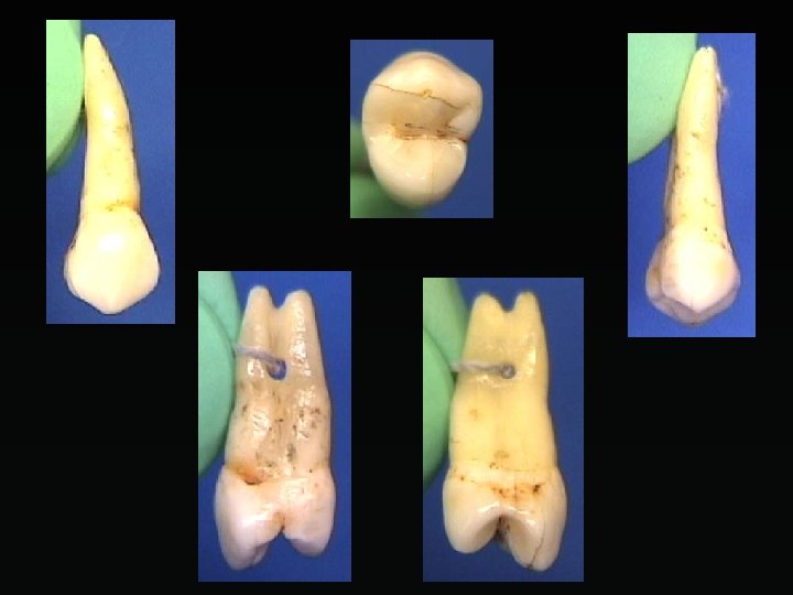
Item 3 #5 Facial Mesial Lingual Occlusal Distal
Item 4 #26 Facial Mesial Lingual Occlusal/Incisal Distal
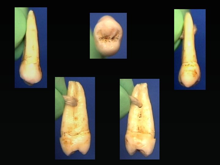
Item 5 #13 Facial Distal Lingual Occlusal Mesial
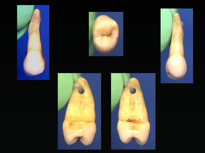
Item 6 #5 Facial Mesial Lingual Occlusal Distal
Item 7 #8 Facial Mesial Lingual Occlusal/Incisal Distal
Item 8 #11 Facial Mesial Lingual Occlusal/Incisal Distal
Item 9 #6 Facial Mesial Lingual Occlusal/Incisal Distal
Item 10 #22 Facial Mesial Lingual Occlusal/Incisal Distal
Item 11 #9 Facial Mesial Lingual Occlusal/Incisal Distal