Permanent Mandibular Molars Permanent Mandibular Molars are the

Permanent Mandibular Molars

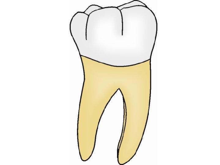
• Permanent Mandibular Molars are the largest group of teeth in the mandibular arch and are three in number on either side of the arch; 1 st, 2 nd and 3 rd molars. Like maxillary molars, these teeth are also non-successor teeth. Permanent mandibular molars help in mastication of food, to maintain proper vertical dimension of face, maintaining continuity of dental arches and also to provide fullness to the cheek. Unlike maxillary molars, all the mandibular molars are wider mesiodistally than bucco-lingually and have two roots.

General characteristics of Mandibular Molars • Shorter than other mandibular teeth but greater in other dimensions • Crown is broader mesio-distally than buccolingually • Crown tapers distally and lingually • Crown tilts distally and lingually on the root base • Lingual cusps are relatively of same size. • Two roots are present: one mesial and distal • Root trunk is shorter.

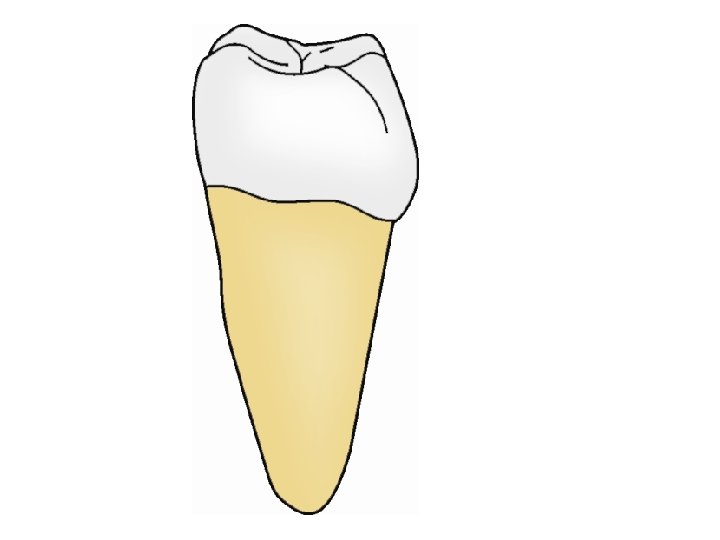
permanent Mandibular first Molars • The mandibular first molars are the largest and strongest of all the mandibular teeth and have the widest crown of all teeth in the dentition. • The mandibular first molar is the first permanent tooth to erupt into the oral cavity and is referred to as the “six-year-molar” as it erupts at 6 years. • It normally erupts slightly before the maxillary first molar and is considered as the key of occlusion. • Normally there are five functioning cusps on the occlusal surface of this tooth.

Buccal Aspect

M. B D

Crown • Shape of buccal aspect • Shape is roughly trapezoidal with the cervical and occlusal outlines representing the uneven sides. • Crown is broader mesio-distally than cervicoocclusally.

Outline of buccal aspect • Mesial outline is relatively straight or slightly concave from the cervical line to the contact area which is located at the junction of occlusal and middle third • Distal outline is straight or slightly convex from cervix to the contact area which is at the middle of middle third beneath the distal cusp. • Cervical line is nearly straight, regular and curves slightly to the root.

• Occlusal outline is represented by the buccal cusps and the cusp slopes. • three buccal cusps can be seen; the mesio-buccal cusp, disto-buccal cusp and a distal cusp. • The mesio-buccal cusp is the longest and widest, followed by disto-buccal cusp which is smaller and shorter and a distal cusp which is the smallest and the pointed than the other buccal cusps.

• The smallest cusp on the buccal aspect is called the distal cusp because the major portion of the cusp is located on the distal part of the crown and only a small portion is seen on the buccal aspect. • From the buccal aspect, a portion of mesiolingual and disto-lingual cusps are also seen, as they are longer than the buccal cusps.

Buccal surface • Buccal surface of first molars is smooth and convex and shows two developmental grooves. • The groove that separates the mesio-buccal and disto-buccal cusp is the mesio-buccal groove which extends up to the middle third and ends in a pit. • The disto-buccal groove separates disto-buccal and distal cusp, which ends at the cervical third without a distinct pit. • The cervical portion of buccal aspect may show a prominent ridge running in a mesio-distal direction which is referred to as buccal cervical ridge

Roots • Mandibular first molar has two roots; mesial and a distal root. • The level of bifurcation is 3 mm below the cervical line. • Since the bifurcation is closer to the cervical line the root trunk is short. • The mesial root is the wider and the stronger of the two. The mesial and distal roots show a distal tilt. • The tip of mesial root is almost in line with the mesio-buccal cusps and of the distal root is often in line or distal to the distal surface of crown.

Lingual Aspect

Crown Shape of lingual aspect • From the lingual aspect the tooth shows a convergence lingually, making a part of mesial and distal surfaces visible. • The degree of lingual convergence is more prominent distally. • Tooth also tapers to the cervical region.

Outlines of lingual aspect Mesial outline is slightly convex. With the crest of contour located at the junction of occlusal and middle third. Distal outline is relatively straight with the crest of curvature located on the distal surface of the distal cusp. Cervical line is slightly irregular and relatively flat.

Occlusal outline is represented by the cusps and the cusp slopes. On this aspect mainly two cusps are seen: mesio-lingual and disto-lingual. Because of the lingual convergence the distal portion of the distal cusps may be visible from this aspect. The mesio-lingual cusp and the disto-lingual cusp are the longest and the sharpest of the five cusps. The mesio-lingual cusp is longer than the disto-lingual cusp and the width may be equal or slightly more than that of the disto-lingual cusp.

Lingual surface • is smooth and convex at the coronal 1/3 rd and almost flat at the cervical region. • Lingual developmental groove extends on to a short distance on to the lingual surface demarcating both the lingual cusps.

Root • From this aspect both the mesial and distal roots are seen which show a lingual convergence. • The root trunk appears to be longer because of the occlusal placement of cervical line. • The level of bifurcation is 4 mm above the cervical line.
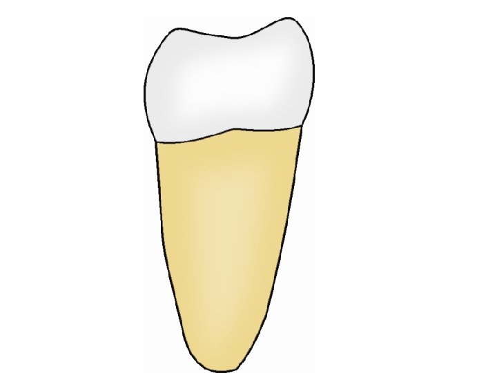

Mesial Aspect Crown Shape of Mesial aspect • Shape is rhomboidal with the crown tilted lingually on the root axis (arch trait). • A greater bucco-lingual measurement of the crown and the root can be appreciated from this aspect.

Outline of Mesial aspect • Buccal outline is noticeably convex at the cervical third where the crest of convexity is located, in the region of the buccal cervical ridge. As the buccal outline continues occlusally it becomes less convex and shows a lingual inclination. • Lingual outline is relatively straight in the cervical third, becomes convex at the middle third where the crest of convexity is located. • Cervical line is irregular and slightly convex towards the occlusal aspect. The cervical line on the lingual surface is at a higher level than the buccal by about 1 mm. This difference in the level of cervical line can be appreciated from the mesial aspect.

Occlusal outline • is represented by the cusps and the marginal ridges. • Two cusps can be seen from mesial aspect: the mesio -buccal and mesio-lingual cusp. • Mesio-lingual cusp is longer and sharper and is in line with the lingual surface of mesial root. • A well developed mesial marginal ridge is seen which is slightly concave and is placed occlusally.

Mesial surface • Mesial surface is smooth and relatively convex except for a slight concavity cervical to the contact area.

Root • Only mesial root is visible from this aspect because the broad mesial root superimposes the narrower distal root. The outline of mesial root is relatively straight up to the junction of cervical and middle third and from there it tapers to a blunt apex. The apex is located directly below the mesio-buccal cusp.

Distal Aspect Crown • The general morphology of distal aspect is similar to that of mesial aspect. The crown is shorter distally than mesially. Due to the distal convergence of the crown, a part of buccal and lingual surface is also seen from this aspect. The distal convergence of the buccal surface is more pronounced than that of the lingual surface. • Curvature of cervical line is less than on mesial aspect.

Differences are • 1. The distal marginal ridge is short, curved and is more cervically located than the mesial marginal ridge. • 2. Because of the distal tilt of the crown and cervical placement of the marginal ridge most of the occlusal surface and all cusps are seen from this aspect.

• Root • The distal root and a part of the mesial root are visible from this aspect. The distal root is narrower than the mesial root and ends in a pointed apex.

Occlusal Aspect

Shape • The occlusal aspect is roughly quadrilateral in shape • the mesio-distal dimension more than bucco-lingual with a difference of 1 mm or more. • The lingual and the distal convergence of the crown can be well appreciated. • Because of the lingual tilt when tooth is viewed from occlusal aspect much of buccal surface also can be seen. • The mesial outline is slightly convex and the contact area is centered in a bucco-lingual direction. • The distal contact area is located buccal to the centre point of distal marginal ridge.

BUCCAL D M E S I A L I S T A L LINGUAL

BUCCAL D M E S I A L I S T A L LINGUAL

• The occlusal surface • Occlusal surface shows various anatomic landmarks such as cusps, ridges, fossae, pits and grooves.

BUCCAL D. B M. B D M E S I A L D I S T A L M. L D. L LINGUAL

Cusps • Mandibular first molar has five cusps: three buccal cusps and two lingual cusps. • The buccal cusps are mesio-buccal, disto-buccal and distal cusps. • Of the three buccal cusps the mesio-buccal cusp is the largest followed by disto-buccal and the distal cusp. • The distal cusp is the smallest and the sharpest and is located at the disto-lingual line angle.

• Lingually there are two cusps: the mesiolingual and disto-lingual cusps. • The mesio-lingual cusp and the disto-lingual cusp are the longest and the sharpest of the five cusps. • The mesio-lingual cusp is longer than the disto -lingual cusp and the width may be equal or slightly more than that of the disto-lingual cusp.

Ridges • a. Triangular ridges are seen extending from the tips of all five cusps towards the central part of occlusal surface. Triangular ridges of lingual cusps are longer than that of buccal cusps. • b. Transverse ridge: The triangular ridge of the mesio-buccal cusp meets the triangular ridge of the mesio-lingual cusp to form a transverse ridge. Similarly a transverse ridge is also formed by the triangular ridges of both the disto-buccal and distolingual cusps. •

• c. Mesial marginal ridge: forms the mesial boundary of the occlusal aspect and is located more occlusally than the distal marginal ridge. It is placed 1 mm below the level of the cusp tips • d. Distal marginal ridge: is located at distal margin of occlusal aspect. It is shorter, concave and more cervically placed. • e. Cusp ridges: forms the buccal and the lingual boundaries of the occlusal aspect

BUCCAL D. B M. B D M E S I A L D I S T A L M. L D. L LINGUAL
 and two minor")
Fossae • Three fossae can be seen; one major (central fossa) and two minor (mesial and distal triangular fossae).

• 1. Central fossa is bounded by the distal slope of the mesio-buccal cusp, mesial and distal slope of the disto-buccal cusp, mesial slope of the distal cusp, triangular ridges of distal and disto-lingual cusps, mesial slope of distolingual cusp, distal slope of mesio-lingual cusp and the transverse ridge.
")
• 2. Mesial triangular fossa is a triangular shaped depression located inner (distal) to the mesial marginal ridge. • 3. Distal triangular fossa is less distinct and is located inner (mesial) to distal marginal ridge.

BUCCAL D. B M. B D M E S I A L D I S T A L M. L D. L LINGUAL

BUCCAL D. B M. B D M E S I A L D I S T A L M. L D. L LINGUAL

Pits are present as small pinpoint depression at the deepest part of all fossae, where the developmental grooves converge. The pits are named according to the fossa in which they are located: central pit, mesial pit and distal pit.
 • Developmental grooves • 1. Central groove:")
Grooves (Developmental and supplemental grooves are seen) • Developmental grooves • 1. Central groove: is the major groove seen on the occlusal aspect and is centrally located dividing occlusal surface into buccal and lingual halves. It starts from the central pit and runs in a mesial direction between the mesio-buccal and disto-lingual cusp to end in the mesial triangular fossa. The distal extension of the central groove runs between the disto-buccal and disto-lingual cusps to end in distal triangular fossa. The central groove follows a zigzag pattern

• 2. Mesio-buccal groove: this groove starts from the central fossa, slightly mesial to the origin of central groove and traverse in a buccal direction between the mesio-buccal and disto-buccal cusps and extend on to the buccal surface. • 3. Disto-buccal groove: starts from the distal portion of the central groove and traverse in a buccal direction between the disto-buccal and distal cusp and extends on to the buccal surface. • 4. Lingual groove: starts in the central pit, extends lingually between the two lingual cusps and on to the lingual surface.

BUCCAL D. B M. B D M E S I A L D I S T A L M. L D. L LINGUAL

BUCCAL Mesio Buccal Gv D. B M. B D M E S I A L D I S T A L Central GV M. L Mesio Buccal Gv D. L LINGUAL

Supplementary grooves • In addition to the developmental groove there are supplementary grooves in triangular fossae extending to a buccal and lingual direction. • Supplementary grooves are less distinct in the distal triangular fossa.

Variations • 6 th cusp may be present on the distal marginal ridge called as tuberculum sextum. • If 6 th cusp is present between the lingual cusps, it is called tuberculum intermedium.

Diff. between Maxillary and Mandibular First Molars • Maxillary first molars • 1. Crown is bucco-lingually broader than mesio-distally • 2. Have four major cusps: two buccal and two lingual cusps. • 3. One accessory cusp i. e. ‘Cusp of Carabelli’ is also present and is seen lingual to mesiolingual cusp. • 4. Lingual cusps are of different size; large mesio-lingual and a smaller disto-lingual cusp • • Mandibular first molars • Crown is mesio-distally broader than bucco-lingually • Have five cusps: three buccal and two lingual cusps • No such cusp is seen • Lingual cusps are nearly of equal size

• 5. Buccal surface is relatively flat • 6. Occlusal aspect is rhomboidal in shape • 7. Occlusal aspect shows buccal convergence • 8. A prominent oblique ridge is seen on occlusal aspect extending from mesiolingual to disto-buccal cusp. • 9. Occlusal aspect has four fossae: two major and two minor fossae • Buccal surface is convex and inclined lingually. • Occlusal aspect is quadrilateral • Occlusal aspect shows a lingual convergence • No such oblique ridge is seen on occlusal aspect • Occlusal aspect has only three fossae: one major and two minor fossae

Mandibular Second Molar

• Mandibular Second Molars are two in number, one on either side of the arch, situated distal to the mandibular first molars. • They supplement the first molar in function. • Although the second molar resembles a first molar in its general morphology, few differences can be observed.

Major differences of Second molar from first molar are a. Crown is smaller and symmetrical from all aspects. b. Only four cusps are present – 2 buccal, 2 lingual c. Only one buccal groove d. Roots are narrow and not well separated and root trunk is longer

Buccal aspect

Buccal Aspect • Shape is roughly trapezoidal with the cervical and occlusal outlines representing the uneven sides. Tooth is wider mesiodistally than the crown length. The degree of cervical convergence is less therefore the tooth appears to be wider at the cervix. Crown tilts distally so the distal side appears to be shorter. • Outline • Mesial outline is straight with the contact area located at the junction of middle and occlusal 1/3. • Distal outline is more convex and the contact area is at the middle of middle 1/3 rd.

• Occlusal outline is represented by the buccal cusps and the cusp slopes. On this aspect mainly 2 buccal cusps can be seen: the mesiobuccal cusp and disto-buccal cusp. Lingual cusps are also visible because they are longer than the buccal cusps. • Cervical line is relatively straight or may curve sharply towards the root.

• Occlusal outline is represented by the buccal cusps and the cusp slopes. On this aspect mainly 2 buccal cusps can be seen: the mesiobuccal cusp and disto-buccal cusp. Lingual cusps are also visible because they are longer than the buccal cusps. • Cervical line is relatively straight or may curve sharply towards the root.

Buccal surface • is smooth and convex. • The buccal groove extends between the mesio -buccal and disto-buccal cusps which ends at the middle third of the surface in a pit. The cervical portion of buccal aspect may show a prominent ridge running in a mesio-distal direction which is referred to buccal cervical ridge (sometimes called as buccal cingulum)

ROOT • Two roots are present; mesial and distal roots. The level of bifurcation is more apical when compared to that of first molar. Both mesial and distal roots are usually closer together, nearly parallel and ending in a pointed tip.

Lingual aspect

Lingual Aspects • Tooth shows convergence lingually but to a lesser extent than that of first molar. • Mesial and distal outlines are more convex. • Occlusal outline is represented by the lingual cusps and the cusp slopes. Two lingual cusps are seen; mesio-lingual cusp and a disto-lingual cusp. The mesio-lingual cusp is slightly wider and longer of the two. • Cervical line is regular.

• Lingual surface is smooth and convex. The lingual groove extends between the mesiolingual and disto- lingual cusps which is shorter than the buccal groove, • ROOT • Two roots, mesial and distal roots are seen which end in a pointed apex.

Mesial aspect

Mesial Aspect • From this aspect second molar resembles that of first molar except for the differences in measurement. • Outlines • Buccal outline is noticeably convex at the cervical third (crest of convexity) in the region of the buccal cervical ridge. As the buccal outline continues occlusally it becomes less convex and shows a lingual inclination. • Lingual outline is nearly straight or slightly convex with crest of convexity at the middle third. • Cervical line is regular and straight with a slight curvature occlusally.

• Occlusal outline is represented by the cusps and the marginal ridge. Two cusps are seen: mesio-lingual and mesio-buccal cusps. Mesiolingual cusp is longer. The mesio-buccal cusp tip is lingual to the buccal outline of mesial root. Mesial marginal ridge is concave and more occlusally placed. • Mesial Surface is smooth and convex

Root • Only mesial root is visible from this aspect because the mesial root is broad enough to hide the distal root.

Distal aspect

Distal aspect • General morphology of distal aspect resemble that of mesial aspect. Differences are • The distal convergence of the tooth makes a portion of buccal and lingual surfaces visible from this aspect. • In addition to disto-buccal and disto-lingual cusps, a part of mesial cusps are also seen. • The distal marginal ridge is concave and more cervically located. • Distal tilt of the crown and cervically located marginal ridge allows most of the occlusal aspect also to be visible from this aspect.

Occlusal aspect

Occlusal Aspect • The tooth when viewed from occlusal aspect has a roughly rectangular shape which is wider in a mesio-distal direction than the bucco-lingual. • The occlusal outline shows a distal and lingual convergence. The extent of the lingual convergence is lesser than the first molar • Mesial outline of the tooth is straight while distal outline is convex. • Because of the lingual tilt when tooth is viewed from occlusal aspect much of buccal surface also can be seen. The mesiobuccal portion of the buccal surface shows a prominent bulge, representing the cervical ridge.

• The occlusal surface shows various anatomic landmarks such as cusps, ridges, fossae, pits and grooves

Cusps • Four cusps are present; the mesio-lingual, disto-lingual, mesio-buccal and disto-buccal. • The mesio-buccal and mesio-lingual cusps are larger than disto-buccal and disto-lingual cusps. • Unlike the mandibular first molars, distal cusp is absent in second molar

Ridges • Triangular ridges are seen extending from the tips of all the four cusps towards the central part of occlusal surface • Transverse ridges – triangular ridges of mesio-buccal and mesio-lingual cusps meet to form a transverse ridge. Similarly a transverse ridge is also formed by the triangular ridges of both the distal cusps. • Mesial marginal ridge – forms the mesial boundary of the occlusal aspect and is located more occlusally than the distal marginal ridge. • Distal marginal ridge – is located at distal margin of occlusal aspect. It is concave and more cervically placed. • Cusp ridges – forms the buccal and the lingual boundaries of the occlusal aspect

Fossae – The central fossa is the largest fossa located at the center of the occlusal aspect. – Mesial triangular fossa is a triangular shaped depression located inner ( distal) to the mesial marginal ridge. – Distal triangular fossa is less distinct and is located inner (mesial) to distal marginal ridge.

Pits • Pits may be present in any of the fossae where the grooves converge.

Grooves • Developmental grooves • Central groove – Begins from central fossa and extends in a mesial and distal direction to end in the mesial triangular fossa and distal triangular fossa respectively. The central groove is relatively straight in second molar when compared to zigzag pattern in first molar. • Buccal groove runs from the central fossa in a buccal direction separating two buccal cusps which also extends to the buccal surface. • Lingual groove extends from central fossa between the two lingual cusps.

Supplementary grooves • There may be many supplementary grooves radiating from the developmental grooves making the occlusal surface irregular. • The developmental grooves arising from central fossa give a criss-cross pattern.

Differences between mandibular first and second molars Second molar First molar • Smaller in all • Larger in all dimensions • Has five cusps, • Has only four cusps, • Three buccal cusps and • Two buccal cusps and two lingual cusps, two lingual cusps Distal cusp is absent • Buccal surface show two buccal grooves • Only one buccal groove

• Less cervical constriction • Crown has a quadrilateral shape from the occlusal aspect • Roots are widely separated • Grooves on occlusal aspect show a zigzag pattern • More cervical constriction • Rectangular • Roots are close together • Grooves on occlusal aspect show a cross pattern

Mandibular 3 rd Molars • Mandibular 3 rd molars are extremely variable in morphology which may resemble a second molar (4 cusps) in most of the cases. Few specimens may also resemble a first molar (5 cusps). It supplements the mandibular second molar in function. Mandibular third molars are most likely to be impacted or congenitally missing.

General features are • Rounded occlusal outline with a narrow occlusal table • Crown is bulbous and is tilted distally on root axis. • Larger and longer mesio-lingual cusp, with short and rounded buccal cusps • Occlusal surface has irregular groove pattern. • Roots are shorter, either fused or separated with more distal tilt.
- Slides: 85