Peritoneum Anatomy functions Acute Peritonitis Intraperitoneal abscess Special

Peritoneum: Anatomy, functions Acute Peritonitis Intraperitoneal abscess Special types of peritonitis Peritoneal neoplasms Dr. Amit Gupta Associate Professor Dept. of Surgery

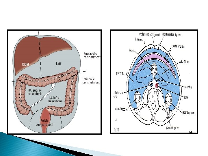
Surgical Anatomy Peritoneum is the largest serous membrane in the body Surface area: approx. 22, 000 cm 2. Divided into parietal and visceral portions Parietal layer lines the abdominal and pelvic cavities and the abdominal surface of the diaphragm. loosely connected with the body wall, separated from it by an adipose layer, tela subserosa Visceral layer covers the abdominal and pelvic viscera and includes the mesenteries. visceral peritoneum is usually tightly attached to the organs it covers.

It does not line the entirety of the abdominopelvic cavity. It is lifted from the body wall, especially posteriorly, by organs located against the wall during embryologic development. This chain of events causes the formation of a retroperitoneal space between the peritoneum and the body wall, with organs situated within the space. An organ that is covered only in part by the peritoneum is referred to as a retroperitoneal organ. An organ that is covered by peritoneum essentially everywhere except for the site of entrance of vessels is referred to as an intraperitoneal organ.

Innervation of peritoneum Parietal peritoneum is sensitive to pain, pressure, temperature & touch Parietal peritoneum is supplied by: T 7 -- T 12, L 1 nerve phrenic nerve. Visceral peritoneum is sensitive to stretch & tearing. It is supplied by autonomic afferent nerves which supply the viscera. NB. Parietal peritoneum of the pelvis is supplied by Obturator nerve.

Functions of peritoneum It suspend the organs within the peritoneal cavity. It fixes some organs within the abdominal cavity. Storage of large amount of fat in the peritoneal ligaments (e. g. . Greater omentum) Peritoneal covering of intestine tends to stick together in infection Greater omentum is called the policeman of abdomen to prevent spread of infection It secretes the peritoneal fluid

Peritoneal fluid is pale yellow fluid rich in leukocytes Mobile viscera glide easily on one another. Peritoneal fluid moves upward towards subphrenic spaces- whatever the position of the body by: ◦ Movements of diaphragm. ◦ Movements of abdominal muscles ◦ Peristaltic movements. Peritoneum is extensive in the region of diaphragm.

Peritonitis

Peritonitis – inflammation of the peritoneum which maybe localised or generalised Peritonism – refers to specific features found on abdominal examination in those with peritonitis Characterised by tenderness with guarding Rebound /percussion tenderness on examination Eased by lying still and exacerbated by any movement Maybe localised or generalised Generalised peritonitis is a surgical emergency – requires resuscitation and immediate surgery

Types Primary: ◦ Not related to intraabdominal abnormality ◦ Also called spontaneous bacterial peritonitis Secondary: ◦ Due to spillage of GI or GU organisms into peritoneal space due to breach of mucosal barrier Tertiary: ◦ clinical peritonitis and systemic signs of peritonitis persist after treatment of secondary peritonitis ◦ No/low virulence organism isolated

Causes Infective – bacteria cause peritonitis most common cause of peritonitis Non-infective – leakage of certain sterile body fluids into the peritoneum can cause peritonitis. Note: although sterile at first these fluids often become infected within 24 -48 hrs of leakage from the affected organ resulting in a bacterial peritonitis

Clinical features Pain Constant and severe Worse on movement Eased by lying still Signs of ileus (generalised peritonitis > localised peritonitis) Distension Vomiting Tympanic abdomen with reduced bowel sounds Signs of systemic shock Tachycardia, tachypnoea, hypotension, low urine output More prominent with generalised than localised peritonitis

Investigations Diagnosis most often made on history and examination If localised peritonitis Bloods tests Chest X Ray ECG Complex investigations are requested depending on suspected diagnosis If generalised peritonitis Surgical emergency – will require emergency operation Following investigations should be performed: Bloods: FBC, U&E, LFT, Amylase!! CRP, clotting, G&S, ABG Chest X ray CT scan

Management ABC Oxygen Fluid resuscitation IV antibiotics Analgesia Surgery

Pelvic Abscess

Ø Rare but the most serious late postop complication Ø Involve one or both residual adnexa (tubo-ovarian abscess) occur almost exclusively in premenopausal women occur despite prophylactic AB Ø Ø often have a latent period of many between surgery and onset of symptoms
 Ø")
Clinical features Ø fever (high spike late in the afternoon or early evening) Ø palpable mass high in the pelvis Ø WBC: around 20, 000/mm Ø ESR

Ultrasonography and CT scan Ø confirm the presence of a mass Ø help to determine whether it is Loculated related to an intraperitoneal structure drainable percutaneously

• Immediate drainage is not mandatory if it is inaccessible AB therapy alone may be successful • isolation of β-lactamase–producing Prevotella species use of clindamycin, metronidazole, or other agents against gram-negative anaerobes

Clindamycin + gentamicin fails to respond drainage Necrosis+infections surgical exploration in some cases Aerobic and anaerobic culture of purulent material or tissue

Primary peritoneal tumours

Defined As Tumors With Primary Manifestation In The Peritoneum In The Absence Of A Visceral Site Of Origin Arise From Mesothelial Cells, Sub Mesothelial Mesenchymal cells, and uncommitted stem cells

Classification

Differential diagnosis

Peritoneal Malignant Mesothelioma Uncommon Malignant Neoplasm Arises From Mesothelial Cells Or Multipotential Subserosal Mesenchymal Cells Account for 6%– 10% of malignant mesotheliomas Diffuse: highly aggressive, are incurable Localized: good prognosis following complete surgical excision

Etiology Exposure To Higher Levels Of Asbestos, Erionite Therapeutic Irradiation Exposure To Simian Virus 40 Chronic pleural or peritoneal irritation Majority in males Median age: 60 years

Clinical presentation Abdominal Pain Or Discomfort Abdominal Distension Increasing Abdominal Girth Nausea, Anorexia Weight Loss Bowel Obstruction Palpable Abdominal or pelvic mass
 scattered over the omental surfaces B: macrolobulated mass with")
A: innumerable tumor nodules (arrows) scattered over the omental surfaces B: macrolobulated mass with foci of intratumoral degeneration and hemorrhage

Imaging Features Nodular Thickening Of The Peritoneum Omental Caking: Fine, Nodular, Soft-tissue Studding Ascites: diffuse to focal, small, loculated collections

Barium examination shows separation of small bowel segments and irregular fold thickening of small bowel segments

A: Axial CT scans: show ascites and omental nodule B: large, heterogeneously enhancing mass in the greater omentum

Treatment The most effective treatment for peritoneal mesothelioma is: Cytoreductive Surgery Hyper thermic Intraperitoneal Chemotherapy (HIPEC)

Primary Peritoneal Serous Carcinoma Epithelial tumor that arises from the peritoneum Almost always occurs in women (mean age, 56– 62 years) Clinical features: ◦ ◦ ◦ abdominal distension Pain, nausea and vomiting Increasing abdominal girth ascites elevated serum levels of cancer antigen CA-125

Multiple nodules on omentum Omental caking Psammoma bodies are commonly present So reffered as psammomacarcinoma

The following criteria have been established to make the diagnosis of primary peritoneal serous carcinoma: ◦ Both ovaries are normal ◦ Involvement of extra ovarian sites must be greater than the involvement on the surface of either ovary ◦ Ovarian involvement is limited to ovarian surface epithelium, either without stromal invasion or involving the cortical stroma with tumor size less than 5 x 5 mm

Imaging Features Ascites Peritoneal nodules and thickening Omental nodules and masses
")
CT scan: showing calcified nodules and soft-tissue caking of the greater omentum (arrowheads)
 Chemotherapy (Cisplatin, Taxol based)")
Treatment Cytoreductive Surgery (optimal < 1 cm residual disease) Chemotherapy (Cisplatin, Taxol based)

Primary Peritoneal Serous Borderline Tumor Rare lesion of low malignant potential Tumor cells do not invade into the submesothelial layers of the peritoneum or omental fat Female patients, 16 - 67 years of age (mean: 33 years) Treated by surgical resection (omentectomy, hysterectomy, and oophorectomy) Have a good long-term prognosis
 Rare, benign entity Innumerable smooth muscle nodules throughout")
Leiomyomatosis Peritonealis Disseminata (diffuse peritoneal leiomyomatosis) Rare, benign entity Innumerable smooth muscle nodules throughout the peritoneal cavity Associated with high estrogen states, caused by pregnancy and oral contraceptive use Spontaneous regression of the leiomyomas or regression following withdrawal of ovarian hormones or oophorectomy
- Slides: 40