Peritoneal Reflections and the Abdominal Cavity Budoor Al

Peritoneal Reflections and the Abdominal Cavity Budoor Al. Qinai

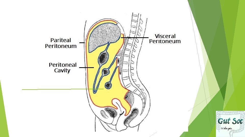
WHAT is the Peritoneum? Thin serous membrane: lines abdominal cavity and viscera within it Parietal Peritoneum Visceral Peritoneum Peritoneal Space: Potential Space between the Parietal and Visceral Peritoneum, contains peritoneal fluid

Basic

Omenta vs. Mesentery Omenta: Doubling up of visceral Peritoneum between stomach and other organs (Lesser and grater Omentum) Mesentery: Where visceral peritoneum doubles up around organ and attaches it to PAW (ex. Transverse Mesocolon) Keep in mind Visceral and Parietal Peritoneum are continuous!

Omenta vs. Mesentery: A Closer Look

Intra vs. Retroperitoneal: Intraperitoneal: Almost completely covered by peritoneal cavity Retroperitoneal: Part of Surface Covered by Peritoneum Pancreas head + body , 2 nd 4 th parts of Duodenum

Intraperitoneal Retroperitoneal Stomach Remainder of Duodenum 1 st part of Duodenum Ascending Colon Jejenum Descending Colon Ileum Rectum (middle 1/3 rd) Cecum Pancreas (except tail) Appendix Transverse Colon Sigmoid Colon Rectum (upper 1/3 rd) Remember: If it has a mesentery it is INTRAPERITONEAL!

Specific Mesentery

SAD PUCKER: Retroperitoneal Organs S = Suprarenal/ Adrenal Glands A = Aorta/IVC D = Duodenum (except for 1 st part) P = Pancreas (except for the tail) U =Ureters C =Colon (Ascending/ Descending Parts) K = Kidneys E = (O) Esophagus R = Rectum

Secondarily Retroperitoneal Became Retroperitoneal during development

Intermediate

Lesser and Greater Omentum Lesser Omentum: connects Stomach to Porta Hepatis of Liver Greater Omentum: connects Greater Curvature of Stomach to Transverse Colon
 Greater")
Lesser and Greater Sac Lesser Sac: Behind Stomach and Liver (shaded in blue) Greater Sac: Makes up most of cavity: starts all the way up in diaphragm

Looking Closely at the Liver Contains Ligaments: Folds of Peritoneum that anchor the liver into place Falciform Ligament: attaches liver to Anterior abdominal wall to inner surface of rectus sheath
 Round ligament: At the base (free margin)")
Looking Closely at the Liver (Cont. ) Round ligament: At the base (free margin) of the falciform ligament Round ligament is a remnant of the umbilical vein : passes in groove between quadrate and left lobe
 for a")
Can you label these? Please see Mc. Minn’s Atlas (or any atlas) for a liver from a specimen as these cannot be shared online as per policy!

Coronary Ligaments


Superior View of the Liver You can see Falciform Ligaments separating Right and Left Lobes Coronary Ligaments coming down from diaphragm- left and right coronary

Inferior/ Posterior Surface of the Liver BARE AREA: In Direct Contact with DIAPHRAGM

Ligamentum Venosum Fissure for Ligamentum Venosum -Fibrous Remnant of Ductus Venosus DIFFERENTIATE BETWEEN TWO LIGAMENTS! Lesser Omentum: From Ligament Venosum and Porta Hepatis LO: connects liver to lesser curvature of stomach and first part of duodenum

Lesser Omentum

Hepatogastric and Hepatoduodenal ligament of LO

Greater Omentum Hangs down In front of loop of small intestines Double Layered Fold of Peritoneum Anterior fold of greater Omentum and posterior foldattaches to transverse colon Lesser sac- B/w these two folds

Margins of Lesser Sac Anterior Wall: • Peritoneum on Caudate Lobe of Liver • Lesser Omentum • Posterior Surface of Stomach • Greater Omentum (Anterior 2 layers) Posterior Wall: • Greater Omentum (Posterior r 2 layers) • Transverse colon +Mesocolon
 Posteriorly: •")
Epiploic Foramen Boundaries Anteriorly: • Free border of LO ( portal triad) Posteriorly: • IVC Superiorly: • Caudate Process of Caudate Lobe of Liver Inferiorly: • 1 st part of Duodenum
 Posteriorly: •")
Epiploic Foramen Boundaries Anteriorly: • Free border of LO ( portal triad) Posteriorly: • IVC Superiorly: • Caudate Process of Caudate Lobe of Liver Inferiorly: • 1 st part of Duodenum

Epiploic Foramen on a Cartoon
")
Epiploic Foramen on a Cadaver Please see Mc. Minn’s Atlas (or any other atlas) for an Epiploic Foramen from a specimen as these cannot be shared online as per policy!
 for")
Greater Omentum on a Cadaver Please see Mc. Minn’s Atlas (or any atlas) for the greater omentum from a specimen as these cannot be shared online as per policy!

Specific Mesentery

End Note: Trick Question: How many Layers?

QUIZ TIME!

What is the Posterior Boundary of the Epiploic Foramen A. Portal Triad B. Caudate Process of Caudate Lobe of Liver C. IVC D. 1 st part of Duodenum

What is the Posterior Boundary of the Epiploic Foramen A. Portal Triad B. Caudate Process of Caudate Lobe of Liver C. IVC D. 1 st part of Duodenum

The Structure Indicated by the Green Rectangle is a Remnant of: Ductus Venosus B. Teres Umbilicus C. Umbilical Vein D. No idea! A.

The Structure Indicated by the Blue Rectangle is a Remnant of: Ductus Venosus B. Teres Umbilicus C. Umbilical Vein D. No idea! A.

True/ False There are 3 layers of Peritoneum Below the Transverse Colon? We just COVERED this GUYS!!

False!!! There are 3 layers of Peritoneum Below the Transverse Colon? 4 layers BELOW!

Summary We have seen how Liver is attached to anterior abdominal wall by falciform ligament How parietal peritoneum surrounds whole peritoneal cavity How peritoneum reflects from inferior surface of diaphragm onto liver to form coronary ligaments Anterior/ superior coronary ligament Posterior/ inferior coronary ligament How peritoneum meets at porta hepatis to form double fold which forms lesser omenta and joins liver to lesser curvature of stomach Stomach entirely encapsulated by fold of peritoneum
 At greater curvature- peritoneum fuses again to form greater omentum which")
Summary (Cont. ) At greater curvature- peritoneum fuses again to form greater omentum which loops down and covers intestinal loops- Then joins on to transverse colon – surrounds it in layer of peritoneum Transverse Upperpart Lower colon attached to PAW by mesentery of mesentery – pass back up to liver part- forms mesentery of small intestine and sigmoid colon
- Slides: 42