Peritoneal carcinomatosis secondary to gastric adenocarcinoma A Axial


.")

.")
.")
 showing thickening of the sigmoid wall ( arrow).")

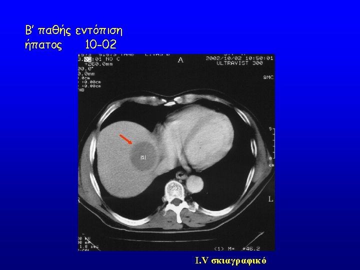
. Multidetector CT (MDCT) postcontrast: cystic liver")

")
 in a gastric mass")
: large exophitic gastric mass with a low density area")


 showing gastric carcinoma")

")

.")


 CT scan 9/14/98 pre-treatment Carboplatin AUC 4 CT scan")

- Slides: 50
Peritoneal carcinomatosis secondary to gastric adeno-carcinoma. A, Axial multi-detector CT (MDCT).
Sagital reformatted MDCT
Postcontrast T 1 -weighted MRI: irregular thickening of the peritoneal surface (arrows).
Coronal T 2 -weighted MRI: cystic implants on the peritoneal surface ( arrow).
Axial multidetector CT (MDCT) showing thickening of the sigmoid wall ( arrow).
Coronal multiplanar reformatted MDCT showing extension of the tumor into the pericolonic fat tissue and infiltration of the uterus (arrow)
Cystic liver metastasis of gastrointestinal stromal tumor (GIST). Multidetector CT (MDCT) postcontrast: cystic liver metastasis of enteric gastrointestinal tumor.
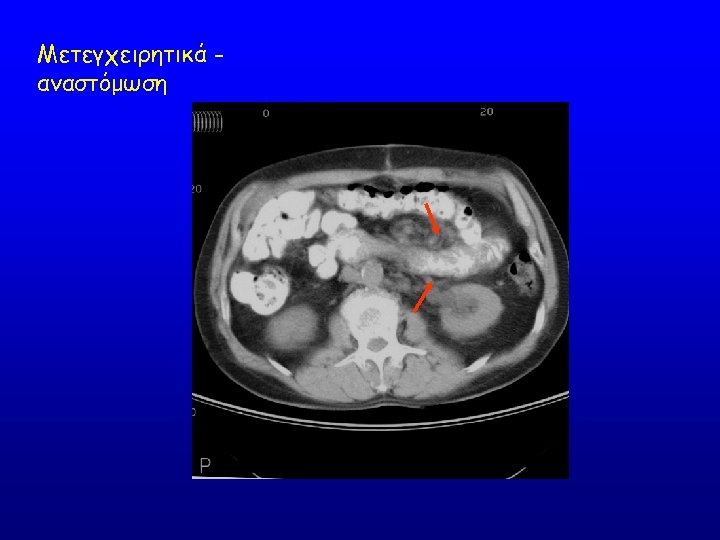
Small bowel carcinoid tumor
T 2 -weighted MRI: high signal in an exophitic gastric mass ( arrow)
T 1 -weighted postcontrast MRI: areas of low signal (necrosis) in a gastric mass (arrow)
Axial postcontrast multidetector CT (MDCT): large exophitic gastric mass with a low density area
MDCT coronal image: large gastric mass with mass effect on adjacent small bowel
Metastatic colon cancer–positive perirectal lymph node. PET-CT shows hypermetabolism on small perirectal lymph nodes
Metastatic gastric cancer peritoneal implant. PET-CT shows hypermetabolism in a small peritoneal implant located in the left paracolic goiter.
Colon cancer local recurrence. PET-CT shows hypermetabolism in a colon cancer local recurrence following sigmoidectomy.
Colon cancer local recurrence. PET-CT shows hypermetabolism in a colon cancer local recurrence following sigmoidectomy.
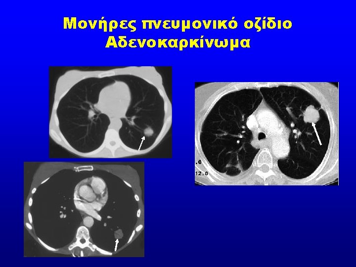
Axial multidetector CT (MDCT) showing gastric carcinoma
Coronal reformatted MDCT.
Coronal, sagittal, and axial images of FDG-PET, CT, and fusion (PET-CT)
Coronal FDG-PET images of the axial body.
Axial images of FDG-PET, CT, and fusion (PET-CT).
Coronal FDG-PET images of the axial body.
Endoscopic ultrasound plays an important role in the staging of gastrointestinal tumors.
This submucosal gastric lesion, hardly seen on routine endoscopy, was diagnosed as a gastrointestinal stromal tumor after an endoscopic ultrasound–guided fine-needle aspiration.
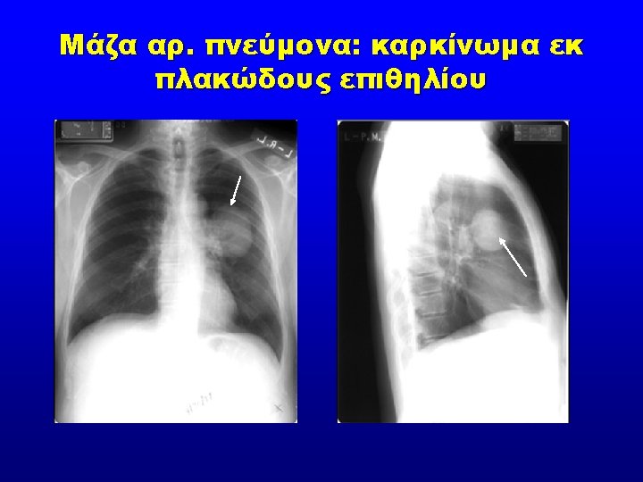
Pancoast tumour
ALIMTA + (400 mg/m 2) CT scan 9/14/98 pre-treatment Carboplatin AUC 4 CT scan 4/1/99 post 8 cycles
Patient Treated with Pemetrexed + Cisplatin Baseline Visit 4