Peripheral Vascular Disease Dr Mahmoud AlAwaysheh General Colorectal


 • Approximately 20% of people aged >60 years in developed")
 • PAD affects the legs eight times more commonly than")
")


 pain indicates severe, multilevel lower limb PAD and is a")


 • In patients with severe lower limb PAD, even")


 VS thrombosis(Venous) Embolus Thrombosis Onset and severity Acute (seconds")
 typically have an")


 ﺗﺠﻠﻂ")

 is prevented by valves which divide the long")
 saphenous vein passes anterior to the medial malleolus at")


 veins often complain of aching")

, or a ‘feeling of swelling’, may be associated with")


 in Legs • The clinical features of DVT depend upon")


- Slides: 62
Peripheral Vascular Disease Dr. Mahmoud Al-Awaysheh General & Colorectal Surgeon Mutah University - Introductory Course 2020
• Greetings • I request the following in my lectures: 1. Silence. 2. No use of mobile phones. 3. No exit-reentry during lectures. 4. You can interrupt me for questions.
What we mean by Peripheral Vascular Disease?
Definition • Peripheral Vascular disease is slow blood circulation disorder that leads to blockage in blood vessels. • The blood is unable to circulate properly due to clots in the arteries & veins. • Claudication, arteriosclerosis obliterans, intermittent claudication, arterial insufficiency of legs are other names to PVD.
Causes of PVD Arterial Disease 1 - Embolism 2 - Trauma 3 - Atherosclerosis Venous Disease 1 - Deep Venous Thrombosis (DVT) ﺗﺠﻠﻂ ﺩﻣﻮﻱ 2 - Superficial Thrombophlebitis 3 - Varicose Vein ﺍﻟﺪﻭﺍﻟﻲ
Risk Factors for PVD • • • 1 - Hypertension 2 - DM 3 - Heart/Kidney Disease 4 - Obesity 5 - Smoking 6 - Unhealthy Lifestyle e. g. Lack of exercise *Modifying risk factors dramatically improves outcomes
Symptoms • PVD Symptoms depend on the affected artery. The lack of blood flow in the artery mostly causes pain in leg calves, thigh & hips. SOCRATES
Symptoms • The pain generally starts, with exercise or regressive physical movements. • Sometimes pain also occurs when you are walking or climbing a stairs
So, Chief complaint for PVD is Mostly? ______
Diagnosis • Diagnosis may be done for measuring pusles in your legs & feets, blood pressure readings in your arm & leg. q Tests could be: • MRI • X-Ray • CT –Scan • Angiography
Treatment • Treatment could be: • Lifestyle changes e. g jogging, losing weight & Balanced diet. Medication- To reduce pain symptoms & increase blood flow. • Surgical procedure
Again……. Causes of PVD Arterial Disease 1 - Embolism 2 - Trauma 3 - Atherosclerosis Venous Disease
PERIPHERAL ARTERIAL SYSTEM *Pulses
Peripheral Arterial Disease (PAD) • Approximately 20% of people aged >60 years in developed countries have PAD but only a quarter of these are symptomatic. The underlying pathology is usually atherosclerosis (hardening of the arteries) affecting large and medium-sized vessels. • PAD, even if asymptomatic, is a powerful marker for premature vascular death. • The first manifestation of PAD may be a life- or limbthreatening complication, e. g. stroke, acute limb ischaemia or ruptured AAA.
Peripheral Arterial Disease (PAD) • PAD affects the legs eight times more commonly than the arms for the following reasons: 1 - The arterial supply to the legs is less well – developed in relation to the muscle mass 2 - The lower limb is more frequently affected by atherosclerosis.
There are four stages of lower limb lack of blood supply (ischaemia)
Asymptomatic ischaemia • Ischemia: lack of blood supply • Haemodynamically significant lower limb ischaemia is defined as an ankle to brachial pressure index (ABPI) <0. 9 at rest. • Most of these patients are asymptomatic, either because they choose not to walk very far, or because their exercise tolerance is limited by other comorbidity. • Although asymptomatic, these patients are at high risk of ‘vascular’ complications and should be assessed and treated medically as if they have intermittent claudication.
Intermittent claudication • Intermittent claudication is pain felt in the legs on walking due to arterial insufficiency and is the most common symptom of PAD. • The pain typically occurs in the calf secondary to femoro-popliteal disease but may be felt in the thigh and/or buttock in proximal (aorto-iliac) obstruction.
Intermittent claudication • Patients describe tightness or ‘cramp-like’ pain which develops after a relatively constant distance; the distance is often shorter if walking uphill, in the cold and after meals. • The pain disappears completely within a few minutes of rest but recurs on walking. The ‘claudication distance’ is how far patients say they can walk before the pain stops them from walking.
Intermittent claudication • There are two other types of claudication • Neurogenic claudication is due to neurological and musculoskeletal disorders of the lumbar spine • Venous claudication is due to venous outflow obstruction from the leg, following extensive DVT. • Neurogenic and venous claudication are much less common than arterial claudication, and can be distinguished on history and examination
Night/rest pain • The patient goes to bed, falls asleep, but is then woken 1– 2 hours later with severe pain in the foot, usually in the instep. • The pain is due to poor perfusion resulting from the loss of the beneficial effects of gravity on lying down and the reduction in heart rate, BP and cardiac output that occurs when sleeping.
Night/rest pain • Patients often obtain relief by hanging the leg out of bed or by getting up and walking around. However, on return to bed, the pain recurs and patients often choose to sleep in a chair. This leads to dependent oedema, increased interstitial tissue pressure, a further reduction in tissue perfusion and ultimately a worsening of the pain.
• Rest (night) pain indicates severe, multilevel lower limb PAD and is a ‘red flag’ symptom that mandates urgent referral to a vascular surgeon as failure to revascularise the leg usually leads to the development of critical limb ischaemia with tissue loss (gangrene, ulceration) and amputation.
• In diabetic patients it may be difficult to differentiate between rest pain and diabetic neuropathy as both may be worse at night. • Neuropathic pain is not usually confined to the foot, is associated with burning and tingling, is not relieved by dependency and is associated with dysaesthesia (pain or uncomfortable sensations sometimes described as burning, tingling or numbness). • Many patients cannot even bear the pressure of bedclothes on their feet
IMPORTANT!
Tissue loss (ulceration and/or gangrene) • In patients with severe lower limb PAD, even trivial injuries to the feet fail to heal. This allows bacteria to enter, leading to gangrene and/or ulceration. This usually progresses rapidly and, without re-vascularisation, often leads quickly to amputation and/or death.
Signs of lower limb PAD • Ischaemic signs include: 1 - absence of hair 2 - thin skin 3 - brittle nails.
• The presence of foot pulses does not completely exclude significant lower limb PAD but they are almost always diminished or absent. • If the history is convincing but pulses are felt, ask the patient to walk until the claudication pain stops him and then recheck the pulses; if they have disappeared then PAD is very likely.
Signs of lower limb PAD
Acute limb ischaemia • The classical features of acute limb ischaemia are the ‘six Ps’ (Box 6. 35). Paralysis (inability to wiggle the toes/ fingers) and paraesthesia (loss of light touch sensation over the forefoot/dorsum of the hand) are the most important and indicate severe ischaemia affecting nerve function.
Acute limb ischaemia • Muscle tenderness is a grave sign indicating impending muscle infarction. A limb with these features will usually become irreversibly damaged unless the circulation is restored within a few hours.
Acute limb ischaemia • The commonest causes of acute limb ischaemia are: • Thromboembolism: usually from the left atrium in association with atrial fibrillation Thrombosis in situ: thrombotic occlusion of an already narrowed atherosclerotic arterial segmen
Acute limb ischaemia: embolus (Arterial) VS thrombosis(Venous) Embolus Thrombosis Onset and severity Acute (seconds or minutes), ischaemia profound (no pre-existing collaterals) Insidious (hours or Embolic source Present (usually atrial fibrillation) Absent Previous claudication Absent Present Pulses in contralateral leg Present Absent Diagnosis Clinical Angiography Treatment Embolectomy and anticoagulation Medical, bypass surgery, thrombolysis days), ischaemia less severe (pre-existing collaterals)
• Patients with critical limb ischaemia (rest pain, tissue loss) typically have an ankle BP <50 mm. Hg and a positive Buerger’s test.
Acute limb ischaemia • Acute arterial occlusion produces intense spasm in the arterial tree distal to the blockage. The limb appears ‘marble white’. Over a few hours, the spasm relaxes and the skin microcirculation fills with deoxygenated blood, leading to light blue or purple mottling, which has a fine reticular pattern and blanches on pressure.
Acute limb ischaemia • As ischaemia progresses, blood coalesces in the skin, producing a coarser pattern of mottling which is dark purple, almost black, and does not blanch. Finally, large patches of fixed staining lead to blistering and liquefaction.
• As ischaemia progresses, blood coalesces in the skin, producing a coarser pattern of mottling which is dark purple, almost black, and does not blanch. Finally, large patches of fixed staining lead to blistering and liquefaction. • Fixed mottling of an anaesthetic, paralysed limb, with muscle rigidity and turgor, indicates irreversible ischaemia; amputation or end-oflife care is the only option
Compartment syndrome • Compartment syndrome occurs where there is increased pressure within the fascial compartments of the limb, most commonly the calf, which compromises perfusion and viability of muscle and nerves.
Compartment syndrome • The two commonest causes are lower trauma, e. g. fractured tibia, and reperfusion following treatment of acute lower limb ischaemia. Failure to recognise and treat compartment syndrome may require limb amputation. • The key symptom is severe pain often unrelieved by opioids and exacerbated by active or passive movement. Peripheral pulses are usually present.
Causes of PVD Arterial Disease Venous Disease 1 - Deep Venous Thrombosis (DVT) ﺗﺠﻠﻂ ﺩﻣﻮﻱ 2 - Superficial Thrombophlebitis 3 - Varicose Vein ﺍﻟﺪﻭﺍﻟﻲ
Venous Disease • Its due to Obstruction in the venous outflow of the leg due to iliofemoral venous occlusion e. g. Thrombosis
Venous Disease • Backward flow (reflux) is prevented by valves which divide the long column of blood from the foot to the right atrium into a series of short low-pressure segments. • As a result, the ‘ambulatory venous pressure’ in the feet in health is usually <20 mm. Hg. The great majority of lower limb venous symptoms and signs are due to failure of the muscle pump and/or valves and the resulting ‘ambulatory venous hypertension’. • Deep veins follow the course of the main arteries; are often paired; and may be affected by primary or postthrombotic (following DVT) valvular insufficiency
• The great (long) saphenous vein passes anterior to the medial malleolus at the ankle, up the medial aspect of the calf to behind the knee, then up the medial aspect of the thigh to join the common femoral vein in the groin at the saphenofemoral junction (Fig. 6. 41). • The lesser (short) saphenous vein passes behind the lateral malleolus at the ankle and up the posterior aspect of the calf. It commonly joins the popliteal vein at the saphenopopliteal junction, which usually lies 2 cm above the posterior knee crease.
Venous Disease • There are numerous intercommunications between the long and short saphenous, and between the deep and superficial venous (via perforating or communicating veins) systems; and the venous anatomy of the leg is highly variable.
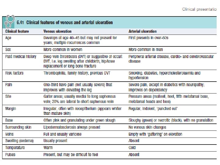
Clinical Presentation • Lower limb venous disease presents in four ways: • Varicose veins • Superficial thrombophlebitis • DVT • Chronic venous insufficiency and ulceration.
Pain • Patients with uncomplicated varicose (dilated, tortuous, superficial) veins often complain of aching leg discomfort, itching and a feeling of swelling. Symptoms are aggravated by prolonged standing and are often worse towards the end of the day. Once established, DVT causes pain and tenderness in the affected part (usually the calf).
Pain • Superficial thrombophlebitis produces a red, painful area overlying the vein involved. Varicose ulceration may be surprisingly painless; if it is painful, this may be relieved by limb elevation (but exclude coexisting arterial disease). Bandaging for a leg ulcer is contraindicated unless there is documented evidence of adequate arterial circulation. Do this by feeling the pulses or by measuring the ABPI.
Swelling • Swelling (or oedema), or a ‘feeling of swelling’, may be associated with lower limb venous disease.
Discoloration • Chronic venous insufficiency is associated with lipo-dermatosclerosis, which results from the deposition of haemosiderin (from the breakdown of extra-vasated blood) in the skin. Lipodermatosclerosis varies in colour from deep blue/black to purple or bright red and usually affects the medial aspect of the lower third of the leg, although it may be lateral if superficial reflux predominates in the lesser saphenous vein.
Venous Ulceration • Chronic venous ulceration usually affects the medial aspect, is shallow; is pink (granulation tissue) or yellow/ green (slough); has an irregular margin; and is always associated with other skin changes of chronic venous insufficiency (varicose eczema, lipodermatosclerosis)
DVT could be in Legs Arms
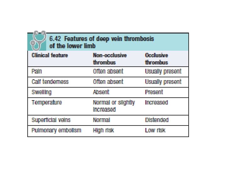
Deep Venous Thrombosis (DVT) in Legs • The clinical features of DVT depend upon its site, extent and whether it is occlusive or not • The so-called ‘classical’ features of DVT relate to well-established occlusive thrombus. • Most patients who die from pulmonary embolism have non-occlusive thrombosis and the leg is normal on clinical examination.
DVT • Non-occlusive DVT poses the greatest threat of pulmonary embolism as the clot lies within a flowing stream of venous blood, is more likely to propagate and has not yet induced an inflammatory response in the vein wall to anchor it in place. • Perform an urgent duplex Doppler ultrasound scan of the leg in any patient with a suspected DVT
DVT in Arms • Axillary subclavian vein thrombosis can occur as a result of repetitive trauma at the thoracic outlet due to vigorous, repetitive exercise. Upper limb DVT may also complicate indwelling subclavian/jugular venous catheters. • Symptoms include arm swelling and discomfort, often exacerbated by activity, especially when holding the arm overhead.
Axillary vein thrombosis due to Trauma • The arm is swollen and the skin is cyanosed and mottled, especially when dependent. Look for superficial distended veins (acting as collaterals) in the upper arm, over the shoulder region and on the anterior chest wall
Risk factors for deep vein thrombosis
Case Scienario • A 40 Years old - Patient had Hip replacement 5 days ago. • Whats your diagnosis? • Mention other differntials • What are the complications?
• What’``s your diagnosis? DVT • Mention other differentials? Cellulites/ chronic venous insufficiency. • What are the complications? • Pulmonary embolism • Ulcers • Ischemia
THANK YOU
Resources • Macleod’s Clinical Examination – Graham Douglas, Fiona Nicol, Colin Robertson.