Peripheral Nerve Injury and Repair Clinical PointsIntroduction Peripheral


- Slides: 28
Peripheral Nerve Injury and Repair
Clinical Points-Introduction �Peripheral nerve injury can be devastating for a patient. A host of factors influence the highly dynamic degenerative processes �The regenerative capacity of peripheral nerves is remarkable �Degradation and regeneration of peripheral nerves is distinct from that of nerves in the central nervous system �Prognosis of peripheral nerve injury is dependant upon age, the nerve injured, the level of the injury, the degree of injury and the timing of repair
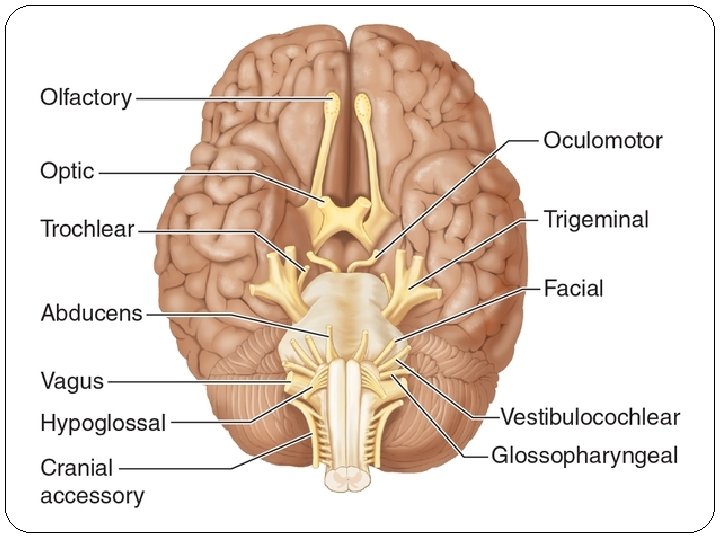
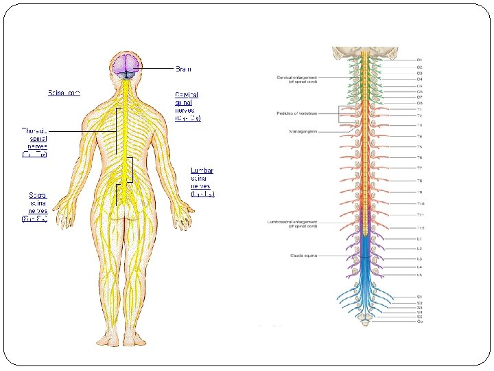
Anatomy of the peripheral nerves - General Features There are 31 pairs of spinal nerves which contain a mixture of sensory and motor fibers. They are formed by fusion of anterior and posterior nerve roots There are 12 pairs of cranial nerves which are concerned with receiving information and controlling activities of the head and neck and, to a lesser extent, the thoracic and abdominal viscera Peripheral nerve fibers have been classified in relation to their conduction velocity, which, in general is proportional to size and function.
Anatomy of the peripheral nerves - General Features The largest myelinated fibers may be motor or proprioceptive, and the smallest, whether myelinated or not, are autonomic or sensory The widest fibers appear to conduct most rapidly
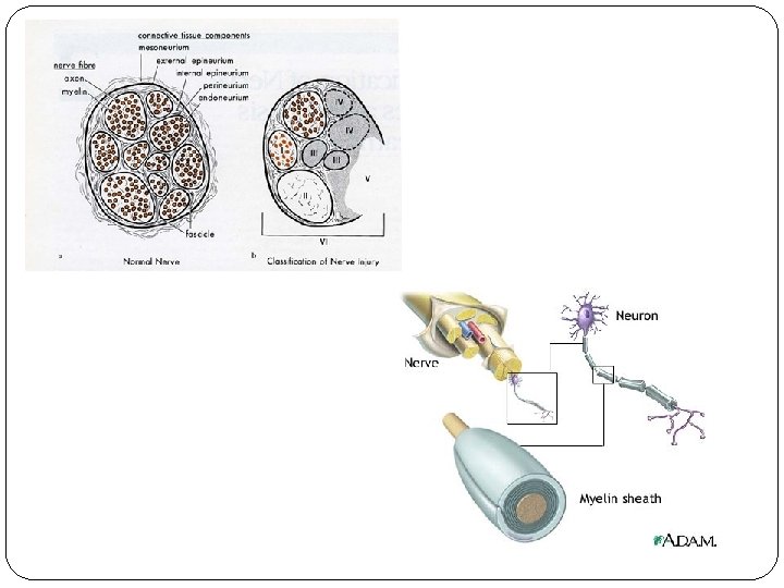
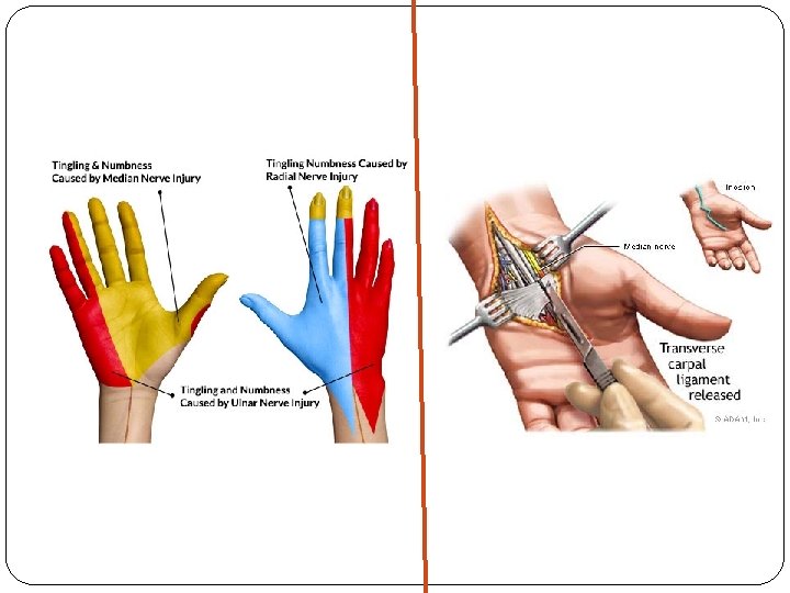
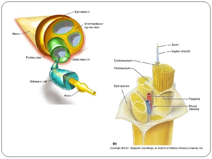
Anatomy of the peripheral nerves - General Features Within a given peripheral nerve, fibers are organized in separate bundles known as fascicles Less than half of the nerves are enclosed within myelin sheaths. The remaining unmyelinated fibers travel in deep gutters along the surface of Schwann cells Each Schwann cell is surrounded by a network of reticular collagenous fibers, the endoneurium. Each fascicle is covered by an epithelium, the perineurium. All of the fascicles are surrounded by epineurium (a loose vascular tissue) which encloses an individual nerve
Classification of Nerve Injury �The two systems used most widely are those developed by Seddon and by Sunderland
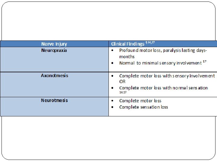
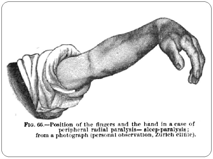
Seddon Classification of Nerve Injury Neuropraxia: Little or no structural damage with no loss of nerve continuity. Symptoms are transient, there is no severance or tearing of the neural elements and there is little or no histological change seen. The effects appear to be reversible, unless ischemia persists for approximately 8 hours. Entrapment neuropathies, such as carpal tunnel syndrome, and Saturday night palsy (wrist drop), a radial nerve paralysis caused by pressure on the arm after the person has fallen asleep, usually during an alcoholic binge Excellent recovery normally within weeks or months
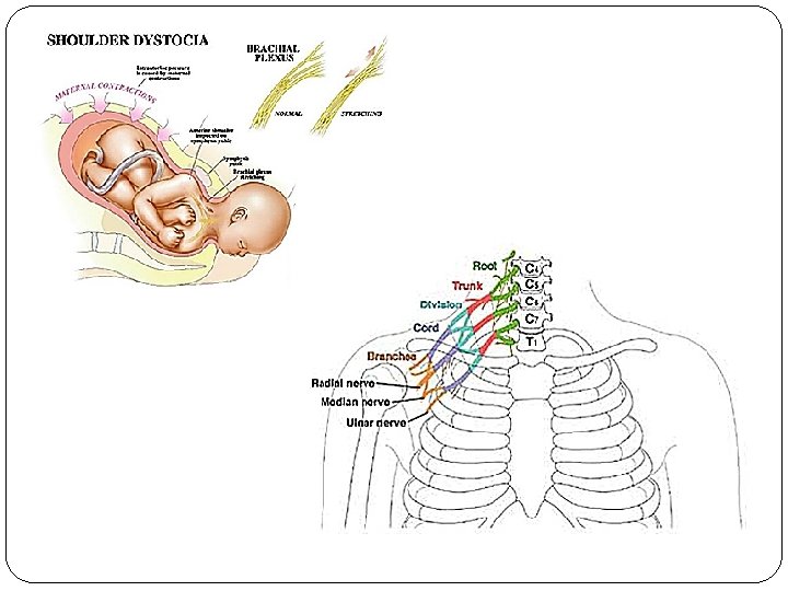
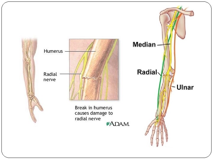
Seddon Classification of Nerve Injury �Axonotmesis: There is complete interruption of the nerve axon and its myelin sheath, but the mesenchymal structures including perineurium and epineurium are either completely or partially intact �Birth-related brachial plexus injury, or in association with fractures such as a radial nerve injury secondary to a humeral fracture. Lacerations, including those caused by broken glass, are also a common type of injury that may cause axonotmesis �Prognosis depends on the extent of injury,
Seddon Classification of Nerve Injury �Neurotmesis: When a nerve, along with its surrounding stroma, becomes completely disconnected. �There is no spontaneous recovery and even after surgery prognosis is poor. �This type of injury is only seen in major trauma
Sunderland Classification of Nerve Injury � 5 different classes � First degree injuries are equivalent to neuropraxia � 2 nd, 3 rd and 4 th degree injuries are equivalent to axonotmesis, the difference being the degree of mesenchymal damage to the nerve � In 2 nd degree injuries recovery is good whilst in 4 th degree injuries recovery is poor �Fifth degree injuries are equivalent to neurotmesis
Management of Nerve Injury With more severe forms of axonotmesis surgery may be required Neurotmesis can easily be detected upon exploratory surgery as the nerve can be seen to be completely transected. In neurotmesis, surgery is indicated as there is no hope of spontaneous recovery The timing of surgical nerve reconstruction is important for optimal recovery. In every case of acute injury, the surgeon must decide whether a primary repair or an early secondary repair is the treatment of choice Timing can be divided into immediate, early (1 month), delayed (3 -6 months), and late (1 -2 years or more) Immediate repair is preferred when the nerve has been lacerated and there has been a clean cut. The nerve ends should also be uninjured
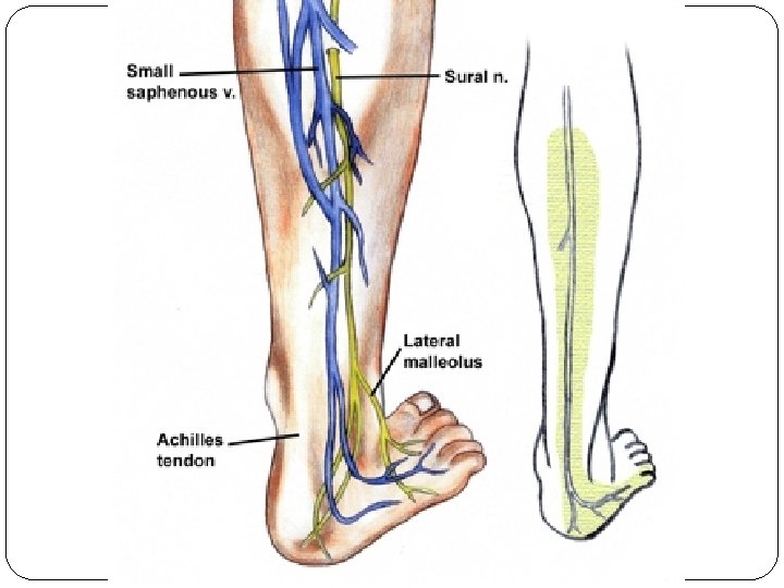
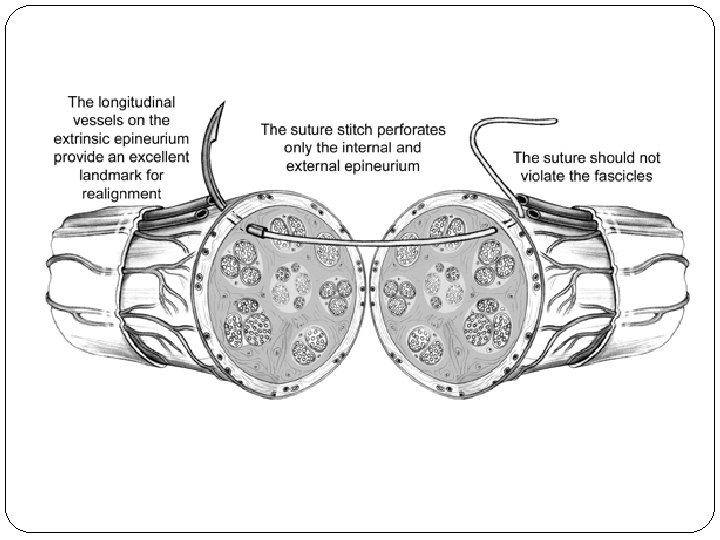
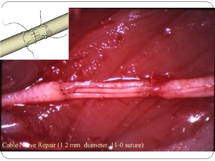
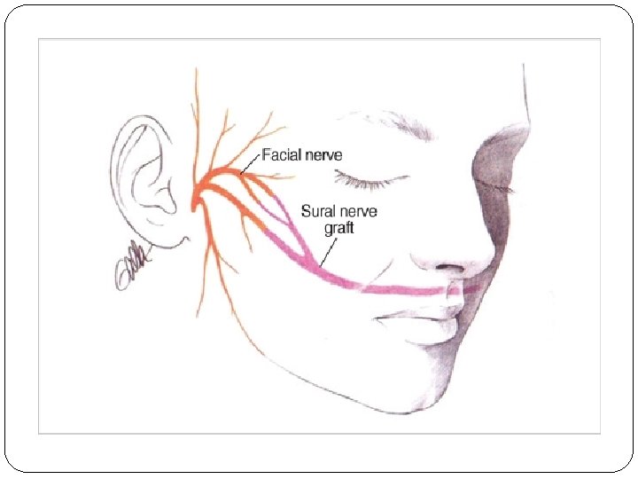
Management of Nerve Injury �Early reconstruction is preferred for injuries caused by blunt trauma or avulsion, which are thought to have caused complete nerve destruction. The graft used is usually from the sural nerve � Delayed reconstruction is preferred when the degree of injury has not yet been ascertained. The quality of motor recovery decreases steadily after a 6 month delay of repair �Late reconstruction is generally only carried out for pain control, such as neuroma resection �Epineural repair with nylon suture
Outcome of Nerve Injury � Sensory recovery appears to be similar for most nerves � Motor function varies according to individual nerves. In one study, the motor recovery in ulnar nerves was 71% lower than that in median nerves � It appears that age (younger patients fare better), site, the nerve injured, and delay, significantly influence prognosis after microsurgical repair � After surgery the affected area should be immobilized for approx. 6 weeks. After this, movement is encouraged and physiotherapy is most useful. Strength exercises may be performed along with the use of electro-
The Future? �Molecular therapy �Neurotrophic factors �Despite a huge amount of studies, treatment for peripheral nerve injury remains largely unchanged
Evolution………