Peripheral nerve injuries Hisham Abdul Aziz Alsanawi Assistant

Peripheral nerve injuries Hisham Abdul. Aziz Alsanawi Assistant Professor Department of Orthopaedics KSU and KKUH

Compression Neuropathy • Chronic condition with sensory, motor, or mixed involvement • First lost light touch – pressure – vibration • Last lost pain - temperature • microvascular compression neural ischemia paresthesias Intraneural edema more microvascular compression demyelination --> fibrosis --> axonal loss

Common systemic conditions leading to compression neuropathy • SYSTEMIC – – Diabetes Alcoholism renal failure Raynaud • INFLAMMATORY – – Rheumatoid arthritis Infection Gout Tenosynovitis • FLUID IMBALANCE – Pregnancy – Obesity • ANATOMIC – – – Synovial fibrosis Lumbrical encroachment Anomalous tendon Median artery Fracture deformity • MASS – Ganglion – Lipoma – Hematoma

Symptoms • • night symptoms dropping of objects clumsiness weakness • Rule out systemic causes

Physical Exam • Examine individual muscle strength --> grades 0 to 5 --> pinch strength - grip strength • Neurosensory testing --> – dermatomal distribution – peripheral nerve distribution

Special Tests • Semmes-Weinstein monofilaments --> – cutaneous pressure threshold --> function of large nerve fibers --> first to be affected in compression Neuropathy – Sensing 2. 83 monofilament is normal • Two-point discrimination → – performed with closed eyes – abnormal → Inability to perceive a difference between points > 6 mm – late finding


Electrodiagnostic testing • EMG and NCS • Sensory and motor nerve function can be tested • Operator dependent • objective evidence of neuropathic condition • helpful in localizing point of compromise • early disease → High false-negative rate

Electrodiagnostic testing • NCSs → – conduction velocity and distal latency and amplitude – Demyelination → ↓conduction velocity + ↑distal latency axonal loss → ↓ potential amplitude • EMG → – muscle electrical activity – muscle denervation → fibrillations - positive sharp waves - fasciculations

Double-crush phenomenon • blockage of axonal transport at one point makes the entire axon more susceptible to compression elsewhere

Median Nerve Compression • Carpal Tunnel Syndrome • Pronator Syndrome • Anterior Interosseos Neuropathy

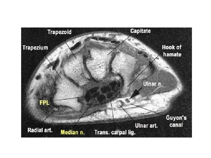
CTS • Most common compressive neuropathy in the upper extremity • Anatomy of the carpal tunnel – Volar → TCL – radial → scaphoid tubercle +trapezium – ulnar → pisiform +hook of hamate – Dorsal → proximal carpal row + deep extrinsic volar carpal ligaments

CTS • Carpal Tunnel Content – median nerve + FPL + 4 FDS + 4 FDP = 10 • Normal pressure → 2. 5 mm Hg • >20 mm Hg → ↓↓ epineural blood flow + nerve edema • 30 mm Hg → ↓↓ nerve conduction

Forms of CTS • Idiopathic → most common in adults • Mucopolysaccharidosis → most common cause in children • anatomic variation – Persistent median artery – small carpal canal – anomalous muscles – extrinsic mass effect

Risk Factor • • • obesity pregnancy diabetes thyroid disease chronic renal failure Others inflammatory arthropathy, storage diseases, vitamin deficiency, alcoholism, advanced age, vibratory exposure during occupational activity

Be aware! • no established direct relationship between → repetitive work activities such as keyboarding and CTS

Acute CTS • causes → – high-energy trauma – hemorrhage – infection • Requires emergency decompression

CTS diagnosis • symptoms → – Paresthesias and pain – often at night – volar aspect → thumb - index - long - radial half of ring • provocative test → carpal tunnel compression test -Durkan test → Most sensitive • Other provocative tests include Tinel and Phalen

CTS diagnosis • affected first → light touch + vibration • affected later → pain and temperature • Semmes-Weinstein monofilament testing → early CTS diagnosis • late findings Weakness - loss of fine motor control - abnormal two-point discrimination • Thenar atrophy → severe denervation

CTS – Electrodiagnostic testing not necessary for the diagnosis of CTS Distal sensory latencies > 3. 5 msec motor latencies >4. 5 msec ↓ conduction velocity and ↓ peak amplitude → less specific • EMG → ↑ insertional activity - sharp waves fibrillation - APB fasciculation • •

CTS - Differential diagnoses cervical radiculopathy brachial plexopathy TOS pronator syndrome ulnar neuropathy with Martin-Gruber anastomoses • peripheral neuropathy of multiple etiologies • • •

CTS Treatment • Nonoperative → – activity modification – night splints – NSAIDs • Single corticosteroid injection → transient relief – 80% after 6 weeks – 20% by 1 year – ineffective corticosteroid injection → poor prognosis → less successful surgery

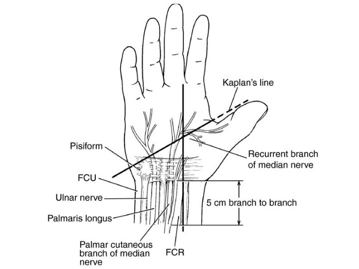
CTS – Operative • Can be → open - mini-open – endoscopic • internal median neurolysis OR flexor tenosynovectomy No benefit • too ulnar surgical approach → Ulnar neurovascular injury • too radial surgical approach → recurrent motor branch of median nerve injury • recurrent motor branch variations – Extraligamentous → 50% – Subligamentous → 30% – Transligamentous → 20%
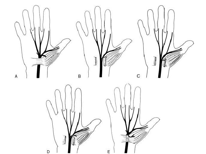

CTS – Endoscopic release • short term: – less early scar tenderness – improved short-term grip/pinch strength – better patient satisfaction scores • Long-term → – no significant difference – May have slightly higher complication rate – incomplete TCL release

CTS – release outcome • pinch strength → 6 weeks • grip strength → 3 months • Persistent symptoms after release → – – – incomplete release iatrogenic median nerve injury missed double-crush phenomenon concomitant peripheral neuropathy space-occupying lesion • revision success → identify underlying failure cause

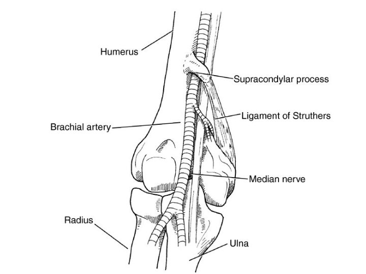
Pronator Syndrome • Median nerve compression @ arm/forearm • Symptoms → – proximal volar forearm pain – sensory symptoms → palmar cutaneous branch • DDx AIN syndrome motor and pain

Ulnar Nerve Compression Neuropathy • Cubital Tunnel Syndrome • Ulnar Tunnel Syndrome

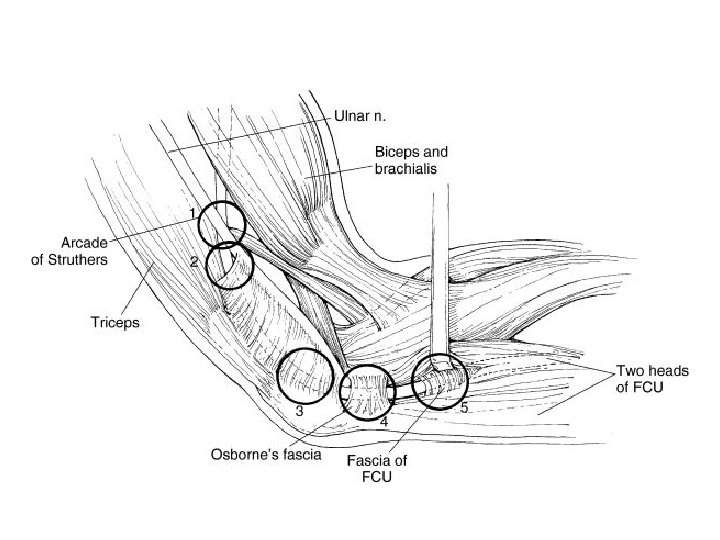
Cubital Tunnel Syndrome • Second most common compression neuropathy of the upper extremity • Cubital tunnel borders: – floor →MCL and capsule – Walls → medial epicondyle and olecranon – Roof → FCU fascia and arcuate ligament of Osborne

Cubital tunnel syndrome • Compression sites many • Symptoms paresthesias of ulnar half of ring finger and small finger • Provocative tests → – direct cubital tunnel compression – Tinel sign – elbow hyperflexion • Froment sign → thumb IP flexion - FPL during key pinch → weak adductor pollicis

Cubital Tunnel Syndrome - Treatment • Electrodiagnostic tests diagnosis and prognosis • Nonoperative treatment – activity modification – night splints → slight extension – NSAIDs

Cubital Tunnel Syndrome - Treatment • Surgical Release Numerous techniques – In situ decompression, Anterior transposition, Subcutaneous, Submuscular, Intramuscular, Medial epicondylectomy • No significant difference in outcome between simple decompression and transposition

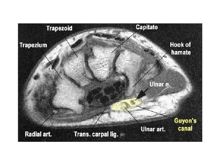
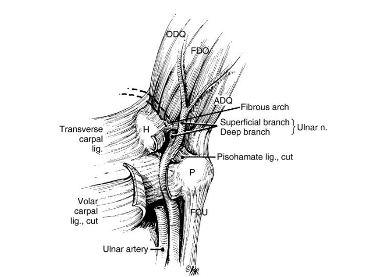
Ulnar Tunnel Syndrome • Compression neuropathy of ulnar nerve in the Guyon canal • Causes ganglion cyst → 80% , hook-ofhamate nonunion, ulnar artery thrombosis

Ulnar Tunnel Syndrome - Invx • CT → hamate hook fracture • MRI → ganglion cyst or other space-occupying lesion • Doppler ultrasonography → ulnar artery thrombosis

Ulnar Tunnel Syndorme • Treatment success → identify cause • Nonoperative treatment – activity modification – splints – NSAIDs • Operative treatment → decompressing by removing underlying cause • ulnar compression @ Guyon + CTS ⇒ CTS release is enough

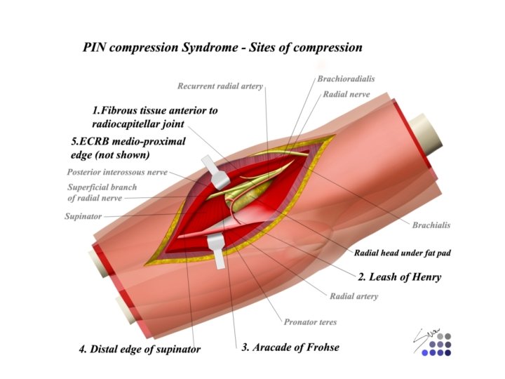
Radial Nerve • Radial nerve compression rarely compressed mainly motor symptoms • PIN compression lateral elbow pain + muscle weakness • Radial Tunnel Syndrome – lateral elbow and radial forearm pain – no motor or sensory dysfunction – Cheiralgia paresthetica superficial sensory radial nerve pain, numbness, paresthesias over dorsoradial hand

Peripheral nerve injuries • causes → – compression – stretch – blast – crush – avulsion – transection – tumor invasion

Peripheral nerve injuries • good prognostic factor for recovery: – young age → most important factor – stretch injuries – clean wounds – after direct surgical repair • poor outcome – crush or blast injuries – infected or scarred wounds – delayed surgical repair.

Classification • Neuropraxia • Axonotmesis • Neurotmesis

Neurapraxia • • • Mild nerve stretch or contusion Focal conduction block No wallerian degeneration Disruption of myelin sheath Epineurium, perineurium, endoneurium intact Prognosis excellent full recovery

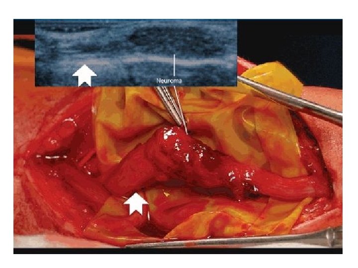
Axonotmesis Incomplete nerve injury Focal conduction block Wallerian degeneration distal to injury Disruption of axons Sequential loss of axon, endoneurium, perineurium • May develop neuroma-in-continuity • Recovery unpredictable • • •

Neurotmesis • • Complete nerve injury Focal conduction block Wallerian degeneration distal to injury Disruption of all layers, including epineurium Proximal nerve end forms neuroma Distal end forms glioma Worst prognosis

wallerian degeneration • Starts in distal nerve segment • degradation products removed by phagocytosis • Myelin-producing Schwann cells proliferate and align form a tube receive regenerating axons • Nerve cell body enlarges increased structural protein production • proximal axon forms sprouts connect to the distal stump migrate @ 1 mm/day

Surgical repair • Best performed within 2 weeks of injury • Repair must be free of tension • Repair must be within clean, well-vascularized wound bed • Nerve length may be gained by neurolysis or transposition

Surgical repair • Repair techniques – Epineurial – Individual fascicular – Group fascicular – No technique deemed superior • nerve conduits → popular for digital nerve gaps >8 mm →polyglycolic acid and collagen based • Larger gaps → grafting

Surgical repair • Autogenous → sural - medial/lateral antebrachial cutaneous - terminal/PIN • Vascularized • Growth factor augmentation → insulin-like and fibroblast → promote nerve regeneration • Chronic peripheral nerve injuries → neurotization and/or tendon transfers • Use of nerve transfers for high radial and ulnar nerve injuries gaining popularity
- Slides: 54