
PERIPHERAL JOINTS PATHOLOGY AND TREATMENT Benjamin Bonte MD
PERIPHERAL JOINTS: PATHOLOGY AND TREATMENT Benjamin Bonte, MD Interventional Pain Fellow Hudson Spine & Pain Medicine 11/8/2017
Outline Shoulder Knee Hip
Shoulder pathology Anatomy Physical Exam Specific pathologies Adhesive capsulitis AC joint sprain/arthritis Rotator cuff pathology Labral pathology Neurogenic thoracic outlet syndrome
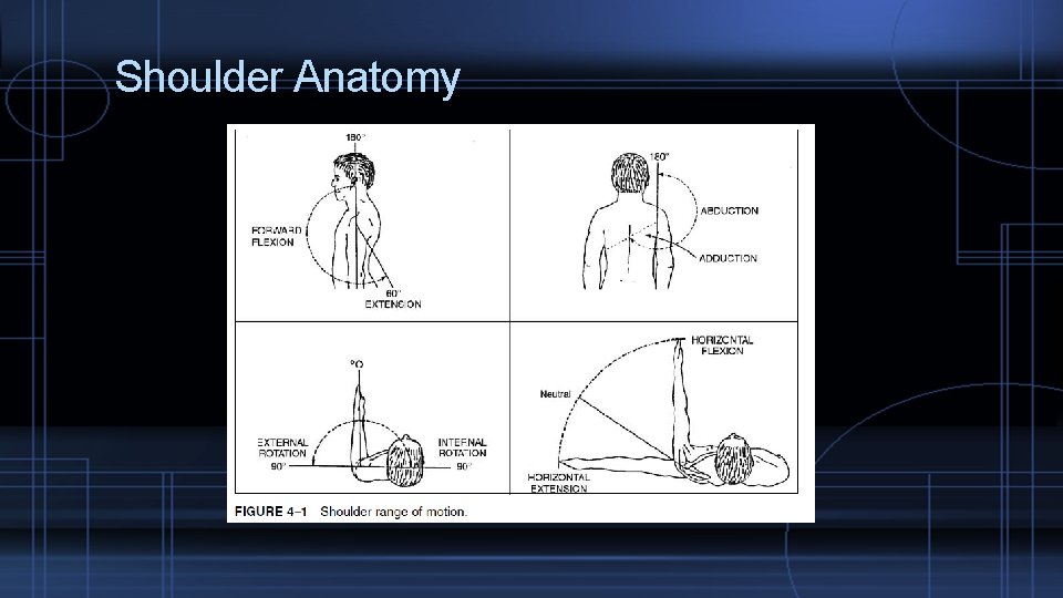
Shoulder Anatomy

Shoulder Anatomy

Shoulder Anatomy
Physical Exam Inspection Swelling, deformities, ecchymosis, rash ROM Palpation Sternoclavicular joint, supraclavicular fossa, AC, biceps tendon, subacromial bursa, supraspinatus/infraspinatus Provocative testing (will discuss with pathologies)
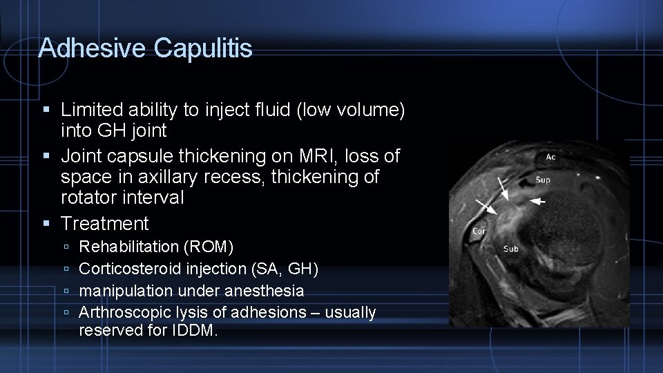
Adhesive Capulitis Limited ability to inject fluid (low volume) into GH joint Joint capsule thickening on MRI, loss of space in axillary recess, thickening of rotator interval Treatment Rehabilitation (ROM) Corticosteroid injection (SA, GH) manipulation under anesthesia Arthroscopic lysis of adhesions – usually reserved for IDDM.
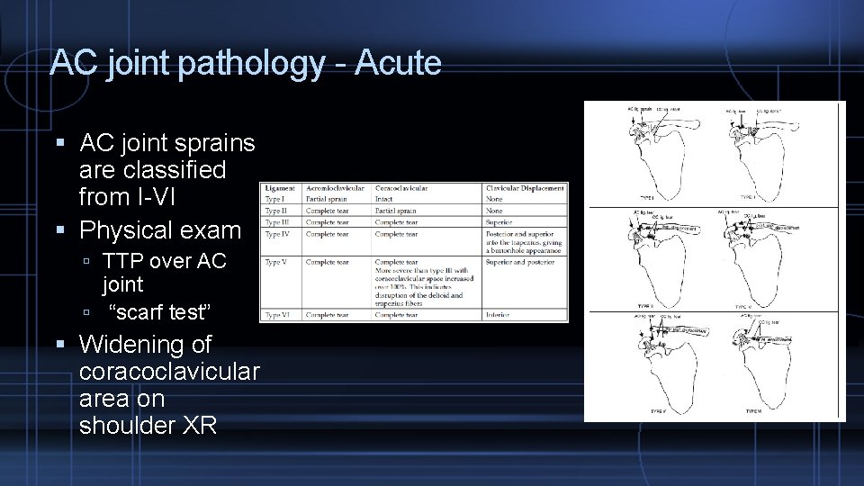
AC joint pathology - Acute AC joint sprains are classified from I-VI Physical exam TTP over AC joint “scarf test” Widening of coracoclavicular area on shoulder XR
AC joint pathology Type I/II – conservative tx Sling for comfort only and not all the time (ROM essential) Return to sport when asymptomatic/full ROM. � 2 weeks for type I � 6 weeks for type II Type III - controversial Surgery for laborers, athletes Type IV-VI – surgery ORIF or distal clavicular resection with reconstruction of the CC ligament
AC joint pathology - chronic Accuracy of blind injection is 60 -67%. Ultrasound guidance can be used
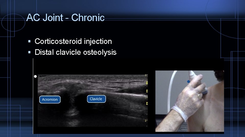
AC Joint - Chronic Corticosteroid injection Distal clavicle osteolysis
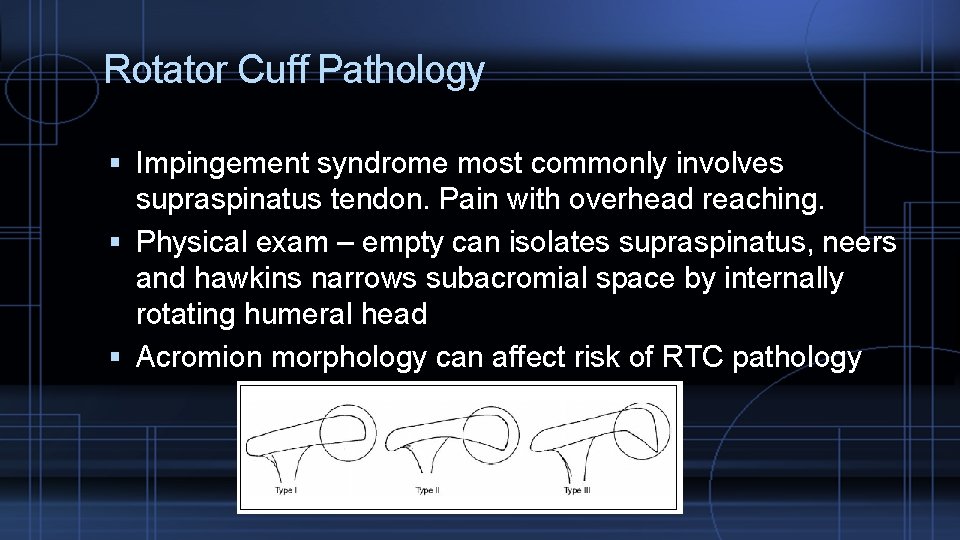
Rotator Cuff Pathology Impingement syndrome most commonly involves supraspinatus tendon. Pain with overhead reaching. Physical exam – empty can isolates supraspinatus, neers and hawkins narrows subacromial space by internally rotating humeral head Acromion morphology can affect risk of RTC pathology
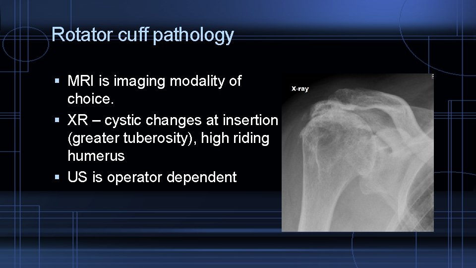
Rotator cuff pathology MRI is imaging modality of choice. XR – cystic changes at insertion (greater tuberosity), high riding humerus US is operator dependent
Rotator Cuff Pathology Rehabilitation ROM, RTC muscle strengthening, scapular stabilizers (rhomboids, levator scapulae, trapezius, serratus anterior) Corticosteroid injections Surgical Tears that fail conservative treatment Aids pain relief, may not always lead to functional improvement Surgeries �anterior acromioplasty, coracoacromial ligament lysis �Excision, repair (if acute athlete, better if repaired within 3 weeks rather than later reconstruction)
OA of shoulder Physical exam: Limited internal rotation of the shoulder XR: cystic changes, joint surface irregularity treatment: NSAIDs, ROM, RTC strengthening, corticosteroid injections accuracy of an anterior approach ranges from 27% to 99%, and a posterior approach ranges from 50% to 91%
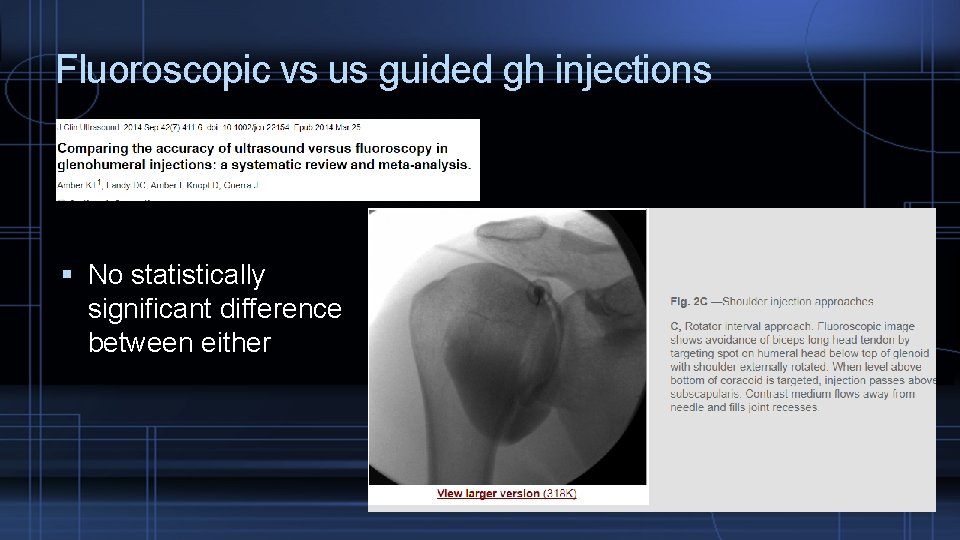
Fluoroscopic vs us guided gh injections No statistically significant difference between either
Labral pathology Repetitive overhead sports, trauma, long head biceps pathology, presents as sharp clicking, locking, and instability Seen on MRI Bankart lesion – anterior dislocation that causes tear of labrum Hill sachs lesion – compression fracture of posterolateral humeral head due to anterior instability (abutment against anterior glenoid) Surgical treatment may be an option when conservative measures fail.
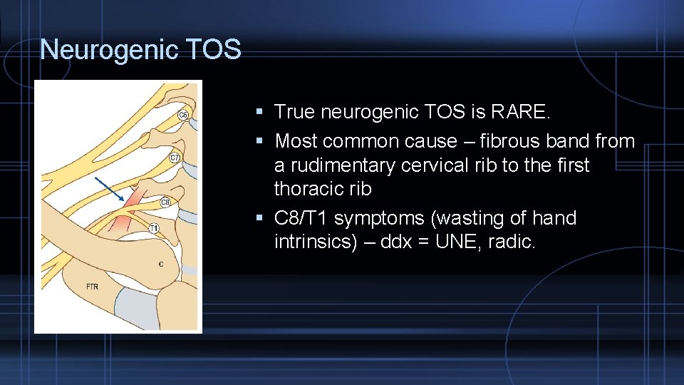
Neurogenic TOS True neurogenic TOS is RARE. Most common cause – fibrous band from a rudimentary cervical rib to the first thoracic rib C 8/T 1 symptoms (wasting of hand intrinsics) – ddx = UNE, radic.
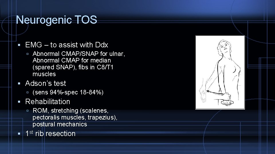
Neurogenic TOS EMG – to assist with Ddx Abnormal CMAP/SNAP for ulnar, Abnormal CMAP for median (spared SNAP), fibs in C 8/T 1 muscles Adson’s test (sens 94%-spec 18 -84%) Rehabilitation ROM, stretching (scalenes, pectoralis muscles, trapezius), postural mechanics 1 st rib resection
Knee pathology Anatomy Physical exam Specific pathologies Osteoarthritis Meniscus ACL PCL Distal ITB syndrome (burisitis) Patellar dislocation/Patellofemoral pain/Patellar tendinopathy Pes anserine bursitis
Knee Anatomy Modified hinge joint Knee ROM: 0 -135 IR/ER of the knee: 10 degrees from neutral
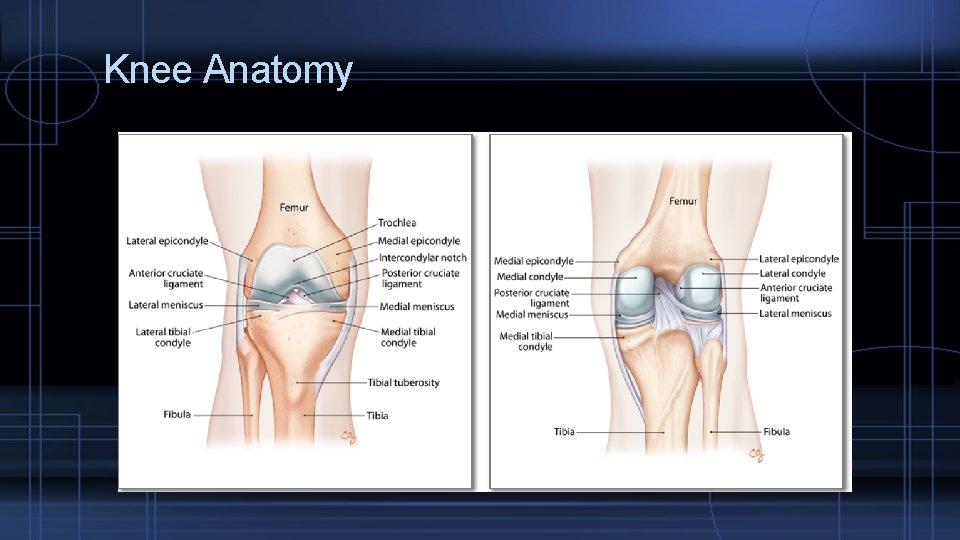
Knee Anatomy
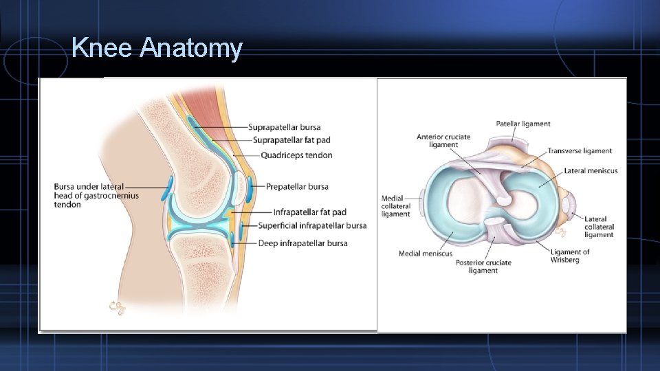
Knee Anatomy
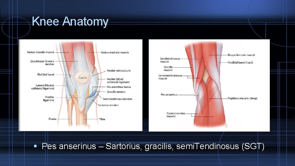
Knee Anatomy Pes anserinus – Sartorius, gracilis, semi. Tendinosus (SGT)
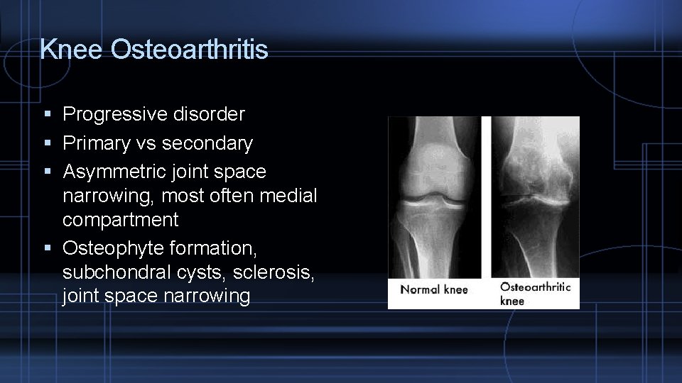
Knee Osteoarthritis Progressive disorder Primary vs secondary Asymmetric joint space narrowing, most often medial compartment Osteophyte formation, subchondral cysts, sclerosis, joint space narrowing
Knee osteoarthritis Conservative treatment, NSAIDs, ice, PT, ice Corticosteroid injections surgery
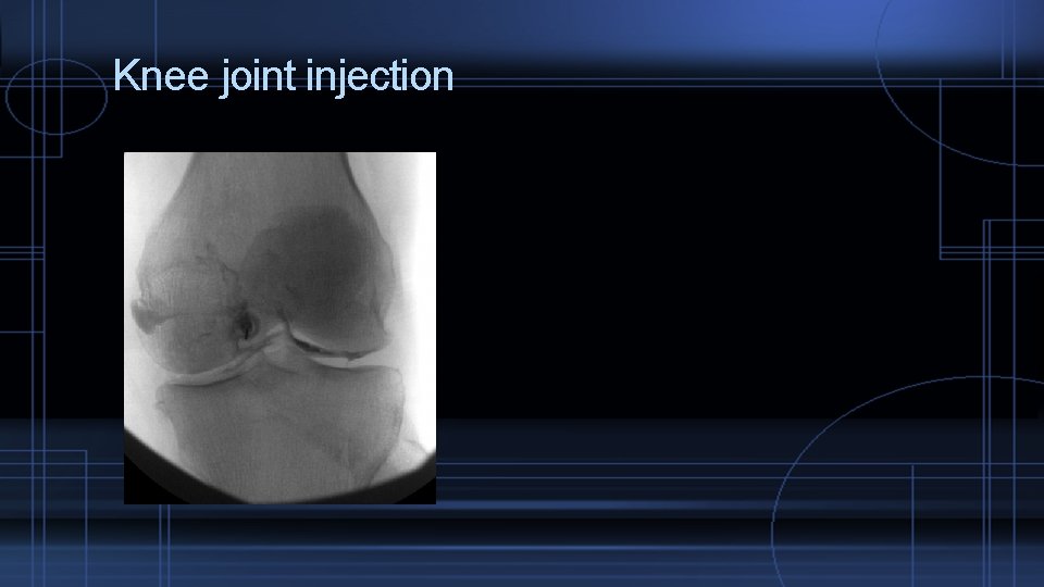
Knee joint injection
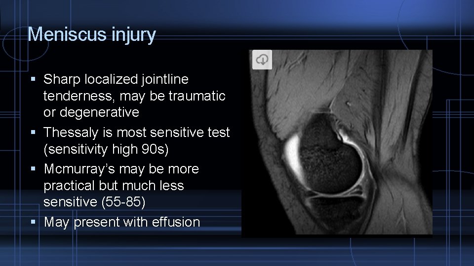
Meniscus injury Sharp localized jointline tenderness, may be traumatic or degenerative Thessaly is most sensitive test (sensitivity high 90 s) Mcmurray’s may be more practical but much less sensitive (55 -85) May present with effusion
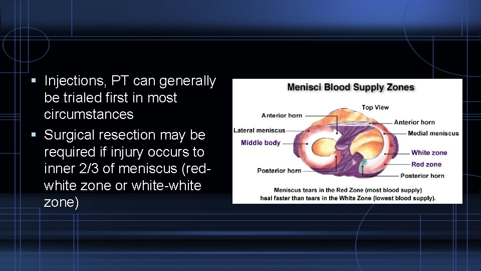
Injections, PT can generally be trialed first in most circumstances Surgical resection may be required if injury occurs to inner 2/3 of meniscus (redwhite zone or white-white zone)
ACL Injury Most commonly injured ligament in athletics Typical story: Internally rotated foot, flexed knee, valgus force Co-occurs with meniscal injuries Lachman’s is most sensitive test however cannot be used acutely due to swelling – also only 90% sensitive Conservative vs surgical treatment based on activity level
PCL injury Impact to tibia with knee flexed Dashboard injury Popliteal tenderness Posterior drawer test Surgical repair of isolated PCL tear is controversial – otherwise conservative management is recommended.
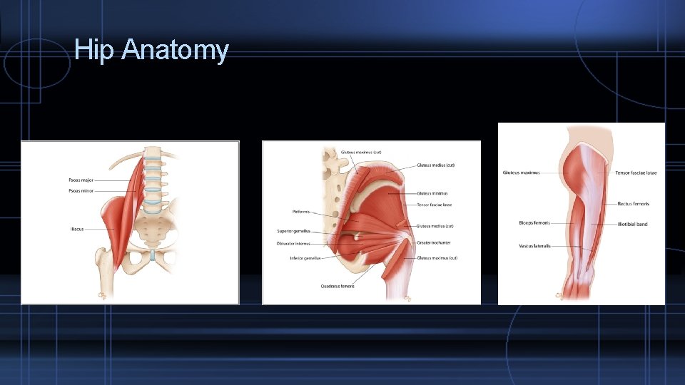
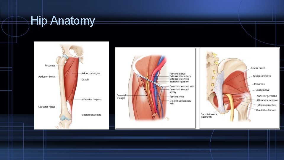
Hip pathology Anatomy Physical exam Specific pathologies Femoral Neck Stress fractures (compression vs tension side) Labral pathology FAI (cam and pincer) Osteoarthritis Internal Snapping hip External Snapping hip GTB syndrome Adductor pathology
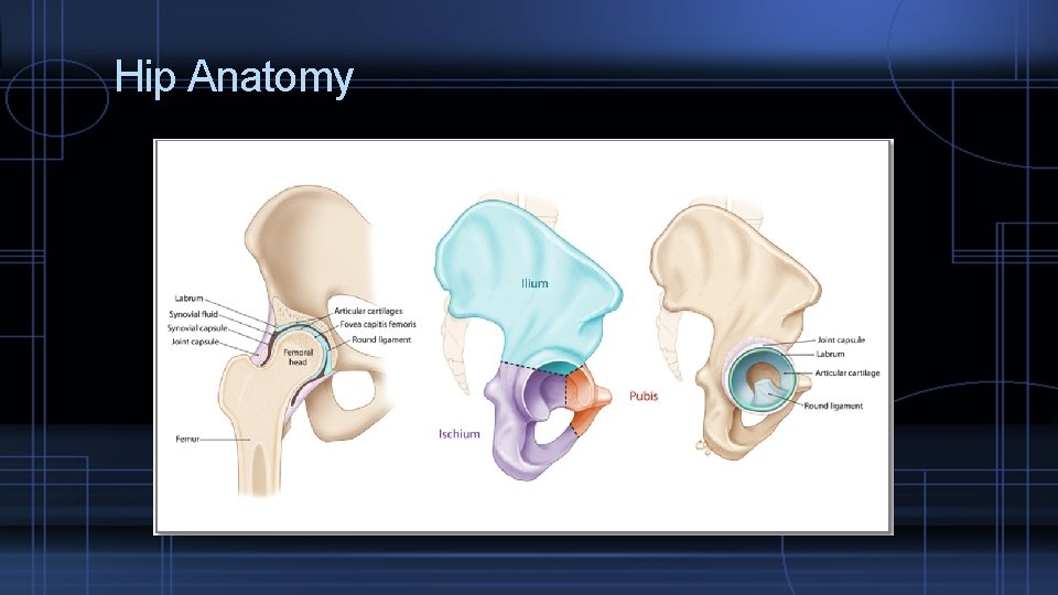
Hip Anatomy
Hip Anatomy
Hip Anatomy
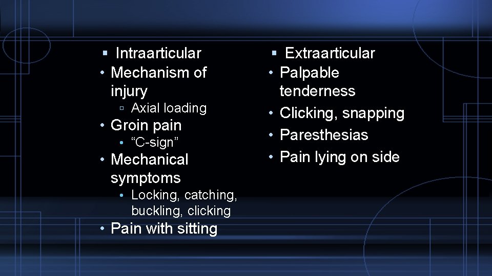
Intraarticular • Mechanism of injury Axial loading • Groin pain • “C-sign” • Mechanical symptoms • Locking, catching, buckling, clicking • Pain with sitting Extraarticular • Palpable tenderness • Clicking, snapping • Paresthesias • Pain lying on side
Patellar dislocation/Patellofemoral pain/Patellar tendinopathy Pes anserine bursitis
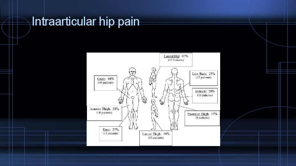
Intraarticular hip pain
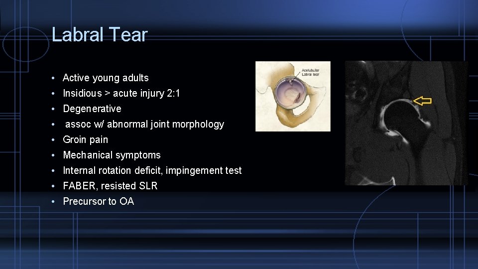
Labral Tear • Active young adults • Insidious > acute injury 2: 1 • Degenerative • assoc w/ abnormal joint morphology • Groin pain • Mechanical symptoms • Internal rotation deficit, impingement test • FABER, resisted SLR • Precursor to OA
Piriformis syndrome Sciatic nerve can be involved Rehabilitation – reduce pain and spasm to recover full hip internal rotation Provocative test is FADIR test (flexion, adduction, internal rotation) Corticosteroid injection can be considerd if more conservative measures fail.
Snapping hip syndrome Pain on hip flexion Ice, NSAIDS, PT, stretching, strengthening. Internal snapping hip – snapping over iliopectineal eminence (with or without pain) External snapping hip – tight ITB snapping over greater trochanter
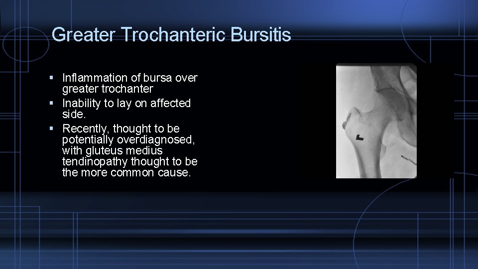
Greater Trochanteric Bursitis Inflammation of bursa over greater trochanter Inability to lay on affected side. Recently, thought to be potentially overdiagnosed, with gluteus medius tendinopathy thought to be the more common cause.
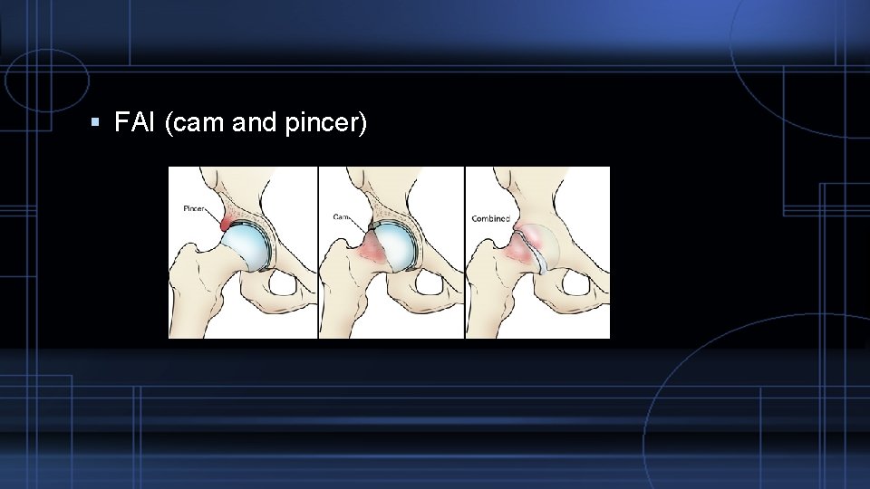
FAI (cam and pincer)
- Slides: 44