PERIOPERATIVE FLUID THERAPY Lecture Objectives Students at the

PERIOPERATIVE FLUID THERAPY

Lecture Objectives Students at the end of the lecture will be able to: -Describe different fluids components -Describe the challenges of Fluid therapy -Answer FAQ
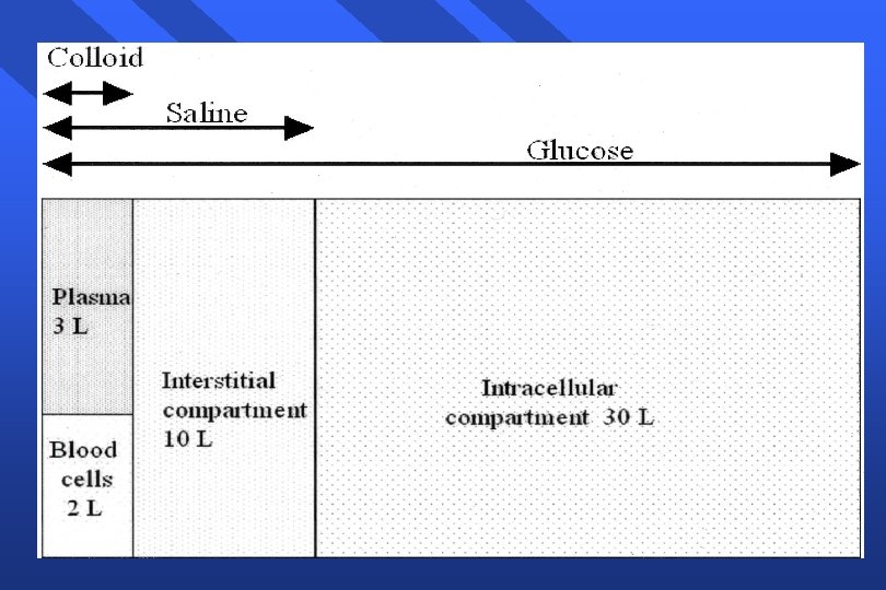
 • • • Varies with age, gender 55% body weight")
Total Body Water (TBW) • • • Varies with age, gender 55% body weight in males 45% body weight in females 80% body weight in infants Less in obese: fat contains little water

Body Water Compartments • • Intracellular water: 2/3 of TBW Extracellular water: 1/3 TBW - Extravascular water: 3/4 of extracellular water - Intravascular water: 1/4 of extracellular water

Final Goals of Fluid resuscitation - Achievement of normovolemia& hemodynamic stability - Correction of major acid-base disturbances - Compensation of internal fluid fluxes - Maintain an adequate gradient between COP&PCWP - Improvement of microvascular blood flow - Prevention of cascade system activation - Normalization of O 2 delivery - Prevention of reperfusion cellular injury - Achievement of adequate urine output

Desirable outcome of fluid resuscitation - No peripheral edema - No ARDS

Fluid and Electrolyte Regulation • Volume Regulation - Antidiuretic Hormone Renin/angiotensin/aldosterone system Baroreceptors in carotid arteries and aorta Stretch receptors in atrium and juxtaglomerular aparatus Cortisol
 Central and")
Fluid and Electrolyte Regulation • Plasma Osmolality Regulation - • Arginine-Vasopressin (ADH) Central and Peripheral osmoreceptors Sodium Concentration Regulation - Renin/angiotensin/aldosterone system Macula Densa of JG apparatus

Preoperative Evaluation of Fluid Status • Factors to Assess: - h/o intake and output blood pressure: supine and standing heart rate skin turgor urinary output serum electrolytes/osmolarity mental status

Orthostatic Hypotension • Systolic blood pressure decrease of greater than 20 mm. Hg from supine to standing • Indicates fluid deficit of 6 -8% body weight - Heart rate should increase as a compensatory measure - If no increase in heart rate, may indicate autonomic dysfunction or antihypertensive drug therapy

Perioperative Fluid Requirements The following factors must be taken into account: 1 - Maintenance fluid requirements 2 - NPO and other deficits: NG suction, bowel prep 3 - Third space losses 4 - Replacement of blood loss 5 - Special additional losses: diarrhea

1 - Maintenance Fluid Requirements • • • Insensible losses such as evaporation of water from respiratory tract, sweat, feces, urinary excretion. Occurs continually. Adults: approximately 1. 5 ml/kg/hr “ 4 -2 -1 Rule” - 4 ml/kg/hr for the first 10 kg of body weight 2 ml/kg/hr for the second 10 kg body weight 1 ml/kg/hr subsequent kg body weight Extra fluid for fever, tracheotomy, denuded surfaces

2 - NPO and other deficits • • • NPO deficit = number of hours NPO x maintenance fluid requirement. Bowel prep may result in up to 1 L fluid loss. Measurable fluid losses, e. g. NG suctioning, vomiting, ostomy output, biliary fistula and tube.

3 - Third Space Losses • • Isotonic transfer of ECF from functional body fluid compartments to non-functional compartments. Depends on location and duration of surgical procedure, amount of tissue trauma, ambient temperature, room ventilation.

Replacing Third Space Losses • • Superficial surgical trauma: 1 -2 ml/kg/hr Minimal Surgical Trauma: 3 -4 ml/kg/hr - head and neck, hernia, knee surgery • Moderate Surgical Trauma: 5 -6 ml/kg/hr - hysterectomy, chest surgery • Severe surgical trauma: 8 -10 ml/kg/hr (or more) - AAA repair, nehprectomy

4 - Blood Loss • • Replace 3 cc of crystalloid solution per cc of blood loss (crystalloid solutions leave the intravascular space) When using blood products or colloids replace blood loss volume per volume

5 - Other additional losses • Ongoing fluid losses from other sites: - • gastric drainage ostomy output diarrhea Replace volume per volume with crystalloid solutions

Example • • 62 y/o male, 80 kg, for hemicolectomy NPO after 2200, surgery at 0800, received bowel prep 3 hr. procedure, 500 cc blood loss What are his estimated intraoperative fluid requirements?
 • • • Fluid deficit (NPO): 1. 5 ml/kg/hr x 10")
Example (cont. ) • • • Fluid deficit (NPO): 1. 5 ml/kg/hr x 10 hrs = 1200 ml + 1000 ml for bowel prep = 2200 ml total deficit: (Replace 1/2 first hr, 1/4 2 nd hr, 1/4 3 rd hour). Maintenance: 1. 5 ml/kg/hr x 3 hrs = 360 mls Third Space Losses: 6 ml/kg/hr x 3 hrs =1440 mls Blood Loss: 500 ml x 3 = 1500 ml Total = 2200+360+1440+1500=5500 mls

Intravenous Fluids: • • Conventional Crystalloids Colloids Hypertonic Solutions Blood/blood products and blood substitutes

Crystalloids • Combination of water and electrolytes - Balanced salt solution: electrolyte composition and osmolality similar to plasma; example: lactated Ringer’s, Plasmlyte, Normosol. - Hypotonic salt solution: electrolyte composition lower than that of plasma; example: D 5 W. - Hypertonic salt solution: 2. 7% Na. Cl.
 -Easy to administer -No")
Crystalloids in trauma Advantages: -Balanced electrolyte solutions -Buffering capacity (Lactate) -Easy to administer -No risk of adverse reactions -No disturbance of hemostasis -Promote diuresis -Inexpensive

Crystalloids contin… Disadvantages: -Poor plasma volume support -Large quantities needed -Risk of Hypothermia -Reduced plasma COP -Risk of edema

Crystalloid solutions Na. Cl Isotonic 0. 9%: 9 g/l , Na 154, Cl 154, Osmolarity: 304 mosmol/l Disadvantages: Hyper-chloremic acidosis

Hypertonic Solutions • • • Fluids containing sodium concentrations greater than normal saline. Available in 1. 8%, 2. 7%, 3%, 5%, 7. 5%, 10% solutions. Hyperosmolarity creates a gradient that draws water out of cells; therefore, cellular dehydration is a potential problem.

Hypertonic saline Advantages: -Small volume for resuscitation. -Osmotic effect -Inotropic effect ( increase calcium influx in sarculima ) -Direct vasodilator effect -Increase MAP, CO -Increase renal, mesenteric, splanchnic, coronary blood flow.

Hypertonic saline Disadvantages: • increase hemorrhage from open vessels. • Hypernatremia • Hyperchloremia. • Metabolic acidosis.

Crystalloids Lactated Ringer's Composition: Na 130, cl 109, K 4, ca 3, Lactate 28, Osmolarity 273 mosmol/l -Sydney Ringer 1880 -Hartmann added Lactate=LR -Minor advantage over Na. Cl Disadvantages: -Not to be used as diluent for blood (Ca citrate) -Low osmolarity, can lead to high ICP

Crystalloids Dextrose 5% Composition: 50 g/l, provides 170 kcal/l Disadvantages: -enhance CO 2 production -enhance lactate production -aggravate ischemic brain injury

Composition

Colloids • • • Fluids containing molecules sufficiently large enough to prevent transfer across capillary membranes. Solutions stay in the space into which they are infused. Examples: hetastarch (Hespan), albumin, dextran.

Colloids Advantages: -Prolonged plasma volume support -Moderate volume needed -minimal risk of tissue edema -enhances microvascular flow

Colloids Disadvantages: Risk of volume overload Adverse effect on hemostasis Adverse effect on renal function Anaphylactic reaction Expensive

Dextran Composition: 40/70 Inhibit platelet aggregation bleeding

Gelatins Ø Derived from hydrolyzed bovine collagen Ø Ø Ø Metabolized by serum collagenase 0. 5 -5 hr Histamine release (H 1 blockers recommended) Decreases Von W factor (VWF) Bovine Spongiform Encephalopathy 1: 1, 000
, 25% (250 g/l) Ø")
Albumins Heat treated preparation of human serum 5% (50 g/l), 25% (250 g/l) Ø Half of infused volume will stay intravascular Ø COP=20 mm. Hg=plasma Ø 25%, COP=70 mm. Hg, it will expand the vascular space by 4 -5 times the volume infused Ø 25% used only in case of hypoalbuminemia Ø

Cochrane studies support mortality following albumin infusion Ø Ø Ø Cardiac decompensation after rapid infusion of 20 - 25% albumin decreased Ionized ca++ Aggravate leak syndrome MOF Enhance bleeding Impaired Na+Water excretion renal dysfunction

Hetastarch 6% Composition: synthetic colloid, 6% preparation in isotonic saline MW 240, 000 D- DS 0. 7 Advantages: low cost, more potent than 5% albumin (COP 30) Disadvantages: Hyperamylesemia, allergy, coagulopathy Dose: 15 -30 ml/kg/day

Pentastarch 10% -MW: 200, 000 D- DS 0. 5 -Low cost -Extensive clinical use in sepsis, burns. . -Low permeability index -Good clinical safety -Decreases PMN-EC activation -Potential to diminish vascular permeability and reduces tissue edema
 MW 130, 000 D- DS 0. 4 Used for volume therapy Dose:")
Tetrastarch (Voluven) MW 130, 000 D- DS 0. 4 Used for volume therapy Dose: 50 ml/kg/day

IVVP Hemod Stability Infusate volume Plasma COP Tissue edema Anaphylaxis Cost Crystalloids Poor Transient Large Reduced Obvious Non-exist Inexpensive Colloids Good Prolong Moderate Maintain Insignific low-mod Expensive

Crystalloids OR Colloids ACS protocol for ATLS: replace each ml of blood loss with 3 ml of crystalloid fluid. 3 for 1 rule. Patient response: Ø Rapid Ø Transient Ø Non-responsive

Clinical Evaluation of Fluid Replacement 1. Urine Output: at least 1. 0 ml/kg/hr 2. Vital Signs: BP and HR normal (How is the patient doing? ) 3. Physical Assessment: Skin and mucous membranes no dry; no thirst in an awake patient 4. Invasive monitoring; CVP or PCWP may be used as a guide 5. Laboratory tests: periodic monitoring of hemoglobin and hematocrit

Summary • • Fluid therapy is critically important during the perioperative period. The most important goal is to maintain hemodynamic stability and protect vital organs from hypoperfusion (heart, liver, brain, kidneys). All sources of fluid losses must be accounted for. Good fluid management goes a long way toward preventing problems.

Transfusion Therapy - 60% of transfusions occur perioperatively. - responsibility of transfusing perioperatively is with the anesthesiologist.
 Why?")
Blood Transfusion (up to 30% of blood volume can be treated with crystalloids) Why? -Improvement of oxygen transport -Restoration of red cell mass -Correction of bleeding caused by platelet dysfunction -Correction of bleeding caused by factor deficiencies

When is Transfusion Necessary? • “Transfusion Trigger”: Hgb level at which transfusion should be given. - Varies with patients and procedures • Tolerance of acute anemia depends on: - Maintenance of intravascular volume - Ability to increase cardiac output - Increases in 2, 3 -DPG to deliver more of the carried oxygen to tissues
 is the oxygen that is")
Oxygen Delivery • • • Oxygen Delivery (DO 2) is the oxygen that is delivered to the tissues DO 2= COP x Ca. O 2 Cardiac Output (CO) = HR x SV Oxygen Content (Ca. O 2): - (Hgb x 1. 39)O 2 saturation + Pa. O 2(0. 003) - Hgb is the main determinant of oxygen content in the blood
 • • Therefore: DO 2 = HR x SV x")
Oxygen Delivery (cont. ) • • Therefore: DO 2 = HR x SV x Ca. O 2 If HR or SV are unable to compensate, Hgb is the major deterimant factor in O 2 delivery Healthy patients have excellent compensatory mechanisms and can tolerate Hgb levels of 7 gm/d. L. Compromised patients may require Hgb levels above 10 gm/d. L.

Blood Groups Blood Group Antigen on erythrocyte Plasma Antibodies A B AB O A B AB None Anti-B Anti-A None Anti-A Anti-B Rh Rh Incidence White African. Americans 40% 27% 11 20 4 4 45 49 42 17

Cross Match • Major: - Donor’s erythrocytes incubated with recipients plasma • Minor: - Donor’s plasma incubated with recipients erythrocytes • Agglutination: - Occurs if either is incompatible • Type Specific: - Only ABO-Rh determined; chance of hemolytic reaction is 1: 1000 with TS blood

Type and Screen • Donated blood that has been tested for ABO/Rh antigens and screened for common antibodies (not mixed with recipient blood). - Used when usage of blood is unlikely, but needs to be available (hysterectomy). - Allows blood to available for other patients. - Chance of hemolytic reaction: 1: 10, 000.

Differential Centrifugation First Centrifugation Closed System Whole Blood Main Bag Satellite Bag 1 First RBC’s Platelet-rich Plasma Satellite Bag 2

Differential Centrifugation Second Centrifugation RBC’s Platelet-rich Plasma Second RBC’s Platelet Concentrate Plasma

Blood Components è Prepared from Whole blood collection è Whole blood is separated by differential centrifugation X X X Red Blood Cells (RBC’s) Platelets Plasma » Cryoprecipitate » Others è Others include Plasma proteins—IVIg, Coagulation Factors, albumin, Anti-D, Growth Factors, Colloid volume expanders

Whole Blood è Storage X 4° for up to 35 days è Indications X Massive Blood Loss/Trauma/Exchange Transfusion è Considerations X X Use filter as platelets and coagulation factors will not be active after 3 -5 days Donor and recipient must be ABO identical

Component Therapy • A unit of whole blood is divided into components; Allows prolonged storage and specific treatment of underlying problem with increased efficiency: - packed red blood cells (p. RBC’s) platelet concentrate fresh frozen plasma (contains all clotting factors) cryoprecipitate (contains factors VIII and fibrinogen; used in Von Willebrand’s disease) albumin plasma protein fraction leukocyte poor blood factor VIII antibody concentrates

Packed Red Blood Cells • • • 1 unit = 250 ml. Hct. = 70 -80%. 1 unit p. RBC’s raises Hgb 1 gm/d. L. Mixed with saline: LR has Calcium which may cause clotting if mixed with p. RBC’s.

RBC Transfusions Administration è Dose X X è Procedure X X è Usual dose of 10 cc/kg infused over 2 -4 hours Maximum dose 15 -20 cc/kg can be given to hemodynamically stable patient May need Premedication (Tylenol and/or Benadryl) Filter use—routinely leukodepleted Monitoring—VS q 15 minutes, clinical status Do NOT mix with medications Complications X X Rapid infusion may result in Pulmonary edema Transfusion Reaction

Platelet Concentrate è Storage X Up to 5 days at 20 -24° è Indications X X X Thrombocytopenia, Plt <15, 000 Bleeding and Plt <50, 000 Invasive procedure and Plt <50, 000 è Considerations X X X Contain Leukocytes and cytokines 1 unit/10 kg of body weight increases Plt count by 50, 000 Donor and Recipient must be ABO identical
 è Storage è X è Indications X")
Plasma and FFP Contents—Coagulation Factors (1 unit/ml) è Storage è X è Indications X è FFP--12 months at – 18 degrees or colder Coagulation Factor deficiency, fibrinogen replacement, DIC, liver disease, exchange transfusion, massive transfusion Considerations X X X Plasma should be recipient RBC ABO compatible In children, should also be Rh compatible Usual dose is 20 cc/kg to raise coagulation factors approx 20%
 èChronic Transfusion Reactions èTransfusion related infections")
Transfusion Complications èAcute Transfusion Reactions (ATR’s) èChronic Transfusion Reactions èTransfusion related infections
 è Febrile Reactions (FNHTR) è Allergic Reactions")
Acute Transfusion Reactions è Hemolytic Reactions (AHTR) è Febrile Reactions (FNHTR) è Allergic Reactions è TRALI è Coagulopathy with Massive transfusions è Bacteremia

Complications of Blood Therapy • Transfusion Reactions: - Febrile; most common, usually controlled by slowing infusion and giving antipyretics - Allergic; increased body temp. , pruritis, urticaria. Rx: antihistamine, discontinuation. Examination of plasma and urine for free hemoglobin helps rule out hemolytic reactions.
 • Hemolytic: Wrong blood type administered (oops). -")
Complications of Blood Therapy (cont. ) • Hemolytic: Wrong blood type administered (oops). - Activation of complement system leads to intravascular hemolysis, spontaneous hemorrhage. Signs: - hypotension, fever, chills dyspnea, skin flushing, substernal pain , Back/abdominal pain Oliguria Dark urine Pallor
 Signs are easily masked by general anesthesia. -")
Complications of Blood Therapy (cont. ) Signs are easily masked by general anesthesia. - Free Hgb in plasma or urine - Acute renal failure - Disseminated Intravascular Coagulation (DIC)
 • Transmission of Viral Diseases: - Hepatitis C; 1: 30, 000")
Complications (cont. ) • Transmission of Viral Diseases: - Hepatitis C; 1: 30, 000 per unit Hepatitis B; 1: 200, 000 per unit HIV; 1: 450, 000 -1: 600, 000 per unit 22 day window for HIV infection and test detection CMV may be the most common agent transmitted, but only effects immuno-compromised patients Parasitic and bacterial transmission very low

Other Complications - Decreased 2, 3 -DPG with storage: ? Significance Citrate: metabolism to bicarbonate; Calcium binding Microaggregates (platelets, leukocytes): micropore filters controversial Hypothermia: warmers used to prevent Coagulation disorders: massive transfusion (>10 units) may lead to dilution of platelets and factor V and VIII. DIC: uncontrolled activation of coagulation system

Treatment of Acute Hemolytic Reactions • • • Immediate discontinuation of blood products and send blood bags to lab. Maintenance of urine output with crystalloid infusions Administration of mannitol or Furosemide for diuretic effect

Massive Blood Transfusion è Massive transfusion is generally defined as the need to transfuse one to two times the patient's blood volume. For most adult patients, that is the equivalent of 10– 20 units

Massive Blood Transfusion è Coagulopathy due to dilutional thrombocytopenia. And dilution of the coagulation factors è Citrate Toxicity does not occur in most normal patients unless the transfusion rate exceeds 1 U every 5 min è Hypothermia è Acid–Base Balance The most consistent acid–base abnormality after massive blood transfusion is postoperative metabolic alkalosis

Massive Blood Transfusion è Serum Potassium Concentration The extracellular concentration of potassium in stored blood steadily increases with time. The amount of eacellular potassium transfused with each unit less than 4 m. Eq per unit. Hyperkalemia can develop regardless of the age of the blood when transfusion rates exceed 100 m. L/min. Hypokalemia is commonly encountered postoperatively, particularly in association with metabolic alkalosis

DIC Type Biological Definition Hemostatic defect without clinical SS Clinical Hemostatic defect+He Diagnosis high D-Dimers and major or minor criteria of platelet consumption same above+microvasc bleeding Complicated +ischemia +organ failure Lab DD≥ 500 ug/l Plat 50 -100, 000 INR 1. 2 -1. 5

Autologous Blood • • • Pre-donation of patient’s own blood prior to elective surgery 1 unit donated every 4 days (up to 3 units) Last unit donated at least 72 hrs prior to surgery Reduces chance of hemolytic reactions and transmission of blood-bourne diseases Not desirable for compromised patients

Administering Blood Products - Consent necessary for elective transfusion Unit is checked by 2 people for Unit #, patient ID, expiration date, physical appearance. p. RBC’s are mixed with saline solution (not LR) Products are warmed mechanically and given slowly if condition permits Close observation of patient for signs of complications If complications suspected, infusion discontinued, blood bank notified, proper steps taken.

Alternatives to Blood Products • • Autotransfusion Blood substitutes

Autotransfusion • • Commonly known as “Cell-saver” Allows collection of blood during surgery for readministration RBC’s centrifuged from plasma Effective when > 1000 ml are collected

Blood Substitutes • • • Experimental oxygen-carrying solutions: developed to decrease dependence on human blood products Military battlefield usage initial goal Multiple approaches: - Outdated human Hgb reconstituted in solution Genetically engineered/bovine Hgb in solution Liposome-encapsulated Hgb Perflurocarbons
 • Potential Advantages: - No cross-match requirements Long-term shelf storage")
Blood Substitutes (cont. ) • Potential Advantages: - No cross-match requirements Long-term shelf storage No blood-bourne transmission Rapid restoration of oxygen delivery in traumatized patients Easy access to product (available on ambulances, field hospitals, hospital ships)
 • Potential Disadvantages: - Undesirable hemodynamic effects: • Mean arterial")
Blood Substitutes (cont. ) • Potential Disadvantages: - Undesirable hemodynamic effects: • Mean arterial pressure and pulmonary artery pressure increases - Short half-life in bloodstream (24 hrs) Still in clinical trials, unproven efficacy High cost

Transfusion Therapy Summary • • Decision to transfuse involves many factors Availability of component factors allows treatment of specific deficiency Risks of transfusion must be understood and explained to patients and patient should be consented Vigilance necessary when transfusing any blood product

What to do? If an AHTR occurs è STOP TRANSFUSION è ABC’s è Maintain IV access and run IVF (NS or LR) è Monitor and maintain BP/pulse è Give diuretic è Obtain blood and urine for transfusion reaction workup è Send remaining blood back to Blood Bank

Blood Bank Work-up of AHTR è Check paperwork to assure no errors è Check plasma for hemoglobin è Repeat crossmatch è Repeat Blood group typing è Blood culture

Monitoring in AHTR è Monitor patient clinical status and vital signs è Monitor renal status (BUN, creatinine) è Monitor coagulation status (DIC panel– PT/PTT, fibrinogen, D-dimer/FDP, Plt, Antithrombin-III) è Monitor for signs of hemolysis (LDH, bili, haptoglobin)

- Slides: 87