PERIOPERATIVE FLUID THERAPY Department of Anesthesiology ICU KKUH


- Slides: 47

PERIOPERATIVE FLUID THERAPY Department of Anesthesiology &ICU KKUH. King Saud University

Total Body Water (TBW) • • • Varies with age, gender 55% body weight in males 45% body weight in females 80% body weight in infants Less in obese: fat contains little water

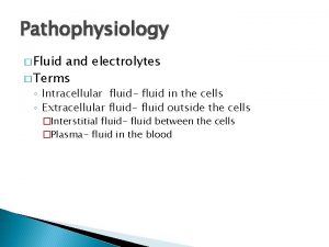
Body Water Compartments • • Intracellular water: 2/3 of TBW Extracellular water: 1/3 TBW - Extravascular water: 3/4 of extracellular water - Intravascular water: 1/4 of extracellular water

Fluid and Electrolyte Regulation • Volume Regulation - Antidiuretic Hormone Renin/angiotensin/aldosterone system Baroreceptors in carotid arteries and aorta Stretch receptors in atrium and juxtaglomerular aparatus Cortisol

Fluid and Electrolyte Regulation • Plasma Osmolality Regulation - • Arginine-Vasopressin (ADH) Central and Peripheral osmoreceptors Sodium Concentration Regulation - Renin/angiotensin/aldosterone system Macula Densa of JG apparatus

Preoperative Evaluation of Fluid Status • Factors to Assess: - h/o intake and output blood pressure: supine and standing heart rate skin turgor urinary output serum electrolytes/osmolarity mental status

Orthostatic Hypotension • Systolic blood pressure decrease of greater than 20 mm. Hg from supine to standing • Indicates fluid deficit of 6 -8% body weight - Heart rate should increase as a compensatory measure - If no increase in heart rate, may indicate autonomic dysfunction or antihypertensive drug therapy

Perioperative Fluid Requirements The following factors must be taken into account: 1 - Maintenance fluid requirements 2 - NPO and other deficits: NG suction, bowel prep 3 - Third space losses 4 - Replacement of blood loss 5 - Special additional losses: diarrhea

1 - Maintenance Fluid Requirements • • • Insensible losses such as evaporation of water from respiratory tract, sweat, feces, urinary excretion. Occurs continually. Adults: approximately 1. 5 ml/kg/hr “ 4 -2 -1 Rule” - 4 ml/kg/hr for the first 10 kg of body weight 2 ml/kg/hr for the second 10 kg body weight 1 ml/kg/hr subsequent kg body weight Extra fluid for fever, tracheotomy, denuded surfaces

2 - NPO and other deficits • • • NPO deficit = number of hours NPO x maintenance fluid requirement. Bowel prep may result in up to 1 L fluid loss. Measurable fluid losses, e. g. NG suctioning, vomiting, ostomy output, biliary fistula and tube.

3 - Third Space Losses • • Isotonic transfer of ECF from functional body fluid compartments to non-functional compartments. Depends on location and duration of surgical procedure, amount of tissue trauma, ambient temperature, room ventilation.

Replacing Third Space Losses • • Superficial surgical trauma: 1 -2 ml/kg/hr Minimal Surgical Trauma: 3 -4 ml/kg/hr - head and neck, hernia, knee surgery • Moderate Surgical Trauma: 5 -6 ml/kg/hr - hysterectomy, chest surgery • Severe surgical trauma: 8 -10 ml/kg/hr (or more) - AAA repair, nehprectomy

4 - Blood Loss • • Replace 3 cc of crystalloid solution per cc of blood loss (crystalloid solutions leave the intravascular space) When using blood products or colloids replace blood loss volume per volume

5 - Other additional losses • Ongoing fluid losses from other sites: - • gastric drainage ostomy output diarrhea Replace volume per volume with crystalloid solutions

Example • • 62 y/o male, 80 kg, for hemicolectomy NPO after 2200, surgery at 0800, received bowel prep 3 hr. procedure, 500 cc blood loss What are his estimated intraoperative fluid requirements?

Example (cont. ) • • • Fluid deficit (NPO): 1. 5 ml/kg/hr x 10 hrs = 1200 ml + 1000 ml for bowel prep = 2200 ml total deficit: (Replace 1/2 first hr, 1/4 2 nd hr, 1/4 3 rd hour). Maintenance: 1. 5 ml/kg/hr x 3 hrs = 360 mls Third Space Losses: 6 ml/kg/hr x 3 hrs =1440 mls Blood Loss: 500 ml x 3 = 1500 ml Total = 2200+360+1440+1500=5500 mls

Intravenous Fluids: • • Conventional Crystalloids Colloids Hypertonic Solutions Blood/blood products and blood substitutes

Crystalloids • Combination of water and electrolytes - Balanced salt solution: electrolyte composition and osmolality similar to plasma; example: lactated Ringer’s, Plasmlyte, Normosol. - Hypotonic salt solution: electrolyte composition lower than that of plasma; example: D 5 W. - Hypertonic salt solution: 2. 7% Na. Cl.

Colloids • • • Fluids containing molecules sufficiently large enough to prevent transfer across capillary membranes. Solutions stay in the space into which they are infused. Examples: hetastarch (Hespan), albumin, dextran.

Hypertonic Solutions • • • Fluids containing sodium concentrations greater than normal saline. Available in 1. 8%, 2. 7%, 3%, 5%, 7. 5%, 10% solutions. Hyperosmolarity creates a gradient that draws water out of cells; therefore, cellular dehydration is a potential problem.

Composition

Clinical Evaluation of Fluid Replacement 1. Urine Output: at least 1. 0 ml/kg/hr 2. Vital Signs: BP and HR normal (How is the patient doing? ) 3. Physical Assessment: Skin and mucous membranes no dry; no thirst in an awake patient 4. Invasive monitoring; CVP or PCWP may be used as a guide 5. Laboratory tests: periodic monitoring of hemoglobin and hematocrit

Summary • • Fluid therapy is critically important during the perioperative period. The most important goal is to maintain hemodynamic stability and protect vital organs from hypoperfusion (heart, liver, brain, kidneys). All sources of fluid losses must be accounted for. Good fluid management goes a long way toward preventing problems.

Transfusion Therapy - 60% of transfusions occur perioperatively. - responsibility of transfusing perioperatively is with the anesthesiologist.

When is Transfusion Necessary? • “Transfusion Trigger”: Hgb level at which transfusion should be given. - Varies with patients and procedures • Tolerance of acute anemia depends on: - Maintenance of intravascular volume - Ability to increase cardiac output - Increases in 2, 3 -DPG to deliver more of the carried oxygen to tissues

Oxygen Delivery • • • Oxygen Delivery (DO 2) is the oxygen that is delivered to the tissues DO 2= COP x Ca. O 2 Cardiac Output (CO) = HR x SV Oxygen Content (Ca. O 2): - (Hgb x 1. 39)O 2 saturation + Pa. O 2(0. 003) - Hgb is the main determinant of oxygen content in the blood

Oxygen Delivery (cont. ) • • Therefore: DO 2 = HR x SV x Ca. O 2 If HR or SV are unable to compensate, Hgb is the major deterimant factor in O 2 delivery Healthy patients have excellent compensatory mechanisms and can tolerate Hgb levels of 7 gm/d. L. Compromised patients may require Hgb levels above 10 gm/d. L.

Blood Groups Blood Group Antigen on erythrocyte Plasma Antibodies A B AB O A B AB None Anti-B Anti-A None Anti-A Anti-B Rh Rh Incidence White African. Americans 40% 27% 11 20 4 4 45 49 42 17

Cross Match • Major: - Donor’s erythrocytes incubated with recipients plasma • Minor: - Donor’s plasma incubated with recipients erythrocytes • Agglutination: - Occurs if either is incompatible • Type Specific: - Only ABO-Rh determined; chance of hemolytic reaction is 1: 1000 with TS blood

Type and Screen • Donated blood that has been tested for ABO/Rh antigens and screened for common antibodies (not mixed with recipient blood). - Used when usage of blood is unlikely, but needs to be available (hysterectomy). - Allows blood to available for other patients. - Chance of hemolytic reaction: 1: 10, 000.

Component Therapy • A unit of whole blood is divided into components; Allows prolonged storage and specific treatment of underlying problem with increased efficiency: - packed red blood cells (p. RBC’s) platelet concentrate fresh frozen plasma (contains all clotting factors) cryoprecipitate (contains factors VIII and fibrinogen; used in Von Willebrand’s disease) albumin plasma protein fraction leukocyte poor blood factor VIII antibody concentrates

Packed Red Blood Cells • • • 1 unit = 250 ml. Hct. = 70 -80%. 1 unit p. RBC’s raises Hgb 1 gm/d. L. Mixed with saline: LR has Calcium which may cause clotting if mixed with p. RBC’s.

Platelet Concentrate • • Treatment of thrombocytopenia Intraoperatively used if platlet count drops below 50, 000 cells-mm 3 (lab analysis). 1 unit of platelets increases platelet count 5000 -10000 cells-mm 3. Risks: - Sensitization due to HLA on platelets - Viral transmission

Fresh Frozen Plasma • Plasma from whole blood frozen within 6 hours of collection. - • Contains coagulation factors except platelets Used for treatment of isolated factor deficiences, reversal of Coumadin effect, TTP, etc. Used when PT and PTT are >1. 5 normal Risks: - Viral transmission Allergy

Complications of Blood Therapy • Transfusion Reactions: - Febrile; most common, usually controlled by slowing infusion and giving antipyretics - Allergic; increased body temp. , pruritis, urticaria. Rx: antihistamine, discontinuation. Examination of plasma and urine for free hemoglobin helps rule out hemolytic reactions.

Complications of Blood Therapy (cont. ) • Hemolytic: - Wrong blood type administered (oops). Activation of complement system leads to intravascular hemolysis, spontaneous hemorrhage. Signs: hypotension, fever, chills, dyspnea, skin flushing, substernal pain. Signs are easily masked by general anesthesia. Free Hgb in plasma or urine Acute renal failure Disseminated Intravascular Coagulation (DIC)

Complications (cont. ) • Transmission of Viral Diseases: - Hepatitis C; 1: 30, 000 per unit Hepatitis B; 1: 200, 000 per unit HIV; 1: 450, 000 -1: 600, 000 per unit 22 day window for HIV infection and test detection CMV may be the most common agent transmitted, but only effects immuno-compromised patients Parasitic and bacterial transmission very low

Other Complications - Decreased 2, 3 -DPG with storage: ? Significance Citrate: metabolism to bicarbonate; Calcium binding Microaggregates (platelets, leukocytes): micropore filters controversial Hypothermia: warmers used to prevent Coagulation disorders: massive transfusion (>10 units) may lead to dilution of platelets and factor V and VIII. DIC: uncontrolled activation of coagulation system

Treatment of Acute Hemolytic Reactions • • • Immediate discontinuation of blood products and send blood bags to lab. Maintenance of urine output with crystalloid infusions Administration of mannitol or Furosemide for diuretic effect

Autologous Blood • • • Pre-donation of patient’s own blood prior to elective surgery 1 unit donated every 4 days (up to 3 units) Last unit donated at least 72 hrs prior to surgery Reduces chance of hemolytic reactions and transmission of blood-bourne diseases Not desirable for compromised patients

Administering Blood Products - Consent necessary for elective transfusion Unit is checked by 2 people for Unit #, patient ID, expiration date, physical appearance. p. RBC’s are mixed with saline solution (not LR) Products are warmed mechanically and given slowly if condition permits Close observation of patient for signs of complications If complications suspected, infusion discontinued, blood bank notified, proper steps taken.

Alternatives to Blood Products • • Autotransfusion Blood substitutes

Autotransfusion • • Commonly known as “Cell-saver” Allows collection of blood during surgery for readministration RBC’s centrifuged from plasma Effective when > 1000 ml are collected

Blood Substitutes • • • Experimental oxygen-carrying solutions: developed to decrease dependence on human blood products Military battlefield usage initial goal Multiple approaches: - Outdated human Hgb reconstituted in solution Genetically engineered/bovine Hgb in solution Liposome-encapsulated Hgb Perflurocarbons

Blood Substitutes (cont. ) • Potential Advantages: - No cross-match requirements Long-term shelf storage No blood-bourne transmission Rapid restoration of oxygen delivery in traumatized patients Easy access to product (available on ambulances, field hospitals, hospital ships)

Blood Substitutes (cont. ) • Potential Disadvantages: - Undesirable hemodynamic effects: • Mean arterial pressure and pulmonary artery pressure increases - Short half-life in bloodstream (24 hrs) Still in clinical trials, unproven efficacy High cost

Transfusion Therapy Summary • • Decision to transfuse involves many factors Availability of component factors allows treatment of specific deficiency Risks of transfusion must be understood and explained to patients Vigilance necessary when transfusing any blood product
 Umass memorial nurse residency program
 Tufts anesthesia residency
 Fluid statics deals with
 Fluid kinematics is part of fluid mechanics in which
 Fluid compartments in the body
 Extracellular fluid and interstitial fluid
 Viscoseal
 Intracellular extracellular fluid
 Ascitic fluid neutrophil count
 Fluid statics deals with fluid at rest
 Interstitial vs intracellular
 Psychoanalytic therapy is to as humanistic therapy is to
 Bioness integrated therapy system occupational therapy
 Psychoanalytic therapy is to as humanistic therapy is to
 Perioperative nursing case study
 Ccs perioperative guidelines
 Preoperative checklist definition
 Perioperative definition
 Preoperative vs perioperative
 Icu transliterator
 Critical care for dummies
 Icu university zambia
 Alfred icu
 Chatecholamine
 Atenolol to carvedilol conversion chart
 Icu layout and design
 Cam icu
 Abcde methode ehbo
 Escala de sedacion
 Emergency drugs in icu
 Icu medical b3108
 List of lecturers at icu zambia
 Icu indication
 Lactated ringers vs normal saline
 Icu case presentation
 Cam icu escala
 Icu unit meaning
 Icu localization
 Icu unicode
 Kp icu jpm
 Shanks algorithm example
 Patient acuity tool
 Pressors icu
 Diet chart for icu patients
 Icu case presentation
 Cheetah nicom interpretation
 Icu orientation
 Icu tersier

