Perineal body and pudendal canal Dr Stuti Tandon

Perineal body and pudendal canal Dr Stuti Tandon Assistant professor Department of anatomy Cims&h

Perineal body • Fibromuscular node situated in the midline at the junction of urogenital triangle and anal triangle. • Lies 1. 25 cm in front of anal margin in males and posterior wall of vestibule of vagina in females.

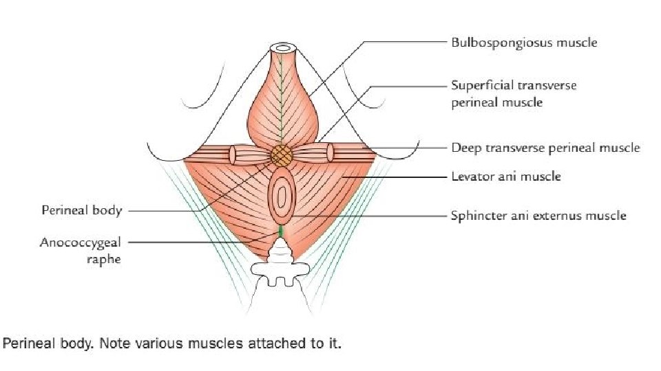

Clinical correlation Damage of perineal body • Perineal body is of extreme importance in females In maintaining integrity of pelvic diaphragm and providing support to pelvic organs. • May get damaged during 1. Difficult childbirth 2. Inadvertently during episiotomy May lead to prolapse of uterus, bladder and rectum.

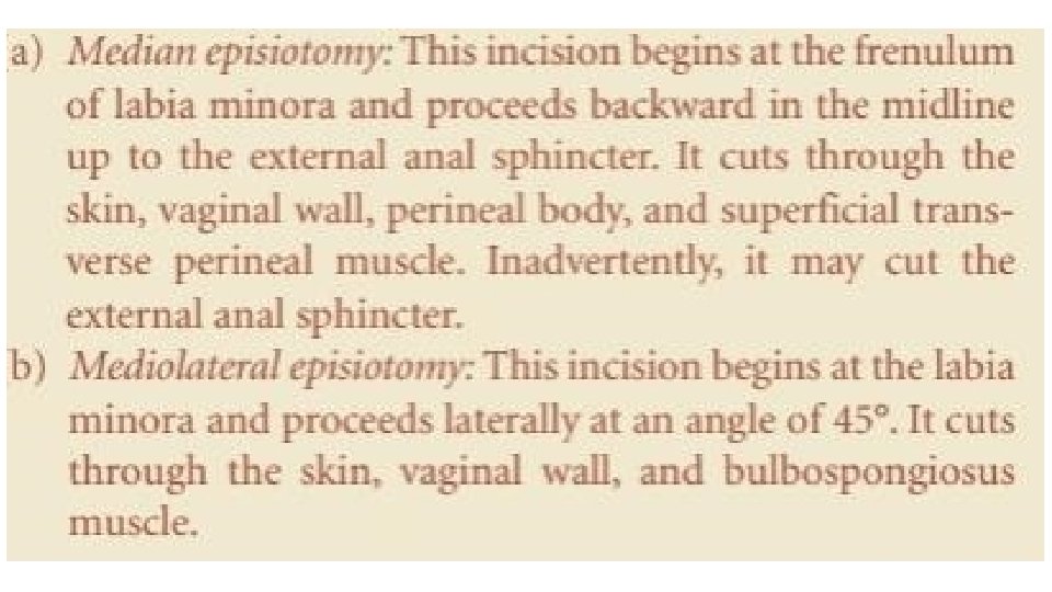
Episiotomy • It is an incision given in the perineum to enlarge the vaginal orifice to facilitate childbirth. 1. Median episiotomy 2. Mediolateral episiotomy

Pudendal canal /Alcock’s canal • It is a fascial canal present in the lateral wall of ischiorectal fossa 2. 5 cm above the ischial tuberosity. • Extends from lesser sciatic foramen to posterior limit of deep perineal pouch. • The pudendal canal in formed either by 1. Splitting of obturator fascia or 2. By separation between fascia lunata and obturator fascia.

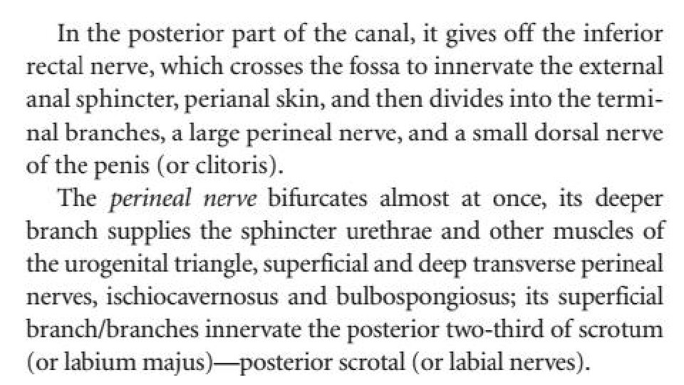
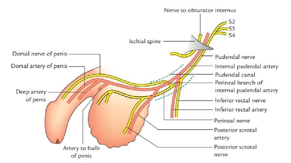
Contents 1. Pudendal nerve which divides within the canal into dorsal nerve of penis and the perineal nerve. 2. Internal pudendal vessels.

Pudendal nerve • It is the chief nerve of perineum. Origin, course and distribution
 traverses the deep perineal pouch")
• The dorsal nerve of penis (or clitoris) traverses the deep perineal pouch then passes through a gap between arcuate pubic and transverse perineal ligaments to reach the dorsum of penis. • It innervates the skin of body and glands of penis.

Pudendal nerve block • The pudendal nerve is infiltrated with a local anaesthetic where it crosses the ischial spine which is palpated through the vagina. • A long needle is inserted through the vaginal wall and guided by the finger to the ischial spine. • The needle can also be inserted through the skin of perineum. • When block is carried out bilaterally 1. There is loss of anal reflex 2. Relaxation of pelvic floor muscles 3. Loss of sensation of vulva and lower 1/3 rd of vagina.

Internal pudendal artery • It is one of the two terminal branches of the anterior division of internal iliac artery. • It leaves the pelvis through greater sciatic foramen below the piriformis. • It crosses the dorsal aspect of ischial spine and enters the perineum through the lesser sciatic foramen • It passes through the pudendal canal in the lateral wall of ischiorectal fossa.

- Slides: 18