Pericardial Diseases Part I Acute Pericarditis 901 1202


- Slides: 55

Pericardial Diseases Part I Acute Pericarditis 9/01 - 12/02 medslides. com 1

9/01 medslides. com 2

Learning Objectives • Understand the diagnosis and treatment of: – Pericarditis • Acute Pericarditis • Pericardial Effusion – Pericardial Compressive Syndrome • Constrictive Pericarditis • Cardiac Tamponade 9/01 medslides. com 3

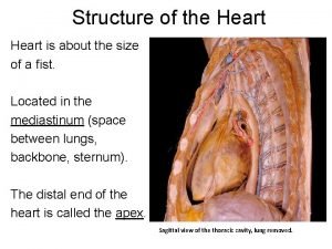
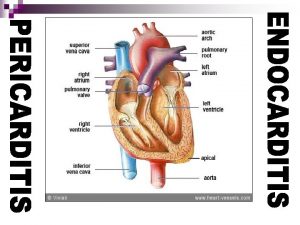
Pericardial Anatomy • Two major components – serosa (viceral pericardium) mesothelial monolayer facilitate fluid and ion exchange – fibrosa (parietal pericardium) fibrocollagenous tissue • Pericardial Fluid – 15 - 50 ml of clear plasma ultrafiltrate • Ligamentous attachments – to the sternum, vertebral column, diaphragm 9/01 medslides. com 4

Pericardial Physiology • not needed to sustain life • physiologic functions – limit cardiac dilatation – limit cardiac displacement – maintain normal ventricular compliance – reduce friction to cardiac movement – barrier to inflammation 9/01 medslides. com 5

Pericardial Disease Clinical Presentation Diseases of the pericardium (either primary or secondary) present clinically in one of three ways: • Acute fibrinous pericarditis; this disorder is sometimes called "dry" pericarditis • Pericardial effusion without major hemodynamic compromise • Pericardial compressive syndrome, either cardiac tamponade or constrictive pericarditis 9/01 medslides. com 6

Pericardial Disease • Other clinical presentations: – Chronic Relapsing Pericarditis – Effusive Constrictive Pericarditis – Restrictive Cardiomyopathy – Localized and Low Pressure Tamponade 9/01 medslides. com 7

Acute Pericarditis 9/01 medslides. com 8

Pericardial Inflammation pathogenesis • Contiguous spread – lungs, pleura, mediastinal lymph nodes, myocardium, aorta, esophagus, liver • Hematogenous spread – septicemia, toxins, neoplasm, metabolic • Lymphangetic spread • Traumatic or irradiation 9/01 medslides. com 9

Pericardial Inflammation pathology • inflammation provokes a fibrinous exudate with or without serous effusion • the normal transparent and glistening pericardium is turned into a dull, opaque, and “sandy” sac • can cause pericardial scarring with adhesions and fibrosis 9/01 medslides. com 10

Acute Pericarditis Diagnostic Clues • History – sudden onset of anterior chest pain that is pleuritic and substernal • Physical exam – presence of two- or three-component rub • ECG – ST elevation (most important clue) – PR segment depression 9/01 medslides. com 11

Chest Pain History pericarditis vs infarction • Common characteristics – retrosternl or precordial with radiation to the neck, back, left shoulder or arm • Special characteristics (pericarditis) – more likely to be sharp and pleuritic – with coughing, inspiration, swallowing – worse by lying supine, relieved by sitting and leaning forward 9/01 medslides. com 12

Heart Murmurs of Pericarditis • Pericardial friction rub is pathognomic for pericarditis • scratching or grating sound • Classically three components: – presystolic rub during atrial filling – ventricular systolic rub (loudest) – ventricular diastolic rub (after A 2 P 2) 9/01 medslides. com 13

Acute Pericarditis: ECG features • ST-segment elevation – reflecting epicardial inflammation – leads I, II, a. VL, and V 3 -V 6 – lead a. VR and V 1 usually shows ST depression • ST concave upward – ST in AMI concave downward like a “dome” • PR segment depression – early stage • T-wave inversion – occurs after the ST returns to baseline 9/01 medslides. com 14

Four Stages of ECG Evolution 1. First hours to days: – diffuse upsloping ST elevation with reciprocal ST depression (a. VR, V 1) – PR depression in the inferolateral leads (II, III, AVF, V 5 -6) – PR elevation in a. VR 2. Normalization of the ST and PR segments 3. Diffuse T wave inversions, generally after the ST segments have become isoelectric. However, this phase is not seen in some patients 4. ECG may become normal or the T wave inversions may persist indefinitely ("chronic" pericarditis) * arrhythmias are uncommon in acute pericarditis, its presence is suggestive of concomitant myocarditis 12/02 medslides. com 15

ECG Features Favoring the Diagnosis of Acute Myocardial Infarction • · The ST segment elevation is more localized, usually convex, may be > 5 mm, often merging with the T wave, often associated with reciprocal ST segment changes Simultaneous ST segment elevation and T wave inversions · Evolving Q waves · Hyperacute T waves · PR segment abnormalities uncommon · Definite QT prolongation 12/02 medslides. com 16

ECG Features Favoring the Diagnosis of Early Repolarization • One-half of normal variants have no ST deviations in the limb leads (whereas diffuse ST elevations in both the limb and precordial leads occur in most cases (47/48 in one study) of acute pericarditis) • Absence of PR deviation • Absence of the ST and T-wave evolution 12/02 Circulation 65, No 5, 1982: 1004 -1009 medslides. com 17

The Differential Diagnosis of Acute Pericarditis from the Normal Variant The ratio of the amplitude of the onset of the ST segment to the amplitude of the T wave in lead V 6 is a reliable discriminator ST/T ratio in V 6 0. 25 T wave in V 6 < 0. 5 m. V 9/01 • ST/T ratio in V 6 0. 25 diagnosed all patients with pericarditis (PPV=1. 0, NPV=1. 0) • ST/T ratio in V 4, V 6, and lead I 0. 25 were also highly suggestive (PPV=0. 90, NPV=0. 88) Circulation 65, No 5, 1982: 1004 -1009 medslides. com 18

Etiology of Inflammatory Pericarditis • Most patients with acute pericarditis have either viral (echovirus and coxsackievirus are most common) or idiopathic pericarditis. Other inflammatory pericarditis include: • Infection • Drug or toxin induced • Radiation • Metabolic • Trauma • Collagen vascular • An extensive initial evaluation is not warranted in uncomplicated patients because the diagnostic yield is low. 9/01 medslides. com 19

Acute Pericarditis common causes • Outpatient setting – usually idiopathic – probably due to viral infections – Coxsackie A and B (highly cardiotropic) are the most common viral cause of pericarditis and myocarditis – Others viruses: mumps, varicella-zoster, influenza, Epstein-Barr, HIV 9/01 medslides. com 20

Acute Pericarditis common causes • Inpatient setting T = Trauma, TUMOR U = Uremia M = Myocardial infarction (acute, post) Medications (hydralazine, procainamide) O = Other infections (bacterial, fungal, TB) R = Rheumatoid, autoimmune disorder, Radiation 9/01 medslides. com 21

Infectious Pericarditis • Any infectious organism (virus, bacterium, tuberculosis, Rickettsia, spirochete, fungus, parasite, or chlamydia) can infect the pericardium • AIDS is unfortunately becoming a leading cause of pericardial disease worldwide • Tuberculous pericarditis (less common in the West) may results in chronic constriction 9/01 medslides. com 22

Viral Pericarditis • The more common viral infections causing pericarditis include coxsackievirus A and B, echovirus, and adenovirus • HIV can infect the pericardium or facilitate infection by other organisms which are ordinarily not virulent • one study of 122 patients with a pericardial effusion admitted to an inner city hospital found that the effusion was associated with HIV infection in 33%, 40% of whom had cardiac tamponade 9/01 Am Heart J 1999 Mar; 137(3): 516 -21 medslides. com 23

Bacterial Pericarditis • any bacteria may infect the pericardium, the notable offenders are Staphylococcus, Pneumococcus, Streptococcus (rheumatic pancarditis), Hemophilus, M. tuberculosis, and Meningococcus • Less common bacteria can invade the pericardium when the bacterial flora have been altered by prolonged antibiotic use and when the immune system is seriously compromised 9/01 medslides. com 24

Bacterial Pericarditis • Rare condition in antibiotic era (steadily decreased over last 40 years) • Typically arises from contiguous spread of intrathoracic infection (pneumonia, empyema, mediastinitis, endocarditis, trauma, surgery) • Usually fatal without adequate treatment • Diagnosis frequently missed • Often lacks characteristic features of acute pericarditis 9/01 medslides. com 25

Purulent Pericarditis (data from 15 patients) • mediastinitis esophageal tear, dental abscess retropharyngeal abscess, chest trauma, post-cardiac surgery infection • endocarditis, prosthetic aortic valve • pneumonia (with empyema) • hepatic abscess (with empyema) • bacterial meningitis • diabetes, leukemia, bone marrow transplant 9/01 Arch Int Med 1996; 156: 1857 # 3 2 3 1 1 3 medslides. com 26

Purulent Pericarditis (possible sources of infection in 19 patients) • Pneumonia • Peridontal infection, mediastinitis floor of mouth abscess • peritonsillar abscess, cervial abscess, mediastinitis • sepsis (skin, oral cavity, colon cancer, parenteral nutrition) • Peritonitis of bile origin • Subphrenic abscess • Possible urinary tract infection • Unknown 9/01 6 (4 with empyema) 3 (2 with empyema) 1 (with empyema) 4 (3 with empyema) 1 1 1 2 JACC 1993; 22: 1661 -5 medslides. com 27

Tuberculous Pericarditis • Incidence of pericarditis in patients with pulmonary TB ranged from 1 -8% • Physical findings: fever, pericardial friction rub, hepatomegaly • TB skin test usually positive • Fluid smear for TB often negative • Pericardial biopsy more definitive 9/01 medslides. com 28

Fungal Pericarditis • In patients with an intact immune system, Histoplasma is the most common cause of fungal pericarditis, especially residents of the Ohio valley. • In the immunocompromised host, important pericardial pathogens include Aspergillus, Candida, and Coccidioides (especially in endemic areas). • Other infections — Rickettsia rickettsii, Chlamydia psittacosi, Borrelia burgdorferi (the agent of Lyme disease), Treponema pallidum, actinomycosis, Mycoplasma pneumoniae, and Nocardia are also infectious causes of pericarditis 9/01 medslides. com 29

Radiation Pericarditis • Radiation exposure can cause acute pericarditis soon after exposure, and late pericardial effusion or constrictive pericarditis • Most cases of radiation pericarditis are secondary to therapy for Hodgkin's disease, or bronchogenic or breast cancer. • Less commonly, radiation exposure in association with accidents at nuclear reactors, or after detonation of a nuclear device 9/01 Int J Radiat Oncol Biol Phys 1995 Mar 30; 31(5): 1205 -11 medslides. com 30

Trauma causing pericarditis may be – blunt, as with a steering wheel injury – sharp, as with bullet or knife wounds. – iatrogenic, include all cardiac invasive diagnostic and therapeutic procedures, and rarely cardiopulmonary resuscitation – cardiac surgery may be the cause of the postpericardiotomy syndrome early or constrictive pericarditis later 9/01 medslides. com 31

Drugs and Toxins The list of drugs that can cause pericarditis is long: • procainamide, hydralazine, isoniazid, and phenytoin which cause induce a lupus-like syndrome • penicillins may cause a hypersensitivity pericarditis with eosinophilia • doxorubicin and daunorubicin • tetracycline or other sclerosing agents, asbestosis, and venom of the highly toxic scorpion fish 9/01 medslides. com 32

Metabolic Disorders Uremic Pericarditis • occurring in 6 -10% of patients with advanced renal failure who are not being dialyzed • 13% of patients on dialysis ( from both inadequate dialysis and/or fluid overload) • in uremic pericarditis, the electrocardiogram does not usually show the typical diffuse ST and T wave elevation 9/01 Semin Dial 1989; 2: 25 medslides. com 33

Metabolic Disorders Severe Hypothyroidism • cardiac contractility, mass, heart rate • peripheral vascular resistance • may cause of pericardial effusion but not usually pericarditis 9/01 Cardiol Clin 1990 Nov; 8(4): 701 -7 medslides. com 34

Metabolic Disorders Severe Hypothyroidism Signs of cardiovascular dysfunction are not common: – Exertional dyspnea and exercise intolerance – Bradycardia – Hypertension (20 -40%) * – Cardiac dysfunction, with poor contractility, dilatation or pericardial effusion – Edema • Patient may appear to have congestive heart failure. However, heart failure due solely to hypothyroidism is rare. 9/01 Cardiol Clin 1990 Nov; 8(4): 701 -7 medslides. com 35

Metabolic Disorders Severe Hypothyroidism • Dyslipidemia is common in hypothyroidism – TC, LDL, VLDL, triglyceride (reduced expression of LDL receptors) – Mayo Clinic evaluated 295 patients with hypothyroidism: hypercholesterolemia 56% hypercholesterolemia and hypertriglyceridemia 34 % hypertriglyceridemia 1. 5 % normal lipid profile 8. 5 % – prevalence of hypothyroidism in patients with hyperlipidemia Among 1509 consecutive patients, hypothyroidism was present in 4. 2 percent, approximately 2 X the incidence in the general population 9/01 Cardiol Clin 1990 Nov; 8(4): 701 -7 medslides. com 36

Malignancy • May be responsible for ~6% of cases of acute pericardial disease • Metastatic involvement of the pericardium most often reflects a primary in the breast, lung, or lymph nodes • Primary tumors of the pericardium are less common. Important among them are the highly malignant mesothelioma, and lipomas which can be extensive • not easy to decide whether the pericardial disease is a manifestation of the neoplasm itself or of treatment by radiation or chemotherapy 9/01 medslides. com 37

Rheumatic and Gastrointestinal • Rheumatic diseases can involve the pericardium: systemic lupus erythematosus and rheumatoid arthritis, progressive systemic sclerosis, mixed connective tissue disease, polyarteritis, giant cell arteritis, and other systemic vasculitides • Gastrointestinal diseases include inflammatory bowel disease (ulcerative colitis and Crohn's disease) and Whipple's disease 9/01 medslides. com 38

Dressler’s Syndrome • Described by Dressler in 1956 • fever, pericarditis, pleuritis (typically with a low grade fever and a pericardial friction rub) • occurs in the first few days to several weeks following MI or heart surgery • incidence of 6 -25% • treat with high-dose aspirin 9/01 medslides. com 39

Chronic Relapsing Pericarditis • occurs in a small % of patients with acute idiopathic pericarditis • steroid dependency requiring gradual tapering over 3 -12 months; NSAIDs, analgesics, and colchicine may be beneficial • pericardiectomy for relief of symptoms is not always effective 9/01 medslides. com 40

Evaluation • Pericarditis is a clinical and, to a lesser extent, electrocardiographic diagnosis • Pericardial effusion on echocardiogram neither confirms nor excludes the diagnosis • Echo should be reserved for patients with clinical indicators of hemodynamic embarrassment 9/01 medslides. com 41

Initial Evaluation • History (emphasizing possible causes of pericarditis) • Physical examination (search for a pericardial friction rub or evidence of tamponade) • ECG, chest x-ray • Antinuclear antibody titer (ANA); Tuberculin skin test (PPD); HIV serology, if the history is appropriate • Blood cultures, if the patient is febrile • Echocardiography if tamponade or purulent pericarditis are suspected, if there is concern about myocarditis, or if there is chest x-ray evidence of cardiac enlargement 9/01 medslides. com 42

Acute Pericarditis : Management • The goals of therapy are relief of pain and resolution of inflammation and effusion • Treat underlying cause • In most patients, therapy should be initiated with aspirin or an NSAID • Follow-up within one week is appropriate 9/01 medslides. com 43

Acute Pericarditis : Management • Anti-inflmmatory agents – ASA 2 to 6 g/day (648 mg q 3 -4 hrs ) preferred in pericarditis associated with MI – NSAID (Indomethacin 25 -50 mg qid, Ibuprofen 400 -800 mg tid-qid, Ketorolac* 15 -30 mg IV/IM q 6 h) – Corticosteroids are symptomatically effective, use for short term only in refractory of relapsing pericarditis • Analgesic agents – codeine 15 -30 mg q 4 -6 hr 9/01 medslides. com 44

Acute Pericarditis Differential Diagnosis • • Acute myocardial infarction Pulmonary embolism Pneumonia Aortic dissection 9/01 medslides. com 45

Case Study 1 A 27 -year-old man presents with chest pain of 3 day duration. He has been unable to achieve any relief. The pain is retrosternal and of somewhat sudden onset. He notes an increased difficulty in his breathing over the past several hours. He has been in good health. Denies smoking, alcohol, and illicit drug use. 9/01 medslides. com 46

Case Study 1 Physical Exam: BP 130/75, HR 124, R 22, T 38 o. C Distant heart sound without murmurs or rubs ECG: PR depression in lead I, II ST elevation in I, II, AVL, V 4 -V 6 9/01 medslides. com 47

Case Study 1 The next step in managing this patient is a. b. c. d. e. 9/01 Administer IV thrombolysis Obtain a stat CT scan of the chest Transfer the patient to the cath lab Obtain an echocardiogram Start heparin and request a V/Q scan medslides. com 48

Case Study 2 A 56 -year-old man develops recurrent chest discomfort 5 days after an anterior myocardial infarction, which was managed initially with tissue plasminogen activator. The pain is sharp and positional, radiating toward both clavicles. It is different from the pain associated with his infarction. 9/01 medslides. com 49

Case Study 2 Physical Exam: Afebrile No pericardial friction rub ECG: mild PR depression in lead 2 no significant change in the evolution pattern of his Q-wave anteroseptal MI 9/01 medslides. com 50

Case Study 2 The most appropriate therapy for this patient is: – Salicylates – Indomethacin – Corticosteroids – Colchicine 9/01 medslides. com 51

Case Study 3 A 36 -year old woman presents to the ER for the second time in a week with pleuritic chest and left shoulder discomfort and a low-grade fever. She had been in an argument with her boy friend 6 days earlier during which he grabbed her by both shoulders and shook her violently. 9/01 medslides. com 52

Case Study 3 HR 82, BP 94/70. Left iris is green, right is blue She is slender, has a straight back, long fingers, high-arched palate, and slight pectus excavatum. A pericardial friction rub is present. 9/01 medslides. com 53

Case Study 3 A chest radiograph shows an increased cardiac silhouette and a small left pleural effusion. ECG shows NSR with diffuse J-point elevation and PR-segment depression in lead 2. 9/01 medslides. com 54

Case Study 3 Which one of the following tests should you order? – An erythrocyte sedimentation rate – A creatine kinase determination – An echocardiogram – An antinuclear antibody – A D-dimer 9/01 medslides. com 55
 Myocardial infarction pain location
 +1316
+1316 Design and fabrication 1202
 Delta flight 1202
 Surface of heart
 5th intercostal space
 Sterno pericardial ligament
 Nursing management of pericarditis
 Superior mediastinum
 What are the 5 heart sounds
 Srijoy mahapatra
 Pericardial fluid
 Oblique pericardial sinus
 Pericardial cavity
 Middle mediastinum: contents mnemonic
 Pericardiatis
 Electrocardiograma pericarditis
 Pericarditis
 Kode icd 10 hydrocele
 Posicion mahometana pericarditis
 Bread and butter pericarditis
 Jugular venous pulse wave
 Pericarditis vs myocarditis
 Signs of myocarditis
 Pericarditis
 Differential diagnosis for pericarditis
 Graves disease mayo clinic
 Pericarditis vs myocarditis
 Tendinous cords
 Ccc901
 Episode 901 review answers
 220-901 dumps questions
 Fns handbook 901
 Qojk
Qojk 901-2
901-2 24 901 miles to km
 Orari 901 torre pellice pinerolo
 Warsaw
 Parts of cocktail bar
 Unit ratio definition
 The phase of the moon you see depends on ______.
 Part part whole
 Part to part variation
 What is a technical description
 Addition symbol
 Chapter 6 musculoskeletal system
 Sarophyte
 Sperm fructose
 Non common communicable diseases
 Iceberg phenomenon of disease
 Types of diseases
 What causes genetic diseases
 Nutritional diseases
 Gingival attachment
 Infulenza b
 Chapter 15 nervous system diseases and disorders
