
PERFORATION Perforation of the esophagus is usually iatrogenic
- Slides: 29
PERFORATION Ø Perforation of the esophagus is usually iatrogenic (instrumental perforations at therapeutic endoscopy) it can be managed conservatively ( not all the time ). Ø Barotrauma’ (spontaneous perforation). is often a life-threatening condition that regularly requires surgical intervention. M. A. Kubtan 3
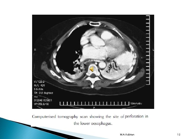
Perforation of the esophagus Ø Potentially lethal complication due to mediastinitis and septic shock. Ø Numerous causes, but may be iatrogenic. Ø Surgical emphysema is virtuall pathognomonic. Ø Treatment is urgent; it may be conservative or surgical, but requires specialised care. M. A. Kubtan 4
Barotrauma (spontaneous perforation) Boerhaave syndrome : Ø This occurs classically when a person vomits against a closed glottis. Ø The pressure in the esophagus increases rapidly, and the esophagus bursts at its weakest point in the lower third sending a stream of material into the mediastinum and often the pleural cavity. Ø Boerhaave syndrome is the most serious type of perforation. This causes rapid chemical irritation in the mediastinum and pleura followed by infection if untreated. M. A. Kubtan 5
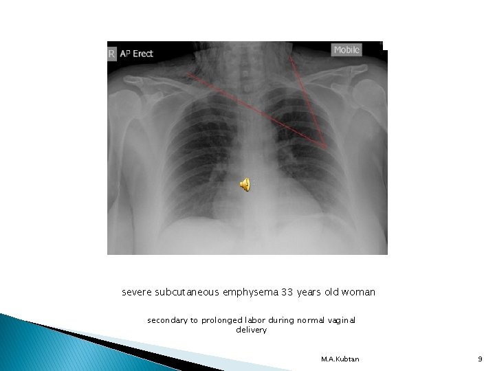
Barotrauma has also been described in relation to other pressure events when the patient strains against a closed glottis (e. g. defaecation, labour, weight-lifting). M. A. Kubtan 6
Diagnosis of spontaneous perforation The clinical history is usually of severe pain in the chest or upper abdomen following a meal or a bout of drinking. Ø Associated shortness of breath is common. � There may be a surprising amount of rigidity on examination of the upper abdomen, even in the absence of any peritoneal contamination. Ø The diagnosis can usually be suspected from the history and associated clinical features. Ø M. A. Kubtan 7
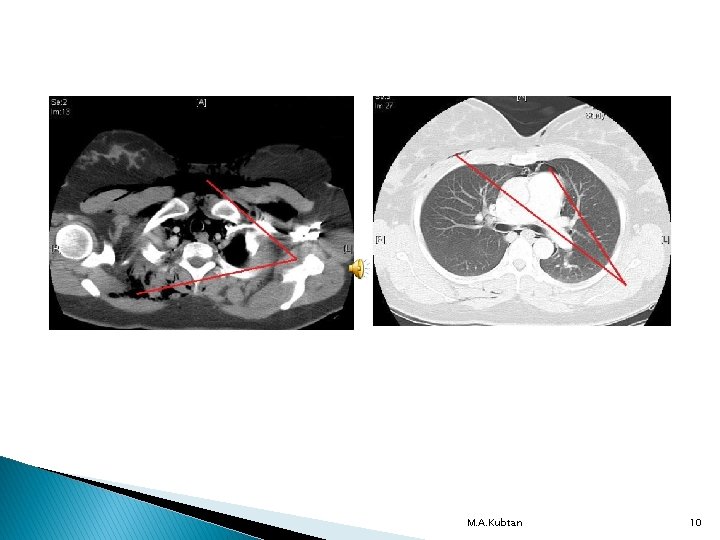
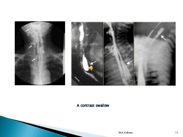
Continue A chest X-ray is often confirmatory with air in the mediastinum, pleura or peritoneum. Ø Pleural effusion occurs rapidly. Ø A contrast swallow or CT is nearly always required to guide management Ø M. A. Kubtan 8
severe subcutaneous emphysema 33 years old woman secondary to prolonged labor during normal vaginal delivery M. A. Kubtan 9
M. A. Kubtan 10
A contrast swallow M. A. Kubtan 11
M. A. Kubtan 12
Pathological perforation Aero digestive fistula is most common and usually encountered in primary malignant disease of the esophagus or bronchus. Ø Erosion into an adjacent structure with fistula formation is more common. Ø Free perforation of ulcers or tumors of the esophagus into the pleural space is rare. Ø Coughing on eating and signs of aspiration pneumonitis may allow the problem to be recognized. Ø M. A. Kubtan 13
Continue Covering the communication with a self-expanding metal stent is the usual solution. Ø Erosion into a major vascular structure is invariably fatal. Ø M. A. Kubtan 14
Penetrating injury Foreign bodies : The esophagus may be perforated during removal of a foreign body. Ø Occasionally, an object that has been left in the esophagus for several days will erode through the wall. Ø Instrumental perforation : Instrumentation is by far the most common cause of perforation. Ø Perforation can occur in the pharynx or esophagus, usually at sites of pathology or when the endoscope is passed blindly. Ø Perforation may follow biopsy of a malignant tumor. Ø M. A. Kubtan 15
Continue The esophagus may be perforated by guide wires, graduated dilators or balloons, or during the placement of self-expanding stents. Ø The risk is considerably higher in patients with malignancy. Ø M. A. Kubtan 16
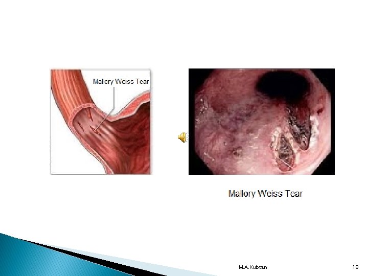
MALLORY–WEISS SYNDROME Forceful vomiting may produce a mucosal tear at the cardia rather than a full perforation. Ø In Mallory–Weiss syndrome, vigorous vomiting produces a vertical split in the gastric mucosa. Ø Tear immediately below the squamocolumnar junction at the cardia in 90% of cases. Ø In only 10% is the tear in the esophagus. Ø M. A. Kubtan 17
M. A. Kubtan 18
Factors influencing Treatment of esophageal perforations Perforation of the esophagus usually leads to mediastinitis. Ø The aim of treatment is to limit mediastinal contamination and prevent or deal with infection. Ø The event causing the perforation (spontaneous vs. instrumental). Ø Underlying pathology (benign or malignant). Ø The status of the esophagus before the perforation (fasted and empty vs. obstructed with a stagnant residue). Ø M. A. Kubtan 19
CORROSIVE INJURY attempted suicide. Ø Accidental ingestion occurs in children and when corrosives are stored in bottles labeled as beverages. Ø All can cause severe damage to the mouth, pharynx, larynx, esophagus and stomach. Ø In general, alkalis are relatively odorless and tasteless, making them more likely to be ingested in large volume. Ø M. A. Kubtan 20
Continue Significant stricture formation occurs in about 50% of patients with extensive mucosal damage. M. A. Kubtan 21
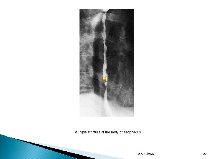
Multiple stricture of the body of esophagus M. A. Kubtan 22
CONGENITAL MALFORMATIONS Most congenital malformations develop during embryonic life between the third and eighth weeks of gestation. M. A. Kubtan 23
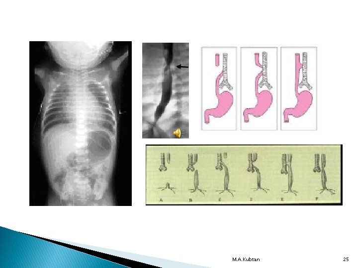
Esophageal atresia A blind proximal pouch with a distal tracheoesophageal fistula is the most common type. Affected infants typically present Ø Soon after birth with frothy saliva. Ø cyanotic episodes, exacerbated by any attempt to feed. Ø The preceding pregnancy may have been complicated by maternal polyhydramnios. M. A. Kubtan 24
M. A. Kubtan 25
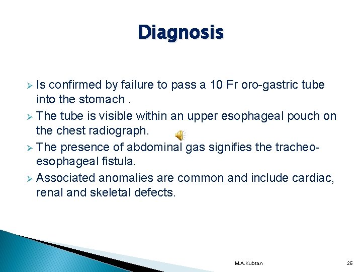
Diagnosis Is confirmed by failure to pass a 10 Fr oro-gastric tube into the stomach. Ø The tube is visible within an upper esophageal pouch on the chest radiograph. Ø The presence of abdominal gas signifies the tracheoesophageal fistula. Ø Associated anomalies are common and include cardiac, renal and skeletal defects. Ø M. A. Kubtan 26
Treatment of Esophageal Atresia Surgical repair : The esophageal ends are anastomosed. Ø Division and repair of tracheo – esophageal tract. Ø M. A. Kubtan 27
Continue Infants with pure esophageal atresia and no tracheoesophageal fistula. Usually best managed by a temporary gastrostomy. Ø Delayed primary repair. Ø Except for very-low-birth weight babies and those with major congenital heart disease, most infants with repaired esophageal atresia have a good prognosis. Ø M. A. Kubtan 28
Potential postoperative complications Anastomotic leak. Ø Stricture. Ø Recurrent fistula formation. Ø Gastro- esophageal reflux. Ø M. A. Kubtan 29