Peptides and Proteins 20 amino acids are commonly


")


 among")
")
 of two or more polypeptide")


 conjugated with nucleic")


- Slides: 15
Peptides and Proteins 20 amino acids are commonly found in protein. These 20 amino acids are linked together through “peptide bond forming peptides and proteins. - The chains containing less than 50 amino acids are called “peptides”, while those containing greater than 50 amino acids are called “proteins”. Peptide bond formation: α-carboxyl group of one amino acid (with side chain R 1) forms a covalent peptide bond with α-amino group of another amino acid ( with the side chain R 2) by removal of a molecule of water. The result is : Dipeptide ( i. e. Two amino acids linked by one peptide bond). By the same way, the dipeptide can then forms a second peptide bond with a third amino acid (with side chain R 3) to give Tripeptide. Repetition of this process generates a polypeptide or protein of specific amino acid sequence.
Peptide bond formation: - Each polypeptide chain starts on the left side by free amino group of the first amino acid enter in chain formation. It is termed (N- terminus). - Each polypeptide chain ends on the right side by free COOH group of the last amino acid and termed (C-terminus).
Examples on Peptides: 1 - Dipeptide ( tow amino acids joined by one peptide bond): Example: Aspartame which acts as sweetening agent being used in replacement of cane sugar. It is composed of aspartic acid and phenyl alanine. 2 - Tripeptides ( 3 amino acids linked by two peptide bonds). Example: GSH which is formed from 3 amino acids: glutamic acid, cysteine and glycine. It helps in absorption of amino acids, protects against hemolysis of RBC by breaking H 2 O 2 which causes cell damage. 3 - octapeptides: (8 amino acids) Examples: Two hormones; oxytocine and vasopressin (ADH). 4 - polypeptides: 10 - 50 amino acids: e. g. Insulin hormone
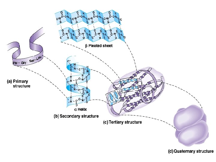
Protein structure: There are four levels of protein structure (primary, secondary, tertiary and quaternary) Primary structure: • The primary structure of a protein is its unique sequence of amino acids. – Lysozyme, an enzyme that attacks bacteria, consists of a polypeptide chain of 129 amino acids. – The precise primary structure of a protein is determined by inherited genetic information. – At one end is an amino acid with a free amino group the (the N-terminus) and at the other is an amino acid with a free carboxyl group the (the C-terminus).
High orders of Protein structure • A functional protein is not just a polypeptide chain, but one or more polypeptides precisely twisted, folded and coiled into a molecule of unique shape (conformation). This conformation is essential for some protein function e. g. Enables a protein to recognize and bind specifically to another molecule e. g. hormone/receptor; enzyme/substrate and antibody/antigen. •
2 - Secondary structure: Results from hydrogen bond formation between hydrogen of –NH group of peptide bond and the carbonyl oxygen of another peptide bond. According to H-bonding there are two main forms of secondary structure: α-helix: It is a spiral structure resulting from hydrogen bonding between one peptide bond and the fourth one β-sheets: is another form of secondary structure in which two or more polypeptides (or segments of the same peptide chain) are linked together by hydrogen bond between H- of NH- of one chain and carbonyl oxygen of adjacent chain (or segment).
• Tertiary structure is determined by a variety of interactions (bond formation) among R groups and between R groups and the polypeptide backbone. a. The weak interactions include: § Hydrogen bonds among polar side chains § Ionic bonds between charged R groups ( basic and acidic amino acids) § Hydrophobic interactions among hydrophobic ( non polar) R groups.
b. Strong covalent bonds include disulfide bridges, that form between the sulfhydryl groups (SH) of cysteine monomers, stabilize the structure.
• Quaternary structure: results from the aggregation (combination) of two or more polypeptide subunits held together by non-covalent interaction like Hbonds, ionic or hydrophobic interactions. • Examples on protein having quaternary structure: – Collagen is a fibrous protein of three polypeptides (trimeric) that are supercoiled like a rope. • This provides the structural strength for their role in connective tissue. – Hemoglobin is a globular protein with four polypeptide chains (tetrameric) – Insulin : two polypeptide chains (dimeric)
Classification of proteins I- Simple proteins: i. e. on hydrolysis gives only amino acids Examples: 1 - Albumin and globulins: present in egg, milk and blood They are proteins of high biological value i. e. contain all essential amino acids and easily digested. 2 - Scleroproteins: They are structural proteins, not digested. include: keratin, collagen and elastin. a- α-keratin: protein found in hair, nails, enamel of teeth and outer layer of skin. • It is α-helical polypeptide chain, rich in cysteine and hydrophobic (non polar) amino acids so it is water insoluble. b- collagens: protein of connective tissues found in bone, teeth, cartilage, tendons, skin and blood vessels.
Conjugated proteins On hydrolysis, give protein part and non protein part and sub classified into: 1 - Phosphoproteins: These are proteins conjugated with phosphate group e. g. Casein of milk 2 - Lipoproteins: These are proteins conjugated with lipids. Functions: a- help lipids to transport in blood b- Enter in cell membrane structure helping lipid soluble substances to pass through cell membranes. 3 - Glycoproteins: proteins conjugated with sugar (carbohydrate) e. g. - Some hormones such as - present in cell membrane structure - blood groups.
4 - Nucleoproteins: These are basic proteins ( e. g. histones) conjugated with nucleic acid (DNA or RNA). e. g. a- chromosomes: are proteins conjugated with DNA b- Ribosomes: are proteins conjugated with RNA 5 - Metalloproteins: These are proteins conjugated with metal like iron, copper, zinc, …… a- Iron-containing proteins: Iron may present in heme such as in - hemoglobin (Hb) b- Copper containing proteins: - Oxidase enzymes such as cytochrome oxidase. c- Zn containing proteins: e. g. Insulin and carbonic anhydrase
Protein Folding • Proteins shape is determined by the sequence of the amino acids • The final shape is called the conformation Denaturation is the process of unfolding the protein – Can be down with heat, p. H or chemical compounds – In the chemical compound, can remove and have the protein renature or refold