Peptic Ulcer Presented by Laith Sorour Peptic Ulcer

Peptic Ulcer Presented by: Laith Sorour

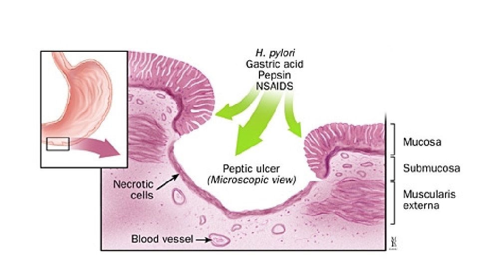
Peptic Ulcer • Erosion: Break <5 mm • Ulcer: Break >5 mm and Histologically demonstrates going through the mucosa into muscularis mucosae. • “Peptic ulcer”: an ulcer in the lower oesophagus, stomach or duodenum, in the jejunum after surgical anastomosis to the stomach or, rarely, in the ileum adjacent to a Meckel’s diverticulum. • *acute chronic fibrosis

Anatomy & Physiology
 • M: F is")
Epidemiology & Statistics • Prevalence: 5%-10% (Decreasing In developed countries) • M: F is 5: 1 -2: 1 for Duodenal Ulcers and 2: 1 For Gastric Ulcers • Chronic Gastric Ulcer is usually Single, 10 -15% of patients have more than one peptic ulcer, in 10% Gastric and Duodenal Ulcers coexist • 90% are situated on the lesser curve within the antrum or at the junction between body and antral mucosa. • Chronic duodenal ulcer usually occurs in the first part of the duodenum and 50% are on the anterior wall

Pathophysiology & Etiology 1. Most common causes: a. Helicobacter pylori infection b. NSAIDs—inhibit prostaglandin production, which leads to impaired mucosal defenses c. Acid hypersecretory states, such as Zollinger–Ellison syndrome 2. Other risk factors associated with higher risk of PUD (have not been shown to be causative) • a. Smoking; confers an increased risk of gastric ulcer and, to a lesser extent, duodenal ulcer. Once the ulcer has formed, it is more likely to cause complications and less likely to heal if the patient continues to smoke. b. Alcohol use c. Emotional stress and dietary factors (such as coffee and spice intake) have not been shown to be associated with higher risk of PUD.

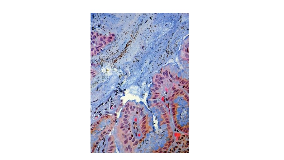
H. pylori is Gram-negative and spiral, and has multiple flagella at one end, which make it motile, allowing it to burrow and live beneath the mucus layer adherent to the epithelial surface. Around 90% of duodenal ulcer patients and 70% of gastric ulcer patients are infected with H. pylori exclusively colonises gastric-type epithelium and is only found in the duodenum in association with patches of gastric metaplasia The cag. A gene product is injected into epithelial cells, interacting with numerous cell-signalling pathways involved in cell replication and apoptosis. vac. A causes increased cell permeability, efflux of micronutrients from the epithelium, induction of apoptosis and suppression of local immune cell activity, especially s 1/ml form of the

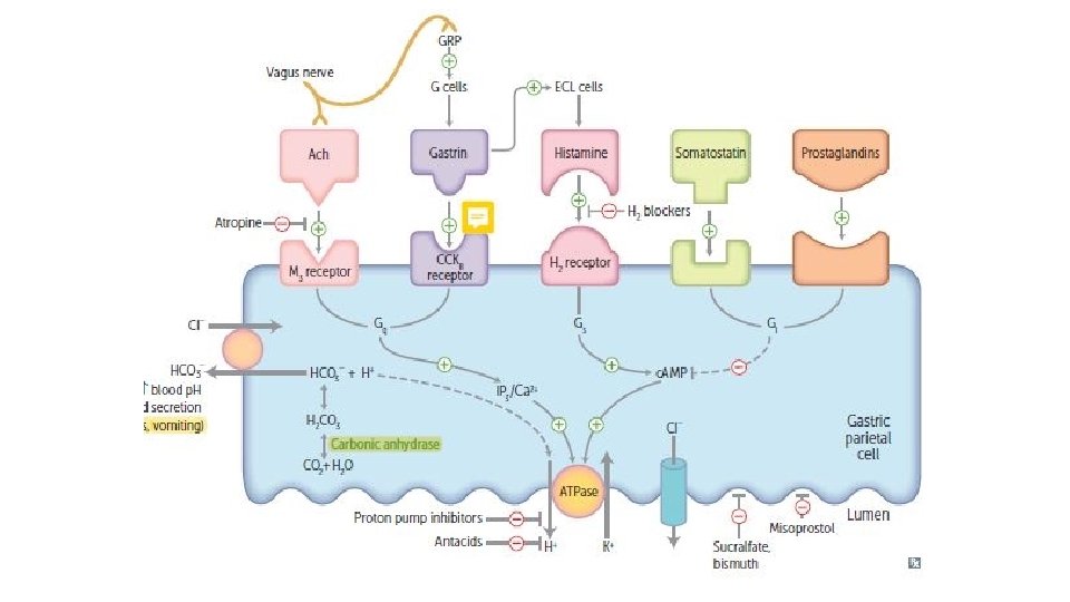
In most people, H. pylori causes localised antral gastritis associated with depletion of somatostatin (from D cells) and increased gastrin release from G cells. The subsequent hypergastrinaemia stimulates increased acid production by parietal cells but, in the majority of cases, this has no clinical consequences. In a minority of patients, this effect is exaggerated, leading to duodenal Ulceration. In Stomach the ulcer probably arises because of impaired mucosal defence resulting from a combination of H. pylori infection, NSAIDs and smoking, rather than excess acid.

In 1%

NSAIDs

Clinical Features Most common presentation is recurrent abdominal pain which has three notable characteristics: 1 -localisation to the epigastrium 2 -relationship to food 3 -episodic occurrence *Duodenal lesions often causes discomfort at night 40% of ulcer subjects have occasional vomiting ; persistent daily vomiting suggests gastric outlet obstruction.

Atypical Presentation • In 1/3 the Hx is less characteristic especially in NSAIDs users and elderly pain may be absent or so slight that it is experienced only as a vague sense of epigastric unease. Occasionally, the only symptoms are anorexia and nausea, or early satiety after meals. • ‘silent ulcers’, presents for the first time with anaemia from chronic undetected blood loss, as an abrupt haematemesis or as acute perforation. • recurrent acute bleeding without ulcer pain.
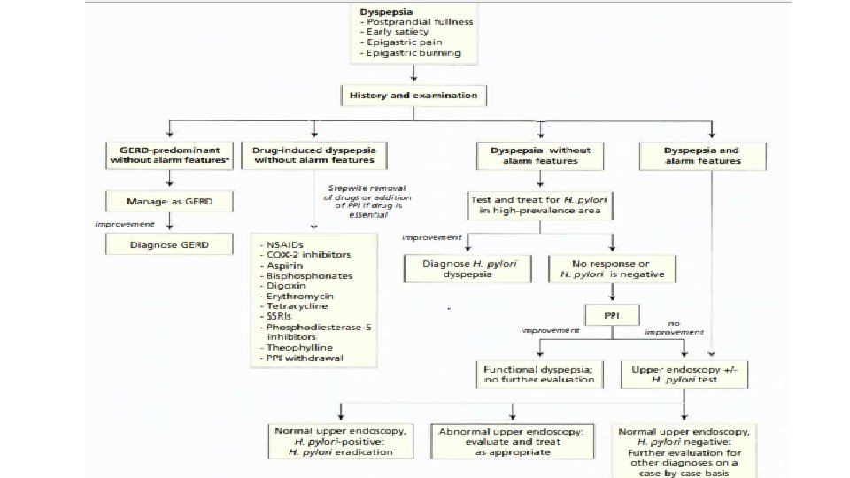

Investigations • Defenitive Dx Endoscopy with Bx Allows Direct visualization to RO malignancy and Definitively Dx H. pylori

Factors favoring Endoscopy • (If you suspect Malignancycomplicationanother different organic Dz. Risk Factors For them Go for endoscopy) • Age >=50 -60 with new onset dyspepsia • >= 1 alarm symptom; unintentional weight loss, early satiety, dysphagia, odynophagia, GI bleeding, IDA, Persistent vomiting, Palpable Abd. Mass, Adenopathy. • Dyspepsia with Family Hx of Upper GI Ca • Childhood in regions with elevated risk of GI Ca (Southeast Asia) • Male sex is controversial

DIAGNOSIS-UPPER ENDOSCOPY Chronic D. U. *Fibrosed Acute ulcer

Management • Aims of management 1 -relieve symptoms 2 -induce healing 3 prevent recurrence. • H. pylori eradication is the cornerstone of therapy for peptic ulcers, as this will successfully prevent relapse and eliminate the need for longterm therapy in the majority of patients. • Modules: -General Measures -Eradicate H. pylori -Maintenance Tx -Surgical Tx

• General measures • Cigarette smoking, aspirin and NSAIDs should be avoided. Alcohol in moderation is not harmful and no special dietary advice is required. • Maintenance treatment: • Continuous maintenance treatment should not be necessary after successful H. pylori eradication. • For the minority who do require it, the lowest effective dose of PPI should be used.

H. Pylori Eradication
")
Triple Therapy: a PPI taken simultaneously with two antibiotics (from amoxicillin, clarithromycin and metronidazole) for 7 days. High-dose, twice-daily PPI therapy increases efficacy of treatment, as does extending treatment to 10– 14 days. Success is achieved in 80– 90% of patients, although compliance, side-effects and antibiotic resistance influence this. Resistance to amoxicillin is rare but rates of metronidazole resistance reach 40% in some countries and, recently, rates of clarithromycin resistance of 20– 40% have appeared. Where the latter exceed 15– 20%, a quadruple therapy regimen, consisting of omeprazole (or another PPI), bismuth subcitrate, metronidazole and tetracycline (OBMT) for 10– 14 days, is recommended. In areas of low clarithromycin resistance, this regimen should also be offered as second-line therapy to those who remain infected after initial therapy, once compliance has been checked. For those who are still colonised after two treatments, the choice lies between a third attempt guided by antimicrobial sensitivity testing, rescue therapy (levofloxacin, PPI and clarithromycin) or long-term acid suppression.

Surgical Compliacations: Dumping syndrome Bile reflux gastritis Diarrhoea and maldigestion Weight loss Anaemia Metabolic bone disease Gastric cancer

Complications • Perforation: • Gastric outlet obstruction • Bleeding *Talk little bit about ZES

THE END • Remember H. pylori has a family too, so suffer in silence
- Slides: 25