Peptic ulcer by Mena Ehab anwer Def PEPTIC

Peptic ulcer by Mena Ehab anwer

Def PEPTIC ULCER Breach in continuity of mucosa of alimentary tract which extends through the muscularis mucosa into the submucosa or deeper

Formation

Formation. con

PROTECTIVE FACTORS PGs Mucous Bicarbonates Blood flow Rapid cell turnover

AGGRESSIVE FACTORS Acid, pepsin, bile Helicobacter pylori NSAIDs & other drugs Smoking, Alcohol, stress Free radicals Oily, spicy, irregular dietary habit Hereditary factors

Peptic ulcer formation

IN VIVO METHODS Pylorus Ligation in Rats • Stress Ulcer Model • Histamine-Induced Gastric Ulcer • Ethanol-Induced Mucosal Damage • Acetic Acid-Induced Gastric Ulcer • Reserpine-Induced Chronic Ulcers • Cysteamine-Induced Duodenal Ulcer • Demaprit-Induced Duodenal Ulcer • Mepirizole-Induced Duodenal Ulcers • Experimental Colitis

H+/K+ - ATPASE INHIBITION ASSAY H+/K+ - ATPase or proton pump --> final step in the synthesis of acid by parietal cells Procedure: • Homogenate of 80 ng Microsomal gastric H+/K+ - ATPase (pig gastric mucosa) incubated with 100µl buffer, 1 m. M ATP and Test compound in microtitre plate for 30 mins at 37° • Reaction is stopped by adding Malachite green (colorimetric agent) • After 10 seconds, 15% sodium citrate is added for 45 minutes • Release of orthophosphate from ATP quantified by colorimeter at 570 nm

Evaluation Percentage inhibition of H+/K+ - ATPase is calculated. Lesser the orthophosphate released, more is the inhibition of H+/K+ - ATPase by test compound.

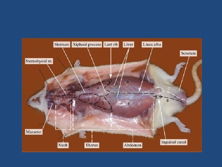
PYLORUS LIGATION IN RATS Shay et al • Pylorus is ligated over a certain period of time • Accumulation of gastric acid causes ulceration Procedure: • Wistar rats weighing 150 -200 grams • Fasting : 48 hours ; water ad libitum. • Housed singly in cages with raised bottoms of wide wire mesh to avoid coprophagy.

Under anaesthesia, a one-inch midline abdominal incision is given below the xiphoid process. • Pylorus is ligated without damaging its blood supply. • Stomach is replaced and abdominal wall closed with sutures. • Test compounds are given either orally or injected s. c. • About 17 -19 hours after pyloric ligation, rats are sacrificed and stomachs are dissected out.
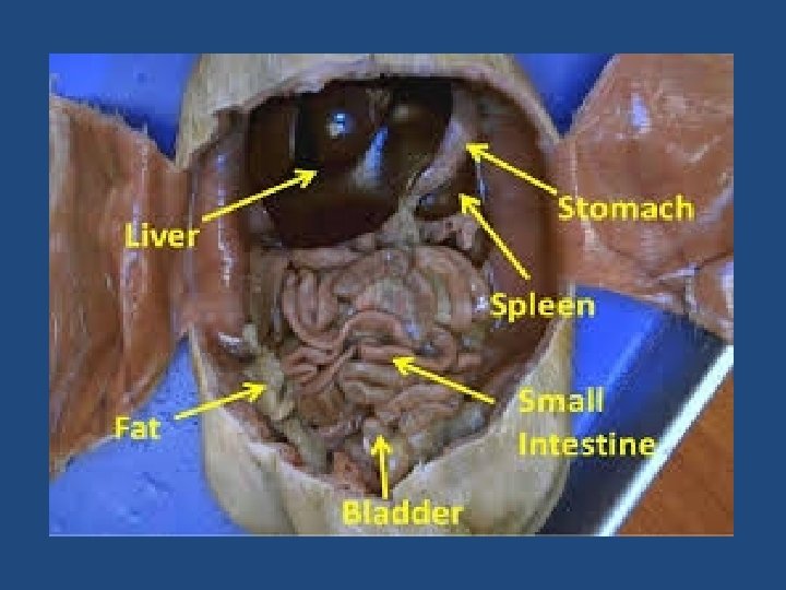

Contents of the stomach are drained into a graduated centrifuge tube and acidity determined by titration with 0. 1 N Na. OH. • Stomach is opened along the greater curvature, pinned on a cork plate. • Its inner surface is examined for ulceration with a binocular microscope. • The Ulcer Index is calculated and the Ulcer Severity graded.

ULCER CLASSIFICATION: SHAY ET AL Grade 4 - Perforation Grade 3 - Hemorrhagic spots with many ulcers Grade 2 - Deeper hemorrhagic spots with few ulcers Grade 1 - Scattered hemorrhagic spots Grade 0 - Normal gastric mucosa

ULCER INDEX Method 1 The ulcer index is calculated as: Ulcer Index = 10 / X Where X = Total mucosal area / Total ulcerated area

Method 2 Ganguly and Bhatnagar extended above criteria for inclusion of petechiae. • Five petechiaes are considered to be equivalent to 1 mm of ulcer area. Ulcer index calculated as described previously.

Method 3 An ulcer index U is calculated as U = Un +Us +Up / 10 Where, Un = Average number of ulcers per animal Us = Average of severity score (graded from 0 to 3) Up = Percentage of animals with ulcer

ULCER SEVERITY SCORE 0 – no ulcer 1 – superficial erosion 2 – deep ulcer 3 – penetrated or perforated ulcer

Other method 0 - Normal stomach 0. 5 - Red coloration 1 - Spot ulcers 1. 5 - Haemorrhagic streaks 2 - Ulcer > 3 mm but < 5 mm 3 - Ulcers > 5 mm

ULCER INCIDENCE & GRADING: WILHELMI AND MENASSE-GDYNIA Quantification of drug induced mucosal damage • Ulcers – necro-haemorrhagic spots > 2 mm diameter 0. 5 – minute, sporadic, punctate lesions • 1 – several small lesions 2 – one large extensive lesion or multiple moderate sized lesion 3 – several large lesion

Srivastava et al. Shedding of epithelium= 10 Petechial & frank hemorrhages= 20 One or two ulcers= 30 More than two ulcers= 40 Perforated ulcers= 50

Inference Ulcer index of test drug compared with control group to detect anti- ulcer effect of test drug.

CRITICAL ASSESSMENT OF THE METHOD The “Shay-rat” has been proven to be a valuable tool to evaluate anti-ulcer drugs with various mechanisms of action.
 , for the first time , described the use")
STRESS ULCER • Selye (1936) , for the first time , described the use of restraints for production of Gastric ulcers Advantages: • Technically simple • Do not require anesthesia or surgery • Lesions located in glandular region of stomach • As psychogenic factors are involved in the pathogenesis of gastric ulcers, psychotropic drugs could be evaluated
 Principle: Stress plays a significant")
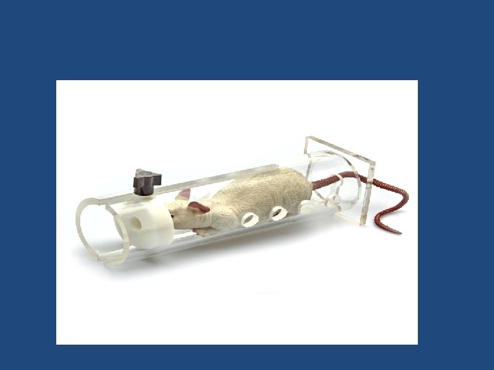
I. RESTRAINT- INDUCED ULCERS (HANSON AND BRODIE , 1960) Principle: Stress plays a significant role in the pathogenesis of gastric ulcers. Procedure: • Albino rats weighing 150 -200 grams are taken • Fasted for 36 hours before experiment • Drug is administered orally or subcutaneously • 30 min later animals are subjected to restraint

• For restraint, the rats are placed in a piece of galvanized steel window screen of appropriate size. • Screen is moulded around the animal and held in place with wire staples. • To restrain the rats, the limbs are put together in pair and tightened with adhesive tape. • Rats are kept under restraint for 24 hours • Then sacrificed & their stomachs dissected out. • Ulcer index and ulcer severity are determined.
 Principle: Exposure of cold conditions")
Cold water immersion induced ulcer (Takagi et al, 1964) Principle: Exposure of cold conditions to restrained animals accelerates the occurrence of gastric ulcers. Shortens the immobilization time. Procedure: • Wistar rats weighing 150 -200 grams are used. • After fasting the animals for 16 hours, the test compound is administered orally

• Rats are then placed individually in restraint cages vertically, and then immersed in water upto the xiphoid process, at 22°C for 1 hour. • Then rats are removed from the cages, dried • Evan’s blue (30 mg/kg) injected i. v. via the tail vein • 10 min later, they are sacrificed • The stomach is removed & ligated at both ends • Filled with formaline & kept overnight • On the next day, the stomach is opened along the greater curvature and examined for ulcerative lesions

III. STRESS & NSAIDS–INDUCED ULCERS • Procedure : • Wistar rats; 150 -200 g • Fasted for 24 -36 hrs • Test agent (in 1% carboxymethyl cellulose) via gastric intubation and a NSAID such as aspirin, indomethacin or diclofenac i. p. • Rats placed in stress cages • Immersed in water upto level of xiphoid process at 230 C for 7 hrs • Animal sacrificed ; Stomach removed ; Evaluated for Ulcer Index • Dose of NSAID required to increase gastric erosion by 100% relative to immobilization is compared with that of NSAID required to produce 100% increase in gastric erosion under the protective effect of test drug

IV. SWIMMING STRESS ULCERS • Procedure : • Albino rats ; either sex • Fasted for 24 hrs; free access to water • Rats forced to swim in deep concrete tube filled with water at 230 C for 5 hrs • Animals removed • Sacrificed & stomach removed ; opened along greater curvature • Severity grading done ; Ulcer Index calculated

0 - Normal lesion 1 - Lesions with diameter < 1 mm 2 - Lesions with diameter 1 – 2 mm 3 - Lesions with diameter 2 – 4 mm 4 - Lesions with diameter > 4 mm
 Principle: Gastric acid secretion is increased when")
HISTAMINE-INDUCED GASTRIC ULCER (Barrett et al, 1955) Principle: Gastric acid secretion is increased when histamine is administered intraperitoneally. Procedure: • Guinea pig weighing 300 -400 grams are taken • Fasted for 36 hours before experiment; water ad libitum • 1 ml of histamine acid sulphate (50 mg base) is administered i. p.

• Promethazine hydrochloride 5 mg is injected i. p. 15 min before and 15 min after histamine to protect the animals against histamine toxicity. • The standard/test drugs are administered p. o. or s. c. 45 minutes before histamine injection. • 4 hours after histamine injection, guinea pigs are sacrificed and stomach dissected out. • The gastric contents are subjected to analysis • Stomach is opened along the greater curvature, ulcers are identified.
 Type 0 : No visible ulcers Type")
ULCER SCORING : (Barrett et al, 1955) Type 0 : No visible ulcers Type 1 : 10 or less small ulcers, 1 -3 mm in diameter Type 2 : 11 or more ulcers, 1 -3 mm in diameter Type 3 : 1 or more ulcers, 4 -6 mm in diameter Type 4 : 1 or more ulcers, 7 mm or more in diameter Type 5 : Perforation of the gastric wall

Advantages: • Produces 100% gastric ulceration • Increased volume of gastric acid secretion
 Principle: Ethanol, being a necrotizing agent, damages")
Ethanol-induced Mucosal damage (Robert et al, 1979) Principle: Ethanol, being a necrotizing agent, damages the superficial epithelial layers & inhibits the release of mucosal prostaglandins. Procedure: • Wistar rats weighing 150 -200 grams are taken • Fasted for 18 hours before experiment; water ad libitum. • Rats are given test drugs or standard drug orally

• 30 mins later 1 ml/200 gm of 99. 80% alcohol is administered orally. • After 1 hour, Rats are sacrificed and stomachs dissected out. • Severity score and ulcer index are calculated Witt et al. (1985) described a method to quantify the extent of ethanol-induced gastric lesions. Using a transmission densitometer to measure the optical density of the photographic negatives of gastric mucosa. Damaged areas have lower optical density values.

Advantages : • Gastric lesions are observed after an hour of administration of ethanol. • Reproducible method to produce gastric lesions in experimental animals.

EVALUATION • The significance of differences in optical density between control and ethanol-treated tissue is evaluated by nonpaired single-tail Student’s t-test.

CRITICAL ASSESSMENT OF THE METHOD • Several prostaglandins provide cytoprotection, particularly in rats, in a dose-range which has no antisecretory activity. • However, clinical experience with prostaglandins showed that ulcer healing is only achieved at antisecretory doses (Lindberg et al. 1990). • Therefore, it seems very likely that the cytoprotective property of a compound in rats has very limited relevance to prediction of its ulcer healing potential in humans if cytoprotection is really separated from its antisecretory potential (Herling and Weidmann 1994).
 A model for inducing chronic gastric")
ACETIC ACID-INDUCED GASTRIC ULCER Takagi et al. (1969) A model for inducing chronic gastric ulcer in rats by means of submucosal injection of acetic acid. (Okabe et al, 1972) New method which involves temporary instillation of acetic acid solution. Principle: • Acetic acid enhances the ulceration in stomach by increasing the acidity of stomach contents.
 • Albino rats used • 0. 05 ml of")
Procedure (Takagi et al, 1969) • Albino rats used • 0. 05 ml of Acetic acid (1 -30%) injected in the submucosal layer of the stomach • Penetrating peptic ulcers : adhered to Liver • Chronic ulcers with repeated healing and reaggravation • Effect of test drug given twice daily for 10 -15 days is noted.
 • Wistar rats weighing 150 -200 grams are taken")
Procedure: (Okabe and Pfeiffer, 1972) • Wistar rats weighing 150 -200 grams are taken • Fasted for 24 hours before experiment. • Pentobarbital anaesthesia • A cylindrical glass tube of 6 mm diameter : tightly placed upon the anterior serosal surface of stomach 1 cm away from the pyloric end. • 50% Acetic acid (0. 06 ml per animal) is instilled into the tube and allowed to remain for 1 minute on the gastric wall. • After removal of Acetic acid solution, the abdomen is closed.

• Animals were caged and fed normally. • Test drugs were given orally on Day 1 twice daily, 4 hours after application of acetic acid and continued upto 10 days after induction of ulcer • Animals were sacrificed after 18 hours of the last dose to assess ulcer size and healing. • Ulcer index and Severity score calculated.

Advantages: • Simple procedure: resulting in ulcers of consistent size and severity at an incidence of 100%. • Resemble human ulcers in terms of both pathological features and healing mechanisms. • Relapse of healed ulcers is frequently observed, just as in peptic ulcer patients. Disadvantages: • Submucosal injection produced ulcers penetrating entire gastric wall & adherence of ulcer base to adjacent organs (mainly Liver).

RESERPINE – INDUCED CHRONIC ULCERS • The mechanism of ulcer formation has been attributed to cholinergic mediated degranulation of gastric mast cells and liberation of histamine. • Procedure : • Female Sprague – Dawley rats; 130 -180 g • Fasted for 48 hrs, Free access to 0. 8% sucrose in 0. 2% Na. Cl w/v • Liquid diet withdrawn 1 hr before starting • Animals injected with Test drug i. p. • ½ hr later, Reserpine(5 mg/kg) or Vehicle injected i. p. • 4 hrs later, Animal sacrificed, Stomach removed, Examined for mucosal lesions
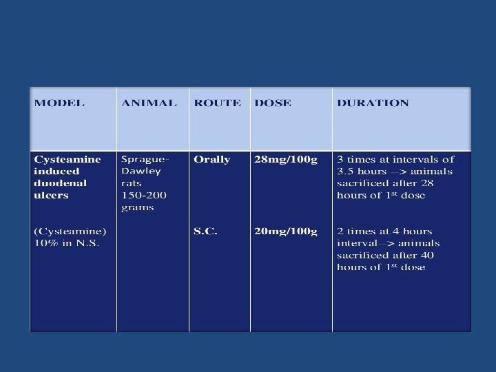
 Cysteamine HCl (β-mercaptoethylamine HCl) Principle: Pathogenesis :")
CYSTEAMINE-INDUCED DUODENAL ULCER Selye and Szabo (1973) Cysteamine HCl (β-mercaptoethylamine HCl) Principle: Pathogenesis : Inhibition of alkaline mucus production Increased gastric acid secretion Increased serum gastrin levels Delayed gastric emptying

Procedure: • Female Sprague-Dawley rats weighing 150 -200 grams are taken • Test drug and the standard drug are administered 45 min prior to Cysteamine administration.

Procedure: • Female Sprague-Dawley rats weighing 150200 grams are taken • Test drug and the standard drug are administered 45 min prior to Cysteamine administration.

Perforating duodenal ulcers are produced ; • Located 2 -4 mm from the pylorus, mainly on the anterior wall of duodenum • Necrotic material and acute inflammatory response is present at ulcer crater

EVALUATION • The intensity of the duodenal ulcer is evaluated using scores from 0 to 3. • 0 = no ulcer • 1 = superficial mucosal erosion • 2 = deep ulcer usually with transmural necrosis • 3 = perforated or penetrated ulcer

CRITICAL ASSESSMENT OF THE METHOD • In view of the development of modern gastric K+/H+- ATPase inhibitors the predictive value of methods using experimental ulcers in the rat for clinical healing rates in man has been challenged (Herling and Weidmann 1994).

Advantages: Ulcerogenesis is seen with one full dose of cysteamine. Easily reproducible Disadvantage: • Ulcers, located on the anterior wall, frequently perforate, resulting in peritonitis, or penetrate into the liver. • A small ulcer is usually present on the posterior wall (“kissing ulcer”) of the duodenum , penetrates the pancreas.
 Principle: H 2 receptor agonist Induced gastric")
DEMAPRIT-INDUCED DUODENAL ULCER del Soldato P (1982) Principle: H 2 receptor agonist Induced gastric erosion in rats after single i. v. dose. Duodenal ulcer in guinea pigs after repeated s. c. dose. Procedure: • Wistar rats weighing 150 -200 g or guinea pigs 250 -300 g are taken • Fasted for 24 hours before experiment; free access to water • Test drug or standard drug is given orally 60 min before injecting Demaprit in rats and 30 min before injecting it in guinea pig.

• Demaprit is given in a dose 100 mg/kg i. v. in rats and 2 mg/kg s. c. every hour for 6 hours in guinea pig. • After 1 hour of Demaprit injection, Animal is sacrificed and stomach dissected out. • Stomach is opened along the greater curvature and examined for ulceration.
 • Model useful for : - Screening of")
MEPIRIZOLE–INDUCED DUODENAL ULCERS Okabe et al(1982) • Model useful for : - Screening of Anti ulcer drugs - Studying pathogenesis of Duodenal Ulcers Procedure : • Male Sprague – Dawley rats (200 -220 g) • Mepirizole(200 mg/kg) in 1% carboxymethyl cellulose solution via gastric intubation is administered • Subsequently, rats kept in cages with raised mesh bottom and deprived of food and water for 24 hr

• Leads to, ulceration in proximal duodenum and erosions in antrum. • Anti ulcer therapy started 24 hrs after Mepirizole administration • 11 th day, Animal sacrificed • Duodenum and Stomach evaluated for ulcer area under microscope • Ulcer or erosion indices are calculated from the sum of area of ulcers & erosions respectively.

EXPERIMENTAL COLITIS • PURPOSE AND RATIONALE Inflammatory bowel diseases, ulcerative colitis and Crohn’s disease, represent chronic alteration of the gastrointestinal tract of unknown etiology perhaps involving immunological events. The immunological parameters have been described as secondary but may possibly be attributed to the chronicity of the disease.

PROCEDURE • A three-step concept is realized to mimic the human disease, using 2, 4, 6 -trinitrobenzene sulfonic acid (TNBS) as a defined hapten: • 1. specific hypersensitivity by active immunization, • 2. local inflammation by local challenge, • 3. chronicity by chronic application of the immunogen.

Female Sprague Dawley rats weighing 150– 200 g are sensitized by intradermal injection of 0. 8% TNBS solution into a shaved area on the back once daily for three consecutive days. After 18 days, the animals receive a further intradermal booster injection. Intradermal challenge of 0. 08% TNBS in 0. 05 ml 0. 9% Na. Cl solution, is given 14 days later in order to determine the type and specificity of the immunological reaction. Ten days after the intradermal challenge, a flexible polyethylene tube of 0. 5 mm diameter is implanted under ketamine (100 mg/kg i. p. ) anesthesia 15 cm proximal to the cecum and emerging at the neck for TNBS or drug administration.

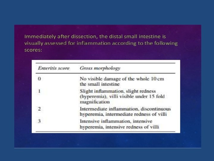
After a 10 -day recovery phase, the animals are treated daily for 3 weeks with 0. 08% TNBS in saline (0. 2 mg/rat) given through the catheter. Control groups receive only saline. Drugs are applied either by gavage twice a day. suspended in carboxymethyl cellulose, or intraluminally once a day, suspended in saline. The animals are sacrificed by CO 2 inhalation 24 h after the last intraluminal application of TNBS. The distal 10 cm of small intestine anterior to the ileo-caecocolic junction (5 cm distance to the open end of the catheter) including Peyer’s patches are dissected, cut open longitudinally and rinsed with saline
 experiments. • Differences")
EVALUATION • Results are expressed as means ± SEM of (n) experiments. • Differences between control and inflamed tissue, and influence of drug treatment are compared. • Statistical significance is calculated by Wilcoxon. Mann. Whitney U-test for unpaired data. • The level of significance is taken as p < 0. 05.

CRITICAL ASSESSMENT OF THE METHODS • The relevance of animal models for the pathogenesis and treatment of human inflammatory bowel disease was reviewed by Dieleman et al. (1997) and by Sartor (1997). • A critical review of in vitro models in inflammatory bowel disease was given by Mc. Kay et al. (1997)

THANK YOU
- Slides: 71