
PEPTIC ULCER AGGRESSIVE FACTORS hydrochloric acid pepsin reverse














- Slides: 48
PEPTIC ULCER
AGGRESSIVE FACTORS • • hydrochloric acid pepsin reverse diffusion of ions of hydrogen products of lipid hyperoxidation
DEFENSE FACTORS • mucus and alkaline components of gastric juice • property of epithelium of mucous tunic to permanent renewal • local blood flow of mucous tunic and submucous membrane
PATHOMORPHOLOGY • Erosion • acute ulcers • chronic ulcers
CLASSIFICATION by Johnson (1965) • I – ulcers of small curvature (for 3 cm higher from a goalkeeper); • II– double localization of ulcers simultaneously in a stomach and duodenum; • III – ulcers of goalkeeper part of stomach (not farther as 3 cm from a goalkeeper)
CLINICAL MANAGEMENT • • Pain Vomiting Heartburn Belching
COMPLICATIONS • • • Penetration Stenosis Perforation Bleeding Malignization
DIAGNOSIS PROGRAM • • • 1. Anamnesis and physical examination. 2. Endoscopy. 3. X-Ray examination of stomach. 4. Examination of gastric secretion by the method of aspiration of gastric contents. 5. Gastric p. H metry. 6. Multiposition biopsy of edges of ulcer and mucous tunic of stomach. 7. Gastric Dopplerography. 8. Sonography of abdominal cavity organs. 9. General and biochemical blood analysis. 10. Coagulogram.
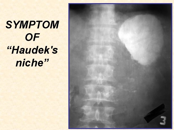
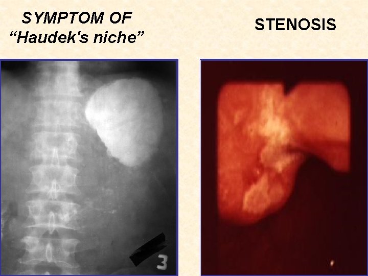
X-Ray examination THE DIRECT SIGNS: • symptom of “Haudek's niche” • ulcerous billow and convergence of folds of mucous tunic. INDIRECT SIGNS: • symptom of “forefinger” (circular spasm of muscles) • segmental hyperperistalsis, • pylorospasm, • delay of evacuation from a stomach • duodenogastric reflux • disturbance of function of cardial part (gastroesophageal reflux).
SYMPTOM OF “Haudek's niche”
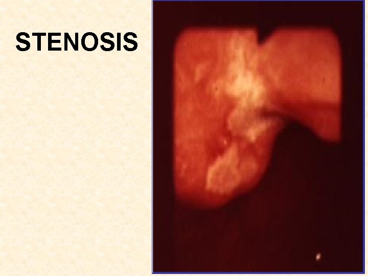
STENOSIS
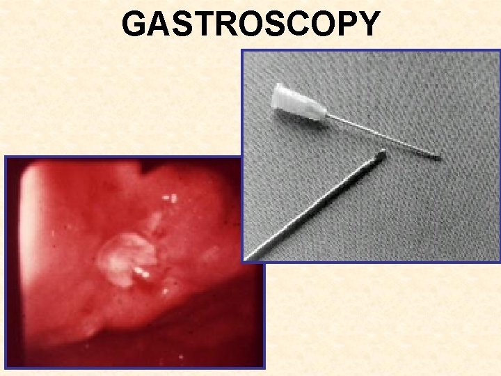
GASTROSCOPY
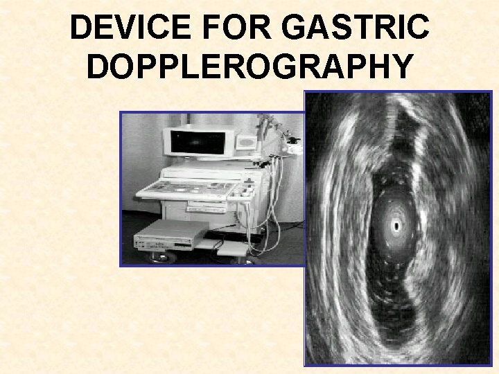
DEVICE FOR GASTRIC DOPPLEROGRAPHY
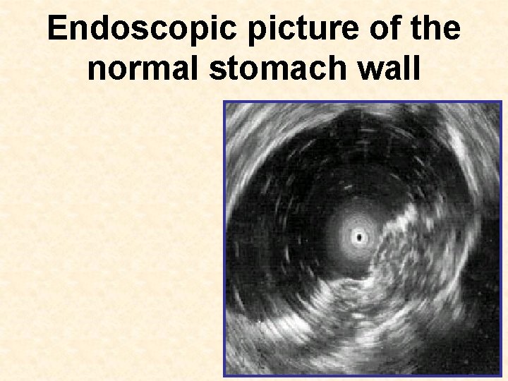
Endoscopic picture of the normal stomach wall
Endoscopic picture of the peptic ulcer
CONSERVATIVE THERAPY a) Omeprazole 20 mg 2 time per day or Н 2 blocker histamine receptor (ranitidine) — 150 mg in the evening, famotidine — 40 mg at night, roxatidine — 150 mg in the evening b) antiacid drugs — in accordance with the results of p. H-metry; c) reparative drugs (dalargin, solcoseryl, actovegin) — for 2 ml 1– 2 times per days d) antimicrobial drugs (clarytromicine 500 mg twice daily, de-nol, metronidazole)
SURGICAL TREATMEN a) at the relapse of ulcer after the course of conservative therapy; b) in the cases when the relapses arise during supporting antiulcer therapy; c) when an ulcer does not heal over during 1, 5– 2 months of intensive treatment, especially in families with “ulcerous anamnesis”; d) ulcer with complications (perforation or bleeding); e) at suspicion on malignization ulcers, in case of negative cytological analysis.
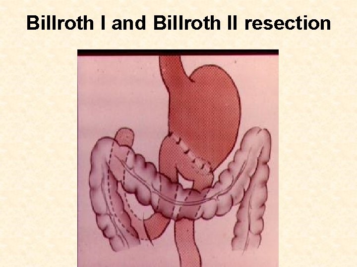
Billroth I and Billroth II resection
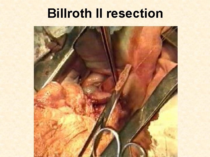
Billroth II resection
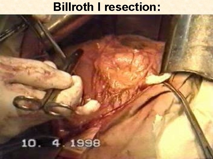
Billroth I resection:
DUODENAL ULCER
CLASSIFICATION I. By etiology: А. True duodenal ulcer. B. Symptomatic ulcers. II. By passing of disease: 1. Acute (first exposed ulcer). 2. Chronic: a) with the rare exacerbation; b) with the annual exacerbation; c) with the frequent exacerbation (2 times per a year and more frequent).
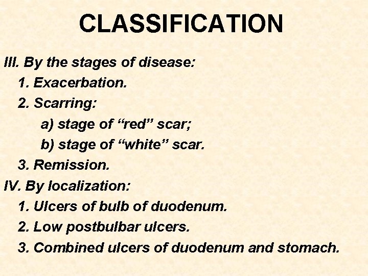
CLASSIFICATION III. By the stages of disease: 1. Exacerbation. 2. Scarring: a) stage of “red” scar; b) stage of “white” scar. 3. Remission. IV. By localization: 1. Ulcers of bulb of duodenum. 2. Low postbulbar ulcers. 3. Combined ulcers of duodenum and stomach.
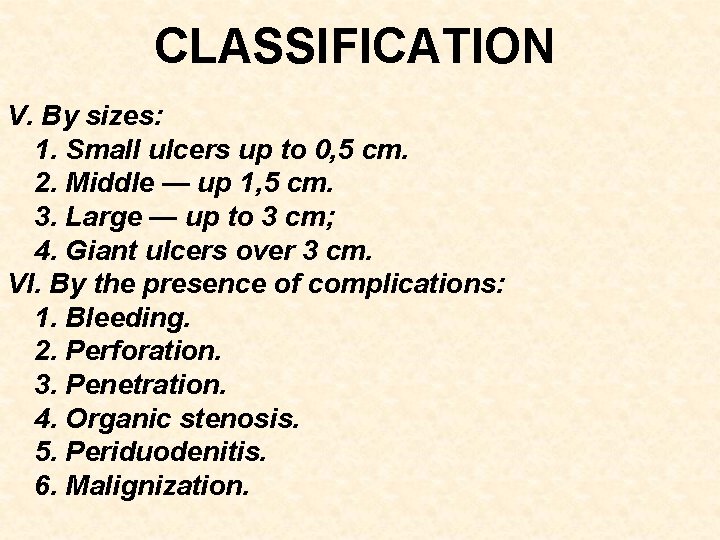
CLASSIFICATION V. By sizes: 1. Small ulcers up to 0, 5 cm. 2. Middle — up 1, 5 cm. 3. Large — up to 3 cm; 4. Giant ulcers over 3 cm. VI. By the presence of complications: 1. Bleeding. 2. Perforation. 3. Penetration. 4. Organic stenosis. 5. Periduodenitis. 6. Malignization.
CLINICAL MANAGEMENT • • Pain Vomiting Heartburn Belching
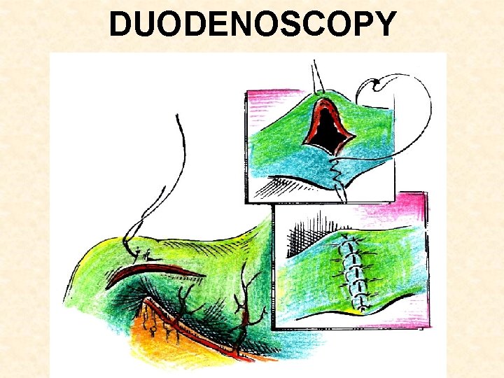
DUODENOSCOPY
SYMPTOM OF “Haudek's niche” STENOSIS
DIAGNOSIS PROGRAM • • • 1. Anamnesis and physical examination. 2. Endoscopy. 3. X-Ray examination of stomach and duodenum. 4. General and biochemical blood analysis. 5. Coagulogram.
CONSERVATIVE THERAPY a) Omeprazole 20 mg 2 time per day or Н 2 blocker histamine receptor (ranitidine) — 150 mg in the evening, famotidine — 40 mg at night, roxatidine — 150 mg in the evening b) antiacid drugs — (almagel, maalox or gaviscon — 1 dessert-spoon in a 1 hour after food intake); c) reparative drugs (dalargin, solcoseryl, actovegin) — for 2 ml 1– 2 times per days d) antimicrobial drugs (clarytromicine 500 mg twice daily, de-nol, metronidazole)
INDICATIONS TO THE ELECTIVE OPERATION • • • 1. Passing of duodenal ulcer with the frequent relapses which could not treated conservatively. 2. Repeated ulcerous bleeding. 3. Stenosis of outcome part of stomach. 4. Chronic penetration ulcers with the pain syndrome. 5. Suspicion for malignization ulcers.
METHODS OF SURGICAL TREATMENT • • • organ-saving operations; organ-sparing operations; resection.
TRUNK VAGOTOMY (Tr. V) 2 4
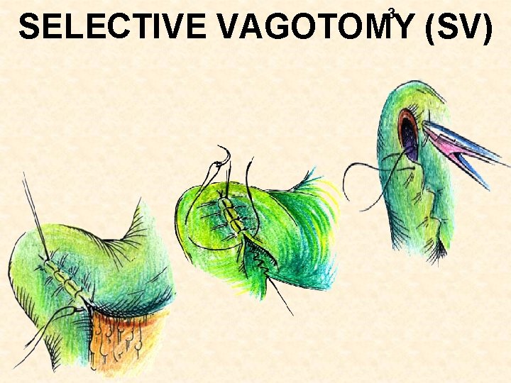
3 SELECTIVE VAGOTOMY (SV)
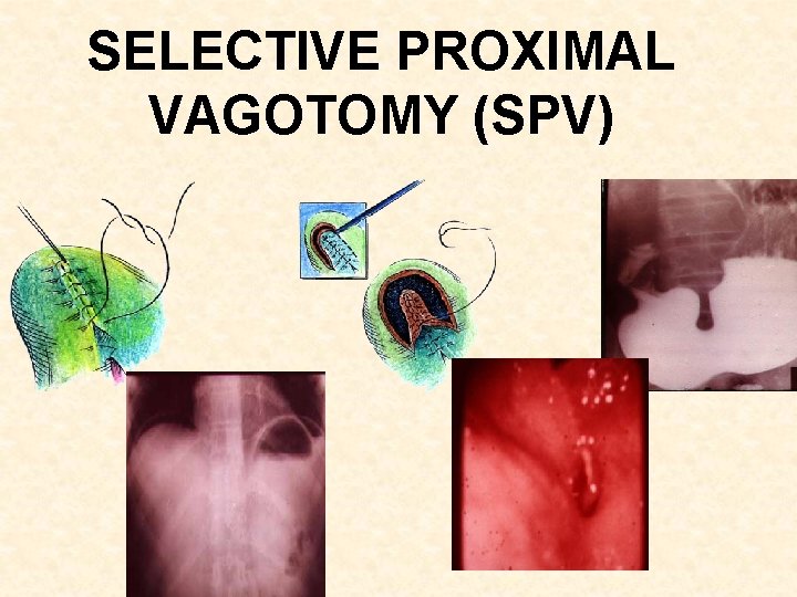
SELECTIVE PROXIMAL VAGOTOMY (SPV)
SELECTIVE PROXIMAL VAGOTOMY (SPV)
Heineke. Mikulicz pyloroplasty
Heineke-Mikulicz pyloroplasty
GASTRODUODENOSTOMY BY JABOULAY
Finney pyloroplasty
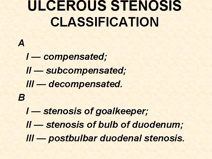
ULCEROUS STENOSIS CLASSIFICATION A I — compensated; II — subcompensated; III — decompensated. B I — stenosis of goalkeeper; II — stenosis of bulb of duodenum; III — postbulbar duodenal stenosis.
DIAGNOSIS PROGRAM • • 1. Complaints of patient and anamnesis of disease. 3. Sounding of stomach and examination of gastric content. 4. Fibergastroduodenoscopy, biopsy. 5. Intragastric р. Н-metry. 6. Study of motility of stomach. 7. Roentgenologic examination of stomach and duodenum (structural features, passage). 8. Sonography.
ULCER STENOSIS
PERFORATED GASTRODUODENAL ULCERS CLASSIFICATION 1. After etiology: • ulcerous; • unulcerous. 2. After localization: • gastric (small curvature, cardial, antral, prepyloric, pyloric) ulcer, front and back walls; • ulcers of duodenum (front and back walls). 3. After passing: • perforated in an abdominal cavity; • covered perforations; • atypical perforations.
DIAGNOSIS PROGRAM • • 1. Anamnesis and physical examination. 2. Global analysis of blood and urine, biochemical blood test, coagulogram. 3. X-Ray examination of abdominal cavity organs for presence of free gas (pneumoperitoneum). 4. Pneumogastrography, contrasting pneumogastrography. 5. Fiber-gastroduodenoscopy. 6. Sonography of abdominal cavity organs.
Perforated ulcer (pneumoperitoneum)
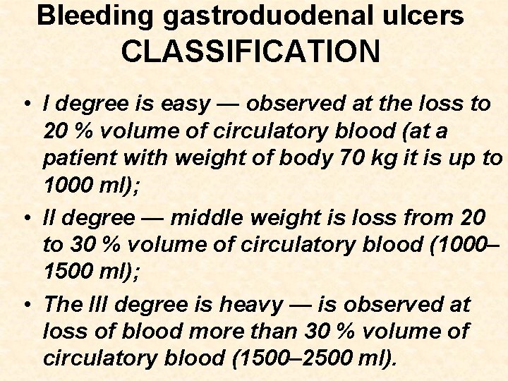
Bleeding gastroduodenal ulcers CLASSIFICATION • I degree is easy — observed at the loss to 20 % volume of circulatory blood (at a patient with weight of body 70 kg it is up to 1000 ml); • II degree — middle weight is loss from 20 to 30 % volume of circulatory blood (1000– 1500 ml); • The III degree is heavy — is observed at loss of blood more than 30 % volume of circulatory blood (1500– 2500 ml).
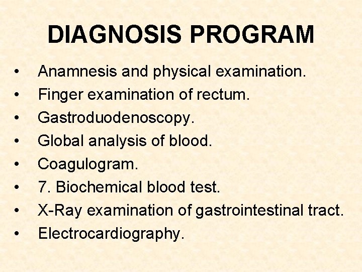
DIAGNOSIS PROGRAM • • Anamnesis and physical examination. Finger examination of rectum. Gastroduodenoscopy. Global analysis of blood. Coagulogram. 7. Biochemical blood test. X-Ray examination of gastrointestinal tract. Electrocardiography.
ENDOSCOPY stopped bleeding