
PEMERIKSAAN RADIOLOGI EXTREMITAS UNTUK DOKTER DI LAYANAN PRIMER


- Slides: 77
PEMERIKSAAN RADIOLOGI EXTREMITAS UNTUK DOKTER DI LAYANAN PRIMER ( SKDI LEVEL 3 DAN 4) Dr. dr. H. Undan. Ruhimat, Sp. Rad (K), . M. H. Kes Departemen Radiologi FK UNPAD /RS Hasan Sadikin Bandung
Principles of Radiologic Interpretation Technical Consideration n Skeletal Anatomy and Physiology n The Categorical approach to bone disease n Radiologic predictor variables n Medicolegal implication n
Technical consideration n Plain Film Radiography n Tomography n Contrast Examination n Radionuclide Imaging n Computed Tomography n Magnetic Resonance Imaging
Skeletal Anatomy and Physiology n Skeletal Development Intramembranous Ossification u Enchondral Ossification n Bone Structure u Epiphyse – Physis – ZPC – Metaphysis – Diaphysis u Cortex – Medulla – Periosteum – Endosteum n Bone Metabolism u Bone mineral - Hormones u
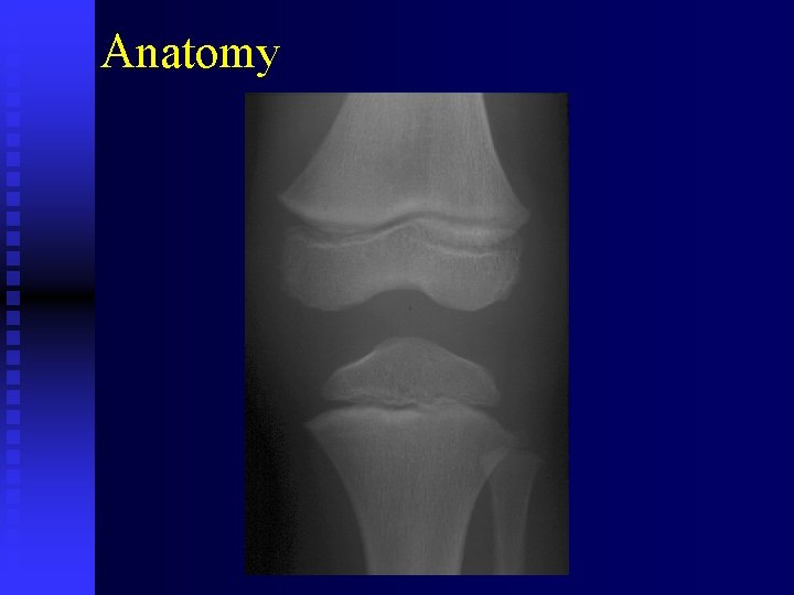
Anatomy
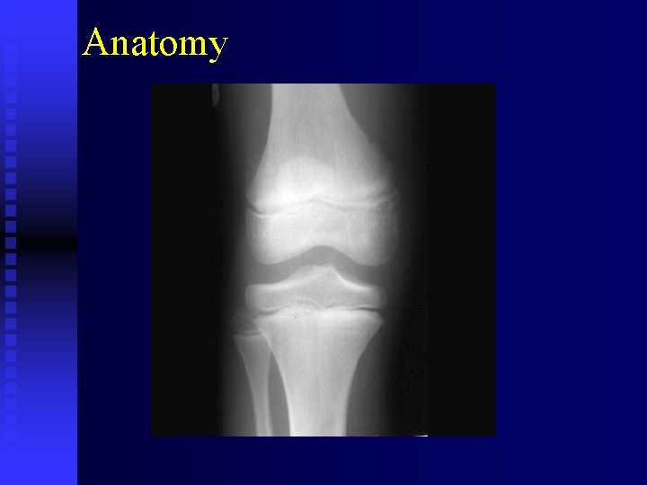
Anatomy
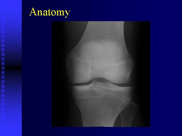
Anatomy
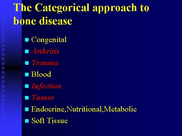
The Categorical approach to bone disease n Congenital n Arthritis n Trauma n Blood n Infection n Tumor n Endocrine, Nutritional, Metabolic n Soft Tissue
Radiologic Predictor Variables n Preliminary Analysis Clinical data u Number of lesions u Symetri of lesions u Determination of Systems Involved u
Radiologic Predictor Variables n Analysis of The Lesions Skeletal Location u Position Within Bone u Site of Origin u Shape u Size u Margination u Cortical Integrity u
Radiologic Predictor Variables n Behavior of Lesions Osteolytic Lesions u Osteoblastic Lesions u Mixed Lesions n Matrix n Periosteal Response u Solid Respons u Laminated Respons u Spiculated Respons u Codmans’ Triangle u
Radiologic Predictor Variables n Soft Tissue Changes n Supplementary Analysis u Other imaging Procedures u Laboratory Examination u Biopsy
TRAUMA Fracture and Dislocation The radiographs should be made n Include at least one joint n Preferably two joints n Two position AP – LAT
TRAUMA Time intervals between Radiographic Study n Initial Diagnostic study n Post reduction and post immobilization n One or Two weeks later, if position has changed n After approximately six eight weeks for Primary callus n After each plaster cast or traction change n Before final discharge of patient
TRAUMA Types of Fracture n Closed fracture u Does not break the skin or communicate with the outside environment u Simple fracture n Open fractur u Penetrates the skin over fracture site u Compound fracture
TRAUMA n. Comminuted fracture Two or more bony fragments have separated n Non Comminuted fracture u Penetrates completely through the bone n Avulsion fracture u Tearing away of a portion of the bone n Impaction fracture u Bone is driven into its adjacent segmen u
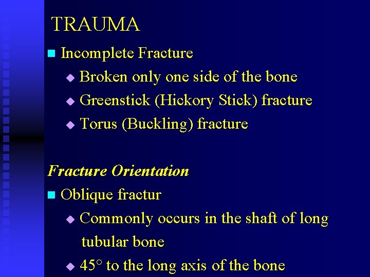
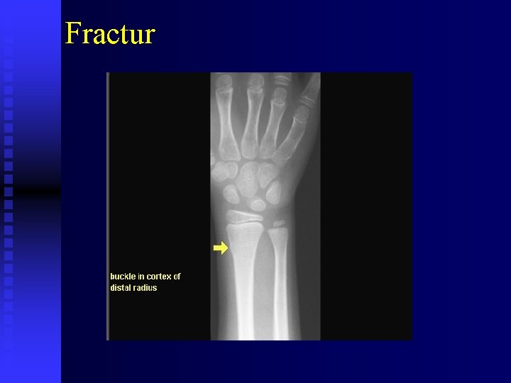
TRAUMA n Incomplete Fracture Broken only one side of the bone u Greenstick (Hickory Stick) fracture u Torus (Buckling) fracture u Fracture Orientation n Oblique fractur u Commonly occurs in the shaft of long tubular bone u 45° to the long axis of the bone
Fractur
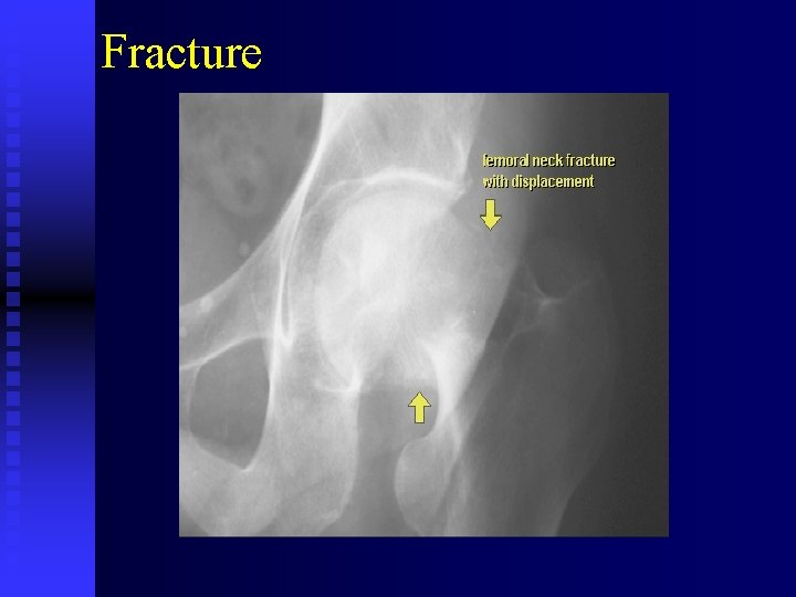
Fracture
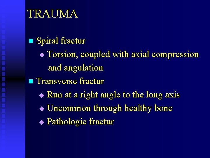
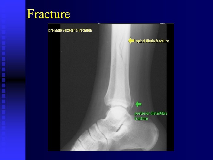
TRAUMA n Spiral fractur Torsion, coupled with axial compression and angulation n Transverse fractur u Run at a right angle to the long axis u Uncommon through healthy bone u Pathologic fractur u
Fracture
TRAUMA Spatial Relationships of Fracture n Aligment u Position of the distal fragment in relation to the proximal fragment n Apposition u Closeness of the bony contact at the fracture site u If the ends are pulled referred to as Distraction
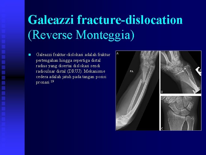
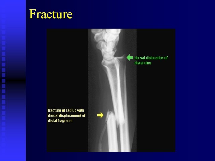
Galeazzi fracture-dislocation (Reverse Monteggia) n Galeazzi fraktur-dislokasi adalah fraktur pertengahan hingga sepertiga distal radius yang disertai dislokasi sendi radioulnar distal (DRUJ). Mekanisme cedera adalah jatuh pada tangan posisi pronasi 19
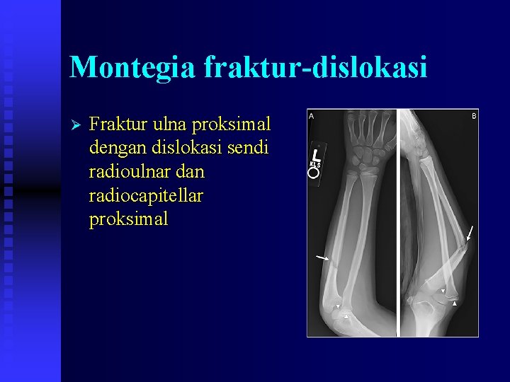
Montegia fraktur-dislokasi Ø Fraktur ulna proksimal dengan dislokasi sendi radioulnar dan radiocapitellar proksimal
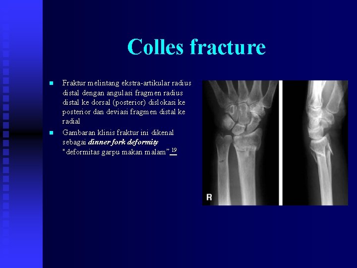
Colles fracture n n Fraktur melintang ekstra-artikular radius distal dengan angulasi fragmen radius distal ke dorsal (posterior) dislokasi ke posterior dan deviasi fragmen distal ke radial Gambaran klinis fraktur ini dikenal sebagai dinner fork deformity "deformitas garpu makan malam” 19
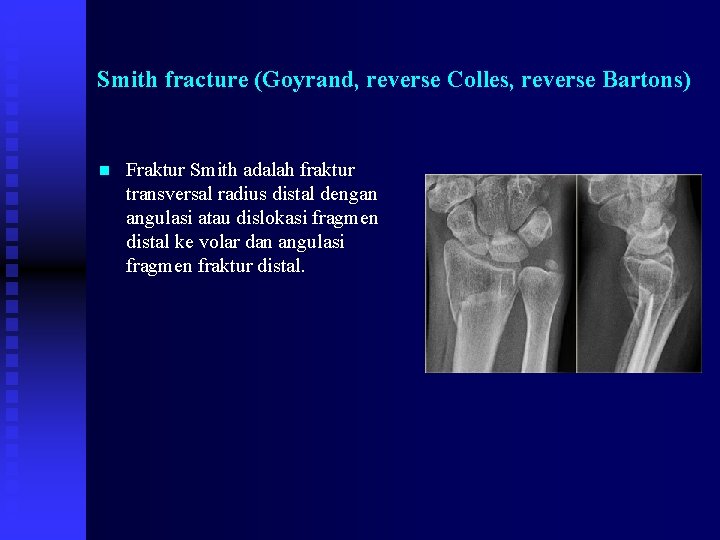
Smith fracture (Goyrand, reverse Colles, reverse Bartons) n Fraktur Smith adalah fraktur transversal radius distal dengan angulasi atau dislokasi fragmen distal ke volar dan angulasi fragmen fraktur distal.
Fracture
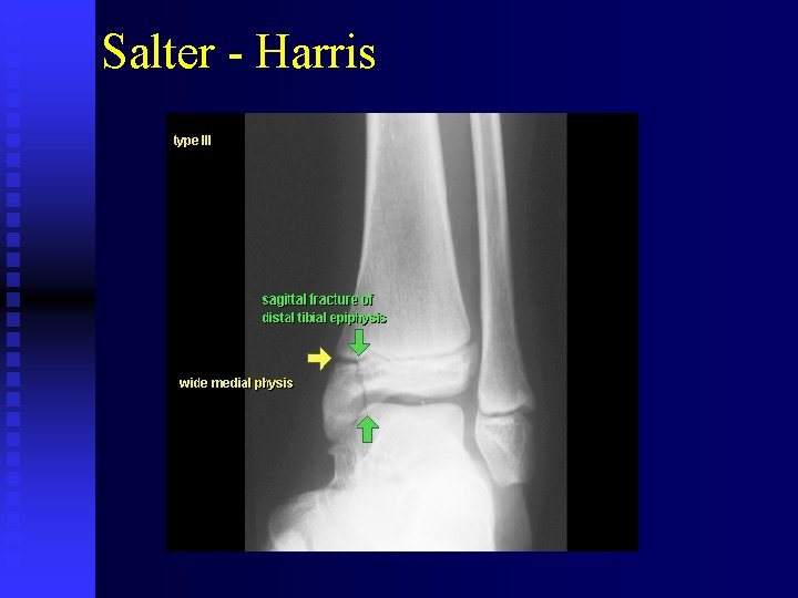
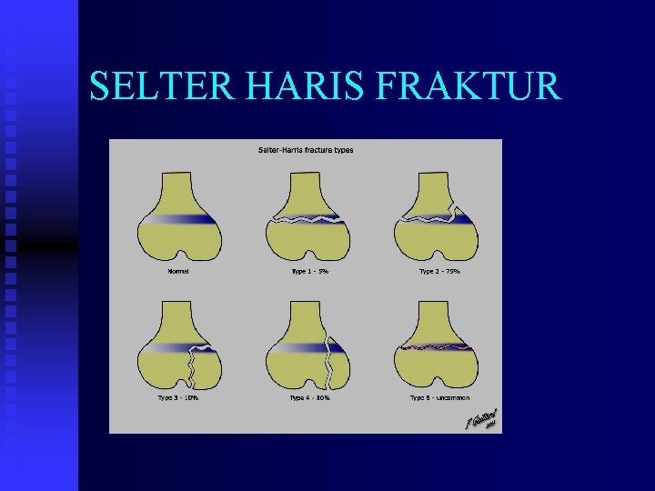
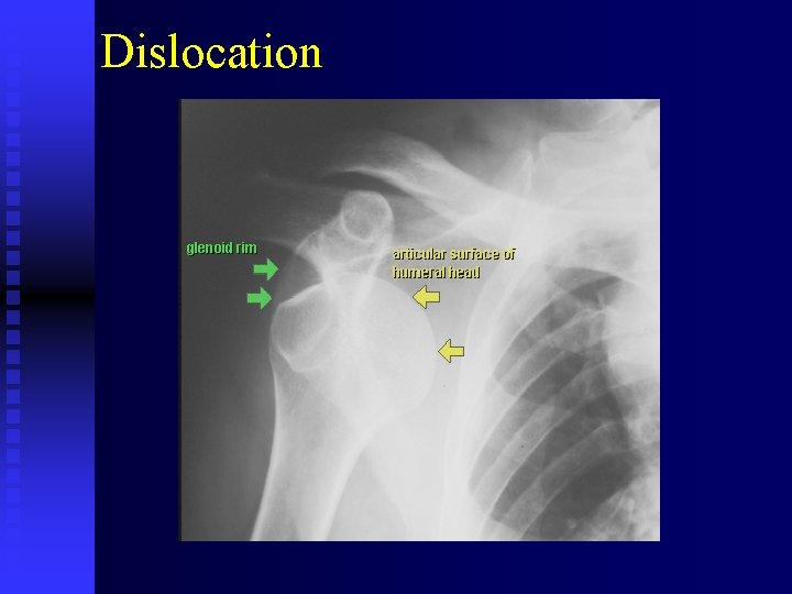
TRAUMA n Rotation Twisting forces on a fractured bone along its longitudinal axis Traumatic Articular Lesions n Subluxation n Dislocation n Diastasis Epiphyseal Fractures n Salter-Harris Classification u
Salter - Harris
SELTER HARIS FRAKTUR
Dislocation
TRAUMA Fracture Healing n Main steps in fracture healing u Formation of hematoma u Organization of hematoma u Formation of fibrous callus u Replacement of fibrous callus by primary bany callus u Absorption primary bany callus Transformation to secondary bony callus u Remodeling
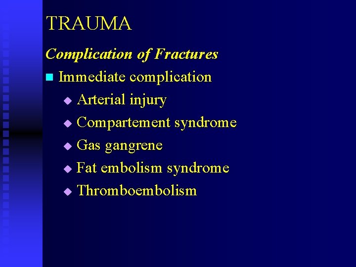
TRAUMA Complication of Fractures n Immediate complication u Arterial injury u Compartement syndrome u Gas gangrene u Fat embolism syndrome u Thromboembolism
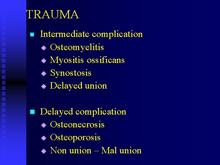
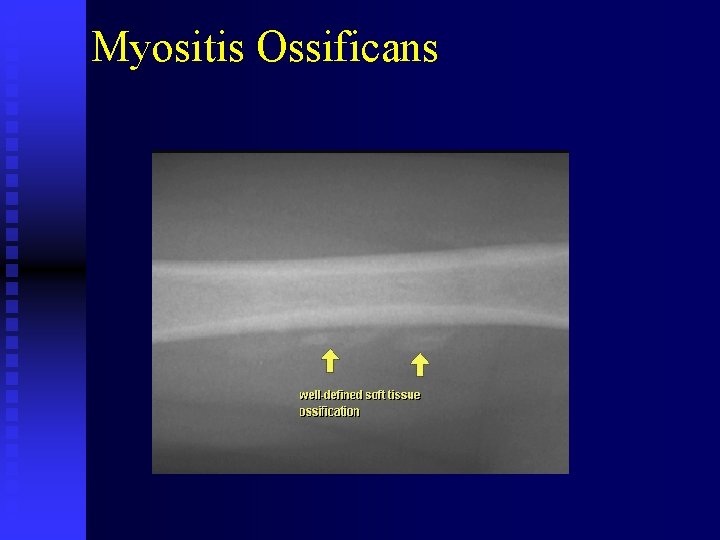
TRAUMA n Intermediate complication u Osteomyelitis u Myositis ossificans u Synostosis u Delayed union n Delayed complication u Osteonecrosis u Osteoporosis u Non union – Mal union
Myositis Ossificans
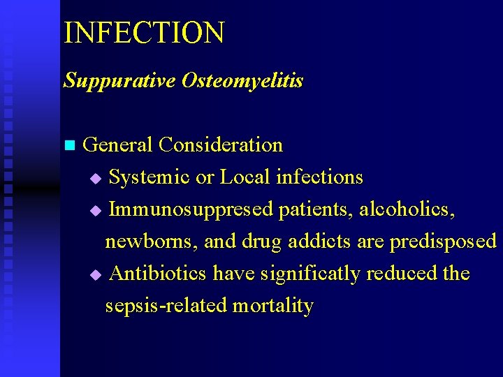
INFECTION Suppurative Osteomyelitis n General Consideration Systemic or Local infections u Immunosuppresed patients, alcoholics, newborns, and drug addicts are predisposed u Antibiotics have significatly reduced the sepsis-related mortality u
INFECTION n Etiology Staphylococcus aureus causes 90% u Pathway for the spread t Hematogenous t Contigunous t Direct Implantation t Postoperative u
INFECTION n Radiologic Features Bone scan are the earliest means of diagnosis u Radiographic latent period for plain film t 10 days for extremities t 21 days for spine u Soft tissue alteration : elevated fat planes, obliterated fat planes, increased density. u
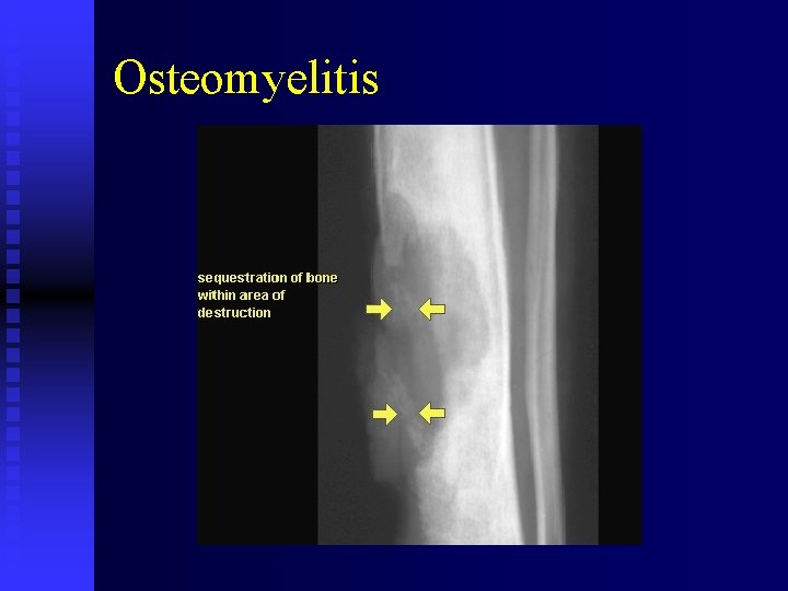
INFECTION u Bone changes : t Moth-eaten bone destruction Usually metaphyseal in origin t Periosteal new bone formation Solid – Laminated – Codman’s Triangle t Sequestrum t Involucrum t Joint space destruction (ankylosis)
0 steomyelitis
Osteomyelitis
INFECTION Septic Arthritis n General consideration u Single joint involvment in the rule u Most common rute is hematogenous or direct traumatic implantation n Etiology u Most frequently is Staphylococcus Aureus
INFECTION n Radiologic Features The knee and hip are the most common sites u Joint effusion leads to distrorsion of the fat folds u Positive Waldenstorm’s sign u Rapid loss of joint space u Bony ankylosis u
INFECTION Nonsuppurative osteomyelitis (tuberculosis) n General Consideration u Found in patients such as prepubertal children, debilitated geriatric, silicosis, AIDS sufferers, Lymphoma patients, Alcoholics, corticosteroid and drug abusers
INFECTION n Etiology Mycobacterium tuberculosis u Two mode of spread t Inhalation t Ingestion u
INFECTION n Radiologic Features u Spinal tuberculosis is most common at L- I u Early sign for spine are : t Lytic endplate destruction t loss of disc height t Anterior “ gouge defect “ t Paraspinal swelling
INFECTION Advanced sign for spinal involvement are: t Vertebral body collaps t Gibbus formation and obliteration of the disc n Tubercular arthritis is common in the hip and knee n Uniform joint space narowing, early destruction of the subchondral cortex, “moth-eaten” bone destruction and juxtaarticular osteoporosis are the cardinal sign of tubercular arthritis u
TUMORS AND TUMORLIKE PROCESSES METASTATIC BONE TUMORS PRIMARY MALIGNANT BONE TUMORS n Multiple myeloma n Osteosarcoma n Ewing’s Sarcoma PRIMARY QUASIMALIGNANT BONE TUMOR n Giant Cell Tumor
TUMORS PRIMARY BENIGN BONE TUMORS n Osteochondroma n Osteoma n Bone island n Osteoid osteoma n Simple bone cyst n Aneurysmal bone cyst
TUMORS Metastatic Bone Tumors n General Consideration u The most common malignant tumors u CNS tumors and basal cell Ca rarely u Life threatening complication n Insidence u 70% are metastatic, 30% are primary u In females 70% from breast Ca In males 60% from prostate Ca
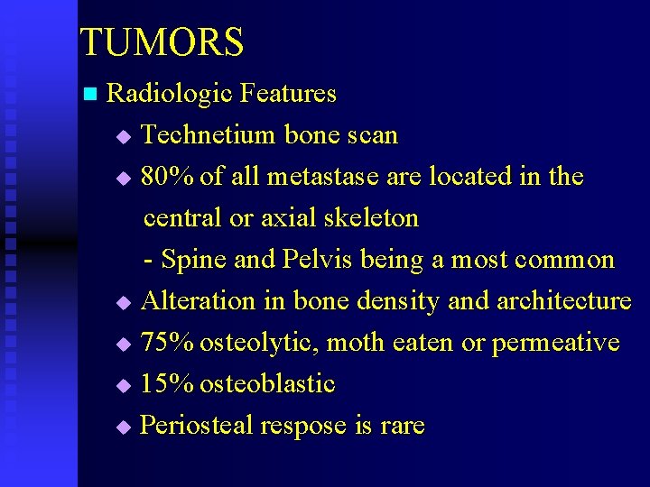
TUMORS n Radiologic Features Technetium bone scan u 80% of all metastase are located in the central or axial skeleton - Spine and Pelvis being a most common u Alteration in bone density and architecture u 75% osteolytic, moth eaten or permeative u 15% osteoblastic u Periosteal respose is rare u
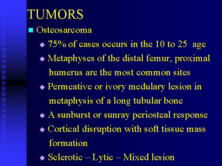
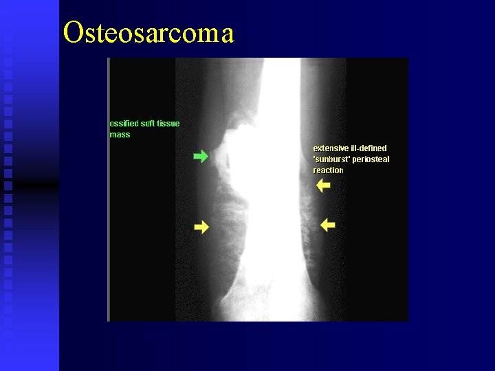
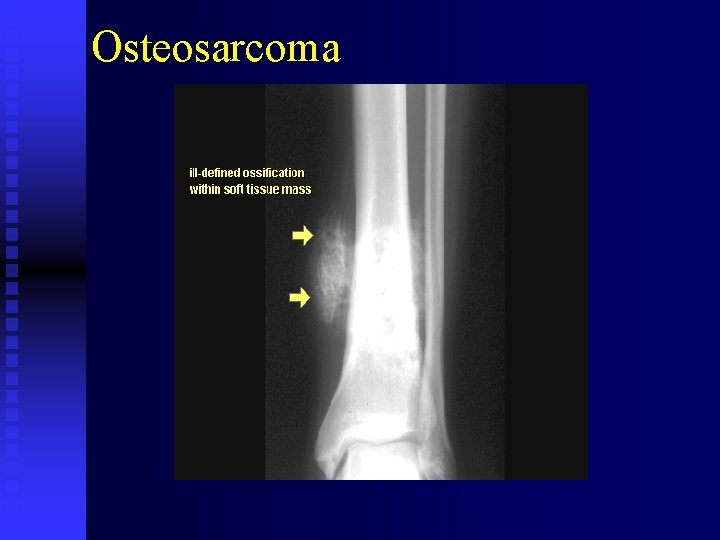
TUMORS n Osteosarcoma 75% of cases occurs in the 10 to 25 age u Metaphyses of the distal femur, proximal humerus are the most common sites u Permeative or ivory medulary lesion in metaphysis of a long tubular bone u A sunburst or sunray periosteal response u Cortical disruption with soft tissue mass formation u Sclerotic – Lytic – Mixed lesion u
Osteosarcoma
Osteosarcoma
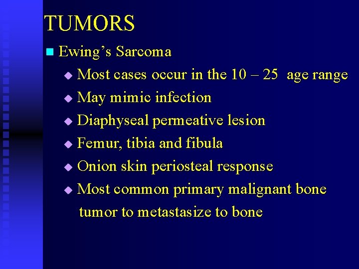
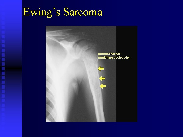
TUMORS n Ewing’s Sarcoma Most cases occur in the 10 – 25 age range u May mimic infection u Diaphyseal permeative lesion u Femur, tibia and fibula u Onion skin periosteal response u Most common primary malignant bone tumor to metastasize to bone u
Ewing’s Sarcoma
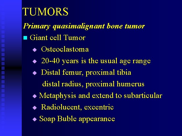
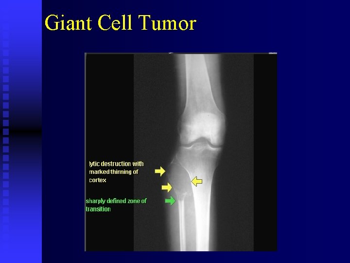
TUMORS Primary quasimalignant bone tumor n Giant cell Tumor u Osteoclastoma u 20 -40 years is the usual age range u Distal femur, proximal tibia distal radius, proximal humerus u Metaphysis and extend to subarticular u Radiolucent, excentric u Soap Buble appearance
Giant Cell Tumor
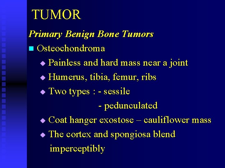
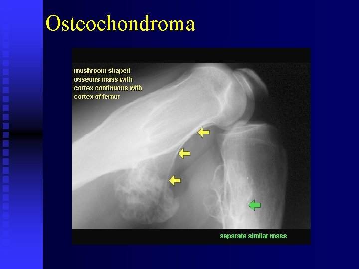
TUMOR Primary Benign Bone Tumors n Osteochondroma u Painless and hard mass near a joint u Humerus, tibia, femur, ribs u Two types : - sessile - pedunculated u Coat hanger exostose – cauliflower mass u The cortex and spongiosa blend imperceptibly
Osteochondroma
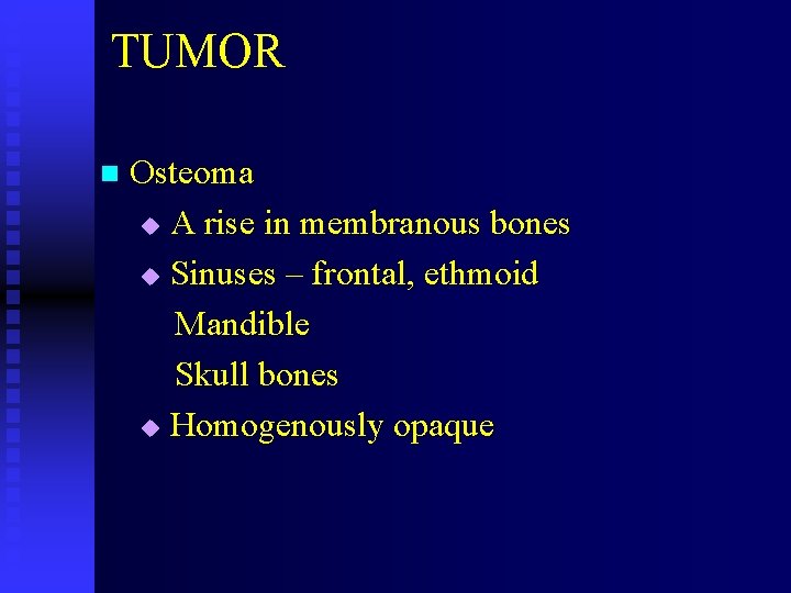
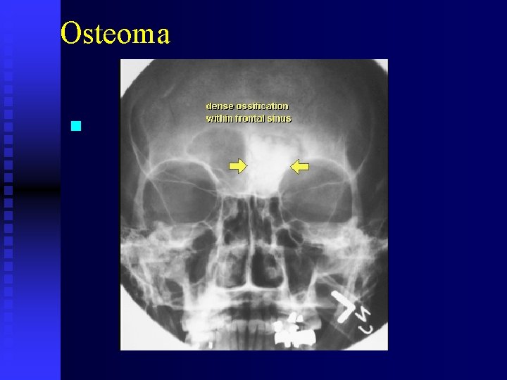
TUMOR n Osteoma A rise in membranous bones u Sinuses – frontal, ethmoid Mandible Skull bones u Homogenously opaque u
Osteoma n
TUMOR n Bone Island Epiphyseal, metaphyseal u Medulary u Round – oval : Long axis oriented Smooth or radiating border Opaque Normal adjecent cortex May change size u
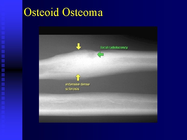
TUMOR n Osteoid osteoma Consists a nidus, thst usually 1 cm or less u Target calsification u Most common location is in the cortex u Radiolucent nidus surrounded by perifocal reactive sclerosis u
Osteoid Osteoma
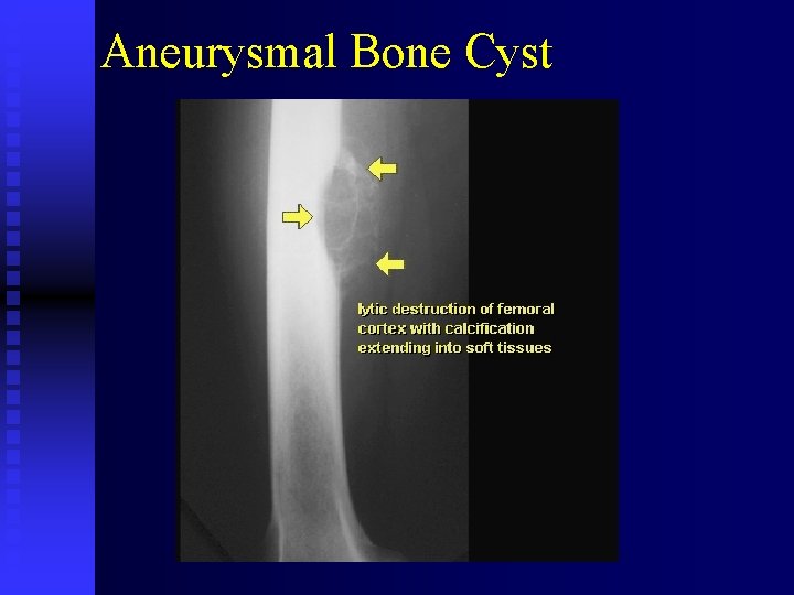
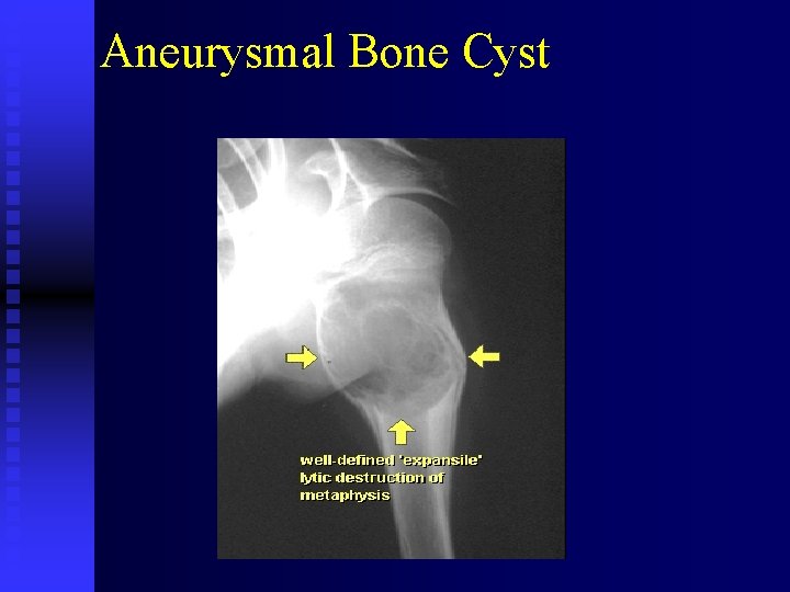
TUMOR n Simple Bone Cyst Expansile radiolucent u Proximal humerus, femur, calcaneus u No periosteal reaction u Pathologic fracture n Aneurysmal Bone Cyst u Some lesion may reach 8 – 10 cm u Cortical ballooning “ blown out app” u
Aneurysmal Bone Cyst
Aneurysmal Bone Cyst
ARTHRITIC DISORDERS Degenerative Disorders u Degenerative Joint Disease u etc Inflamatory Disorders u Rheumatoid Arthritis u etc Metabolic Disorders u Gout u etc
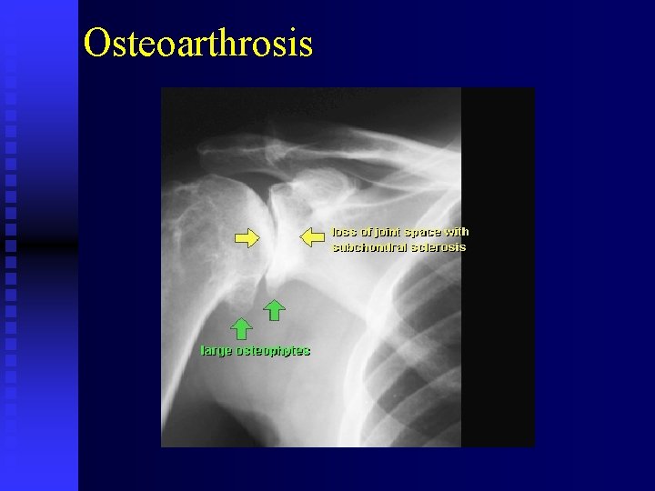
ARTHRITIC n Degenerative Joint Disease Osteoarthritis – Osteoarthrosis u Asimetric distribution u Non uniform loss of the joint space u Osteophytes u Subchondral sclerosis u Subchondral cyst u Loose bodies u Subluxation u
Osteoarthrosis
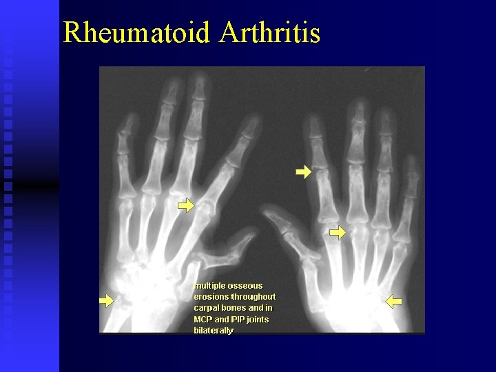
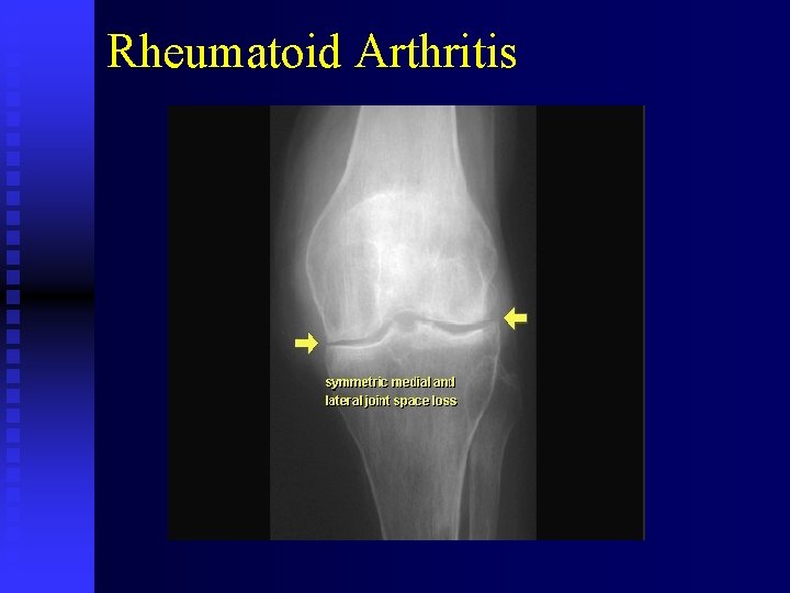
ARTHRITIC n Rheumatoid Arthritis Generalized Connective tissue disorder u Higest insidence among the 40 – 50 year u Symetric peripheral joint pain and swelling u Early : - Soft tissue swelling Marginal erosions Osteoporosis - Periostitis Loss of joint space Late : - Ankylosis u
Rheumatoid Arthritis
Rheumatoid Arthritis
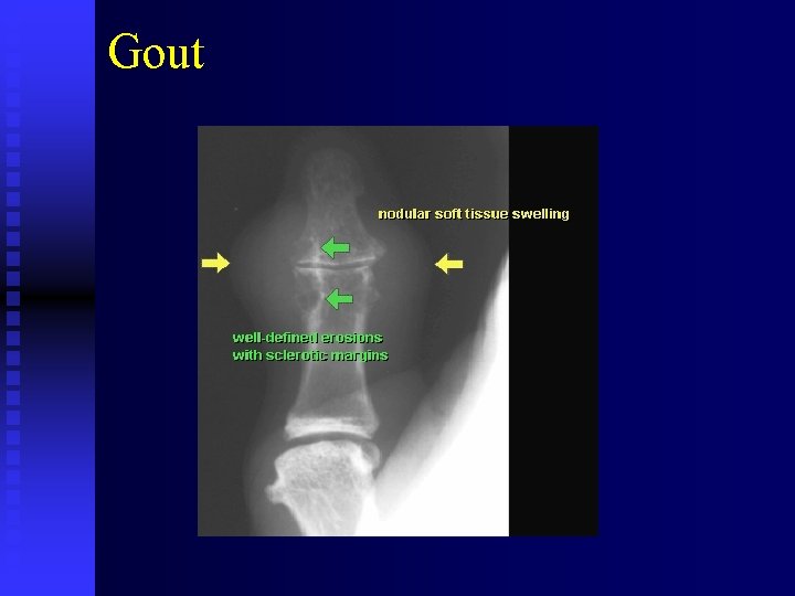
ARTHRITIC n. Gout Disorder of purin metabolism u Deposite of Sodium monourate crystals into cartilage, synovium, periarticular and subcutaneous tissues u Dense soft tissue Tophi, preservation of joint space, Bone erosions (marginal periarticular) “overhanging margin sign” u Metatarsophalangeal joint u
Gout
Sekian TERIMAKASIH Buku ajar fraktur dan dislokssi EGC 2018