Pelvis Perineum Pelvis Lecture Objectives Describe the structure

Pelvis & Perineum

Pelvis

Lecture Objectives • Describe the structure of bony pelvis, perineum, and pelvic diaphragm. • Discuss the nerves and blood vessels of the pelvis. • Describe the radiographic images and the surface landmarks of the pelvis.

Bony Pelvis • Functions – Transmit weight to femur – Supports & protects pelvic viscera – Muscle attachment • Content – 2 hip bones – Sacrum & coccyx
 Girdle • Each coxal (hip) bone consists of three bones that fuse")
Pelvic (Hip) Girdle • Each coxal (hip) bone consists of three bones that fuse together: ilium, pubis, and ischium • The two coxal bones are joined anteriorly by the pubic symphysis (fibrocartilage) • Joined posteriorly by the sacrum forming the sacroiliac joints

The Ilium • Largest of the three hip bones • Ilium is the superior part of the hip bone • Consists of a superior ala and inferior body which forms the acetabulum (the socket for the head of the femur) • Arcuate line separates ala & body

The Ilium • Superior border ‐ iliac crest • Hip pointer ‐ occurs at anterior superior iliac spine • Greater sciatic notch ‐ allows passage of sciatic nerve • Iliac fossa for muscle attachment • Gluteal lines indicating muscle attachment • Sacroiliac joint at auricular surface & iliac tuberosity

Ischium • Ischium ‐ inferior and posterior part of the hip bone – Most prominent feature is the ischial tuberosity, it is the part that meets the chair when you are sitting – ischial spine – lesser sciatic notch – ramus

Pubis • Pubis ‐ inferior and anterior part of the hip bone – Superior and inferior rami • Iliopectineal line – Body • Pubic crest & tubercle
 by age 30")
Sacrum • Union of 5 vertebrae (S 1 ‐ S 5) by age 30 – median sacral crest was spinous processes – sacral ala is fused transverse processes • Sacral promontory (anterosuperior margin) • • • Sacral canal ends at sacral hiatus (laminae don’t meet) Auricular surface & sacral tuberosity of SI joint Anterior and posterior sacral foramina – Ventral and dorsal rami of spinal nerves respectively

Coccyx • Shape. . • Coccygeal cornua • Union of 4 vertebrae (Co 1 ‐ Co 4) by age 30

Orientation of the Pelvis • Front of pubic symphysis of pubis & ASIS at the same vertical plane • The anterior surface of sacrum & pubic symphysis are directed backward & downward

False and True Pelves • Pelvic brim ‐ a line from the sacral promontory to the upper part of the pubic symphysis – Through iliopectineal line • False pelvis ‐ lies above pelvic brim • True pelvis ‐ lies below pelvic brim • Pelvic axis ‐ path of baby during birth

False Pelvis • False pelvis – Lies above the pelvic prim – Boundaries • Lumber vertebrae, iliac fossae & abdominal wall • Part of the abdomen – Little clinical significance – Support abdominal viscera, and uterus during pregnancy
 – Boundaries • Sacral promontory, iliopectineal line")
True Pelvis • Pelvic inlet (pelvic brim) – Boundaries • Sacral promontory, iliopectineal line & pubic symphysis • Pelvic outlet – Boundaries • Coccyx, ischial tuberosities, sacrotuberous ligament & pubic arch • Pelvic cavity – Between inlet & outlet – Short curved canal – Shallow anterior & deep posterior walls

Comparing Male and Female Pelves • Males – Bone are larger and heavier – Pelvic inlet is smaller and heart shaped – Pubic arch is less the 90° • Female – Wider and shallower – Pubic arch is greater than 90° – More space in the true pelvis

Comparing Male and Female Pelves

Comparing Male and Female Pelves

Male pelvis

Female pelvis

Pelvic Walls • • • Bones Ligaments Muscles Fascia Parietal peritoneum

Anterior Pelvic Wall • The shallowest wall • Content – Bodies of the pubic bones – Pubic rami – Symphysis pubis

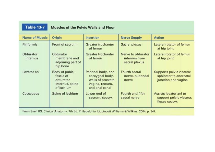
Posterior Pelvic Wall • Sacrum • Coccyx • Piriformis muscle – From sacrum to greater trochanter • Through the greater sciatic foramen

Lateral Pelvic Wall • Hip bones • Obturator membrane – Fibrous sheet that closes the obturator foramen • Obturator canal • Sacrotuberous ligament – From sacrum, coccyx & PIIS to the ischial tuberosity • Sacrospinous ligaments – From sacrum & coccyx to the ischial spine • Obturator internus muscle – From obturator membrane & surrounding bones to the greater trochanter • Through the lesser sciatic foramen

Sacroiliac Joint • Auricular surfaces of the sacrum and the iliac bone • No movement; transmit body weight from vertebral column to pelvis • In elderly people synovial cavity disappear and becomes fibrous joint • Associated ligaments – Posterior sacroiliac ligament – Interosseous sacroiliac ligament (between tuberosities of sacrum and iliac bone) – Anterior sacroiliac ligament • Accessory ligaments – Sacrotuberous ligament – Sacrospinous ligament – Iliolumbar ligament greater sciatic foramen lesser sciatic foramen • Nerve supply: sacral spinal nerves
 • Support the pelvic viscera • Inferior pelvic wall")
Inferior Pelvic Wall (Pelvic Diaphragm) • Support the pelvic viscera • Inferior pelvic wall (pelvic floor) formed by the pelvic diaphragm • The levator ani and coccygeus muscles, along with the fascia which covers them, form the pelvic diaphragm. • The pelvic diaphragm separates the pelvic cavity above from the perineum below • Pelvic diaphragm is deficient anteriorly (urogenital hiatus)

Levator Ani Muscle • Wide thin sheet • Originate laterally from the pubic body, obturator internus fascia & ischial spine • Orientation – downward and medially
 – Inserts")
Levator Anai Muscle: Fibers • Anterior fibers (levator prostatae or sphincter vaginae) – Inserts to the perineal body • Intermediate fibers – Puborectalis – around the anorectal junction – Pubococcygeus • Inserts into the anococcygeal body • Posterior fibers (iliococcygeus) – Inserted into the anococcygeal body & coccyx

Pelvic Fascia • Above is continuous with the fascia lining the abdominal walls • Below is continuous with the fascia of the perineum • Layers – Parietal layer • Lines the walls of pelvis ‐ covers the pelvic diaphragm • Continuous anteriorly with the fascia covering the lower surface of the diaphragm – Visceral layer • Covers & supports the pelvic viscera – Fascial ligaments‐ connects some viscera to the pelvic walls (pubovesical & sacrocervical ligaments) • Parietal peritoneum – Lines the pelvic walls – Covers the viscera and continue as visceral peritoneum

Nerves of the Pelvis • Branches of lumbar plexus – Lumbosacral trunk – Obturator nerve • Sacral plexus – Anterior to periformis muscle – Posterior to internal iliac vessels and rectum • Autonomic nerves

Sacral Plexus • Ventral rami L 4‐S 4 • Anterior to the sacrum and posterior pelvic wall • Form in front of periformis m. • Supplies buttocks, perineum & part of lower limb • Branches exit from the greater sciatic notch • Branches exit inferior to periformis m. except the superior gluteal n.

Autonomic Nerves • Pelvic part of sympathetic trunk – – – Posterior to common iliac vessels Anterior to sacrum Medial to sacral foramina 4‐ 5 ganglia Ganglion impar • Pelvic splanchnic nerves – Parasympathetic (S 2‐S 4) – Inferior hypogastric plexus – Inferior mesenteric plexus • Superior hypogastric plexuses – In front of promontory – Forms right & left hypogastric nerves • Inferior hypogastric plexuses – Hypogastric nerve + pelvic splanchnic nerve – Lateral to rectum, bladder & vagina

Arteries of the False Pelvis • Common iliac a. – Divides at the pelvic prim in front of the sacroiliac joint • External iliac a. – Continue at the brim – Pelvic branches • Deep circumflex iliac • Inferior epigastric – Leave the false pelvis deep to the inguinal lig.

Arteries of the True Pelvis • Internal iliac a. • Superior rectal a. – Continuation of inferior mesenteric a. – Mucus membrane of rectum & upper anal canal • Ovarian a. – From abdominal aorta – L 1 – Cross the external iliac at the brim – Reach the ovary by passing through suspensory lig. – broad lig. – mesovarium • Median sacral a. – Rise at the bifurcation of aorta – Descend anterior to sacrum & coccyx

Internal Iliac Artery • Supply the pelvic viscera & walls, perineum, & buttocks • It divides at the upper margin of the greater sciatic foramen • Divisions – Anterior – Posterior

Internal Iliac Artery Anterior Division • Umbilical a. – Superior vesical a. • Obturator a. • Uterine a. – Cross the ureter superiorly – Ascend on the lateral side of the uterus • Through the broad lig. • Anastomose with ovarian a. • Inferior vesical a. (Vaginal a. ) – Artery to the vas deferens • Middle rectal a. • Internal pudendal a. – Leave via GSF – Come back via LSF – Inter the pudendal canal with the nerve • Inferior gluteal a. – Below piriformis m. via GSF

Internal Iliac Artery Posterior Division • Iliolumbar a. – Ascend posterior to external iliac vessels, psoas & iliacus mm. • Lateral sacral aa. • Superior gluteal a. – Above piriformis m. via GSF

Veins of the Pelvis • External iliac v. – Medial to the external iliac a. – Tributaries • Deep circumflex iliac • Inferior epigastric • Internal iliac v. – Tributaries correspond to the arteries • Median sacral v. – Drain into the left common iliac v. Lymphatics of the pelvis – Lymph drains into the node associated with arteries • External iliac, internal iliac & common iliac lymph nodes

Perineum

Lecture Objectives • Describe the perineum and its boundaries. • Describe the anal triangle including anal canal, levator ani muscles and anal sphincters. • Describe the urogenital triangle.

Perineum – Diamond‐shaped area medial to thighs and buttocks of males and females – The perineum is inferior to the pelvic diaphragm that – Extends from the pubic symphysis anteriorly, to the coccyx posteriorly, and to the ischial tuberosities laterally. – Composed of anal triangle and urogenital triangle – Contains external genitalia and anus

Anal Triangle • Boundaries – Coccyx – posteriorly – Sacrotuberous lig. & ischial tuberosity – laterally • Content – Anal canal & anus – Ischioanal (ischiorectal) fossae

Anal Canal • 4 cm length • Oriented downward & backward • Relations – Posteriorly – anococcygeal body – Laterally ‐ Ischioanal fossae – Anteriorly – perineal body & content of the urogenital triangle • Male • Female

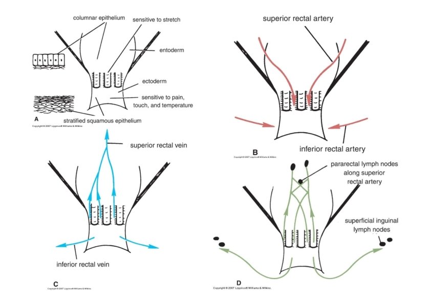
Anal Canal: Structure • Upper half Columnar epithelium Anal columns & valves Blood supply: superior rectal vessels Nerve supply: autonomic hypogastric plexuses – Lymphatics: along the superior rectal a. – pararectal nodes – inferior mesenteric nodes – – Pectinate line • Lower half – Stratified squamous epithelium – Blood supply: inferior rectal vessels – Nerve supply: inferior rectal nerve (somatic) – Lymphatics: superficial inguinal nodes

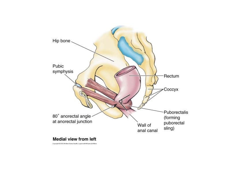
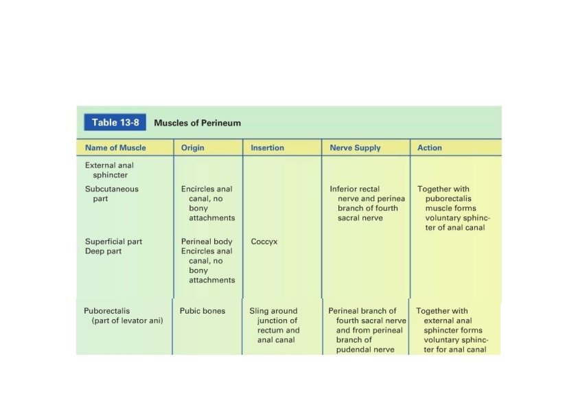
Anal Canal • Muscle coat‐ smooth m. – Outer – longitudinal – Inner ‐ circular • Anal sphincters – Internal sphincter‐ smooth m. • Thickening of the inner circular m. – External sphincter‐ skeletal m. • Subcotaneous part • Superficial part • Deep part – With puborectalis form anorectal ring • Puborectalis m. – Anorectal angle

Ischioanal fossa • Wedge‐shaped space • Boundaries – Obturator internus m. laterally – Levator ani m. & anal canal medially – Skin inferiorly • Content – Filled with dense fat – Pudendal n. & internal pudendal vessels • In pudendal canal on the lateral wall – Inferior rectal n. , a. , v.

Ischioanal fossa • Pudendal nerve – Supply external anal sphincter & muscles & skin of perineum – Branches • Inferior rectal nerve – External anal sphincter – Mucus membrane of lower half of anal canal & perianal skin • Dorsal nerve of the penis (clitoris) • Perineal nerve – Urogenital triangle mm. – Skin of posterior scrotum (labia majora) • Internal pudendal a. – Inferior rectal a. – Branches to the penis (clitoris & labia)

Urogenital Triangle • Boundaries – Pubic arch – anteriorly – Ischial tuberosities – laterally • Content – In male – penis & scrotum – In female – external genitalia & orifices of urethra & vagina • Fascia – Superficial fascia – Superficial perineal pouch • Content • Urogenital diaphragm – Fascia • Deep perineal pouch – content
 – Continuous with the")
Urogenital Triangle Superficial Fascia • Fatty layer (fascia of camper) – Continuous with the fat of the ischioanal fossa – Replaced by dartos muscle in scrotum • Membranous layer (Colles’ fascia) – Attachments • Posteriorly – to urogenital diaphragm & perineal body • Laterally – pubic arch • Anteriorly – continuous with the Scarpa’s fascia • At penis (clitoris) – form tubular sheath

Superficial Perineal Pouch • Enclosed between the Colles’ fascia and the urogenital diaphragm, were they meet: – Posteriorly – at perineal body – Laterally – at pubic arch • Anteriorly – continuous with the potential space between the Scarpa’s fascia and the abdominal mm.

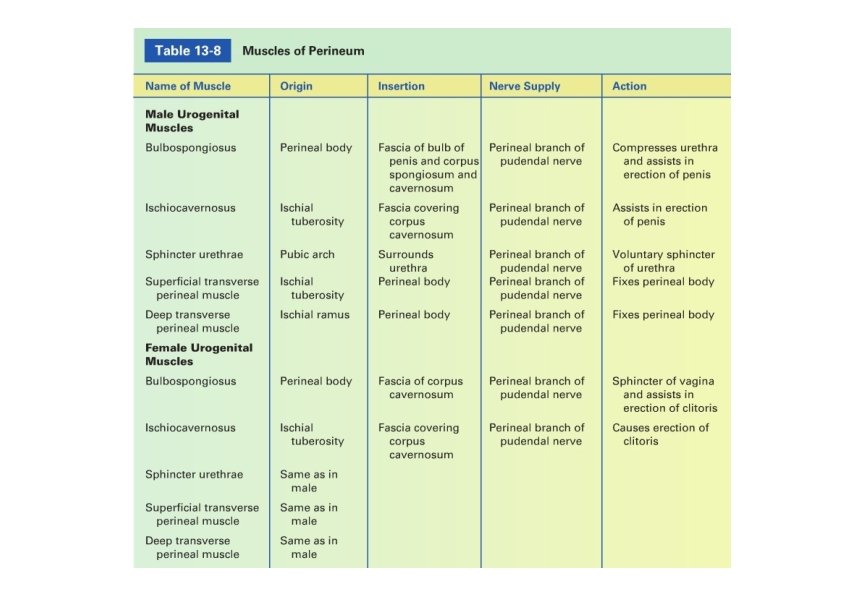
Superficial Perineal Pouch: Content • Root of the penis or clitoris • Muscles – Bulbospongiosus mm. • In male – cover the pulp of penis • In female – cover the pulp of vestibule – Ischiocavernosus mm. • Cover the crura of the penis or clitoris – Superficial transverse perineal mm.

Urogenital Diaphragm Deep Perineal Pouch • Musculofascial triangle at the pubic arch • Muscles – Sphincter urethrae – Deep perineal muscles • Fascia – enclose the mm. – Superior fascial layer – Inferior fascial layer (perineal membrane) – Attachments • Anteriorly – the two layers fuse together • Posteriorly – the two layers fuse & attach to the perineal body • Laterally – pubic arch • Deep perineal pouch – enclosed between the two layers of fascia

Deep Perineal Pouch: Content • Muscles – Sphincter urethrae m. • Surrounds urethra and vagina – Deep transverse perineal mm. • Internal pudendal a. – Branches • Artery of crus • Artery of pulp • Dorsal artery of penis (clitoris) • Dorsal nerve of the penis (clitoris) • Part of urethra – In male – membranous urethra • Part of vagina
- Slides: 58