
PELVIS II FUNCTION TABOOS THE VISCERA Defecation n
PELVIS II: FUNCTION TABOOS (THE VISCERA) Defecation n Urination n Ejaculation n Conception n
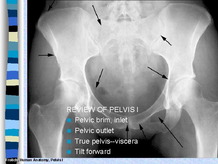
REVIEW OF PELVIS I n Pelvic brim, inlet n Pelvic outlet n True pelvis--viscera n Tilt forward Frolich, Human Anatomy, Pelvis I
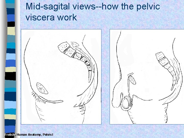
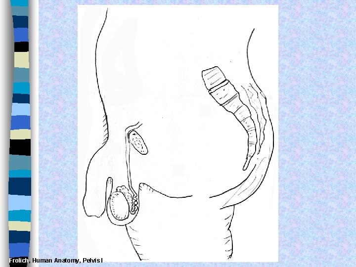
Mid-sagital views--how the pelvic viscera work Frolich, Human Anatomy, Pelvis I
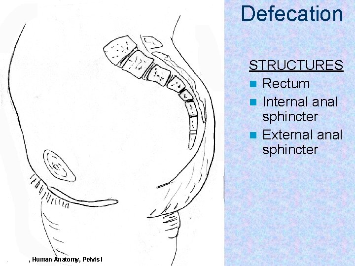
Defecation STRUCTURES n Rectum n Internal anal sphincter n External anal sphincter Frolich, Human Anatomy, Pelvis I
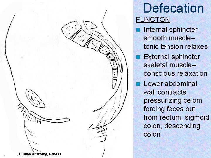
Defecation FUNCTON n Internal sphincter smooth muscle-tonic tension relaxes n External sphincter skeletal muscle-conscious relaxation n Lower abdominal wall contracts pressurizing celom forcing feces out from rectum, sigmoid colon, descending colon Frolich, Human Anatomy, Pelvis I
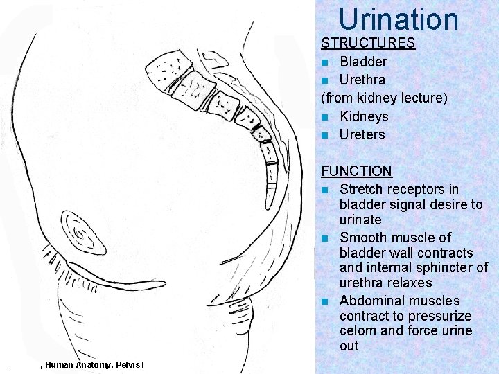
Urination STRUCTURES n Bladder n Urethra (from kidney lecture) n Kidneys n Ureters FUNCTION n Stretch receptors in bladder signal desire to urinate n Smooth muscle of bladder wall contracts and internal sphincter of urethra relaxes n Abdominal muscles contract to pressurize celom and force urine out Frolich, Human Anatomy, Pelvis I
Ejaculation FUNCTION n Sperm mature and collect in STRUCTURES epididymis n Testes n Move through vas deferens by n Vas (ductus) deferens peristalsis of smooth muscle of n Seminal glands wall of vas (vesicles) n Seminal vesicles, prostrate n Prostate contribute to semen n Urethra n Internal urethral sphinchter (at n Corpus spongiosum bladder wall) prevents sperm n Bulbospongiosum m. backflow into bladder n Contractions of urethra move semen to penis n Bulbospongiosus m. (around urethra in penis) contracts to expel semen from penis Frolich, Human Anatomy, Pelvis I
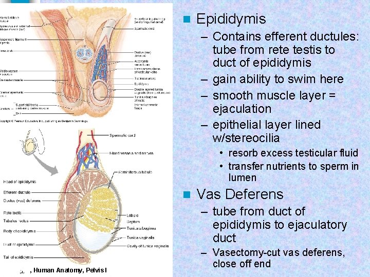
n Epididymis – Contains efferent ductules: tube from rete testis to duct of epididymis – gain ability to swim here – smooth muscle layer = ejaculation – epithelial layer lined w/stereocilia • resorb excess testicular fluid • transfer nutrients to sperm in lumen n Vas Deferens – tube from duct of epididymis to ejaculatory duct Frolich, Human Anatomy, Pelvis I – Vasectomy-cut vas deferens, close off end
Route of sperm is convoluted--testicles to spermatic cord (vas deferens) through inguinal canal around to join urethra at inferior bladder SPERMATIC CORD n Collective name for structures associated with the scrotum n Passes through inguinal canal n Includes – Vas Deferens – Testicular Arteries + Veins – Cremaster Muscle + fibers – Nerves Frolich, Human Anatomy, Pelvis I
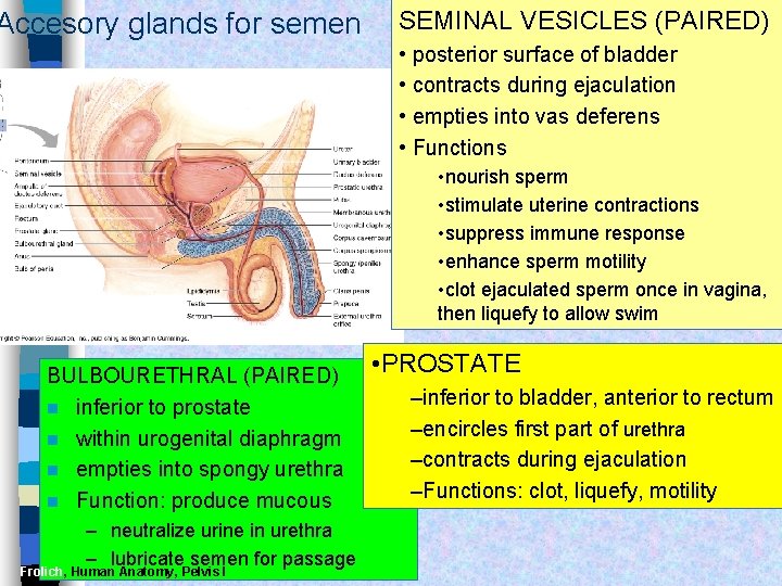
Accesory glands for semen SEMINAL VESICLES (PAIRED) • posterior surface of bladder • contracts during ejaculation • empties into vas deferens • Functions • nourish sperm • stimulate uterine contractions • suppress immune response • enhance sperm motility • clot ejaculated sperm once in vagina, then liquefy to allow swim BULBOURETHRAL (PAIRED) n inferior to prostate n within urogenital diaphragm n empties into spongy urethra n Function: produce mucous – neutralize urine in urethra – lubricate semen for passage Frolich, Human Anatomy, Pelvis I • PROSTATE –inferior to bladder, anterior to rectum –encircles first part of urethra –contracts during ejaculation –Functions: clot, liquefy, motility
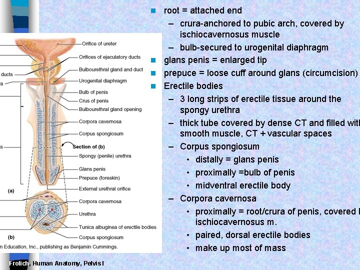
root = attached end – crura-anchored to pubic arch, covered by ischiocavernosus muscle – bulb-secured to urogenital diaphragm n glans penis = enlarged tip n prepuce = loose cuff around glans (circumcision) n Erectile bodies – 3 long strips of erectile tissue around the spongy urethra – thick tube covered by dense CT and filled with smooth muscle, CT + vascular spaces – Corpus spongiosum • distally = glans penis • proximally =bulb of penis • midventral erectile body – Corpora cavernosa • proximally = root/crura of penis, covered b ischiocavernosus m. • paired, dorsal erectile bodies • make up most of mass n Frolich, Human Anatomy, Pelvis I
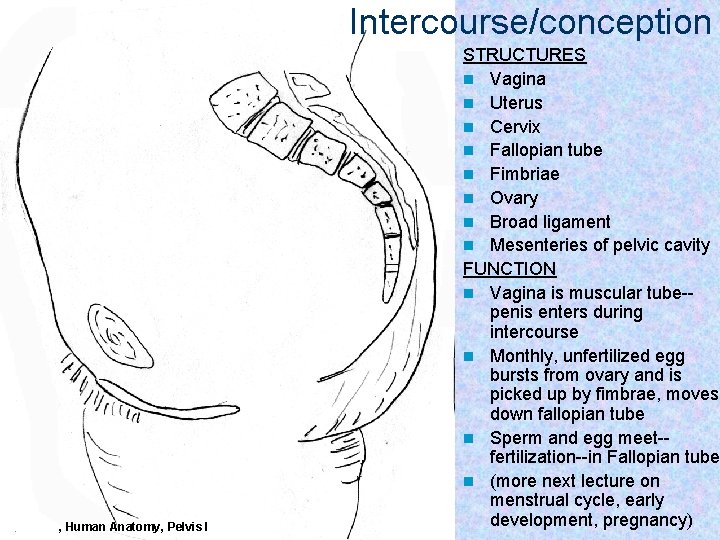
Intercourse/conception Frolich, Human Anatomy, Pelvis I STRUCTURES n Vagina n Uterus n Cervix n Fallopian tube n Fimbriae n Ovary n Broad ligament n Mesenteries of pelvic cavity FUNCTION n Vagina is muscular tube-penis enters during intercourse n Monthly, unfertilized egg bursts from ovary and is picked up by fimbrae, moves down fallopian tube n Sperm and egg meet-fertilization--in Fallopian tube n (more next lecture on menstrual cycle, early development, pregnancy)
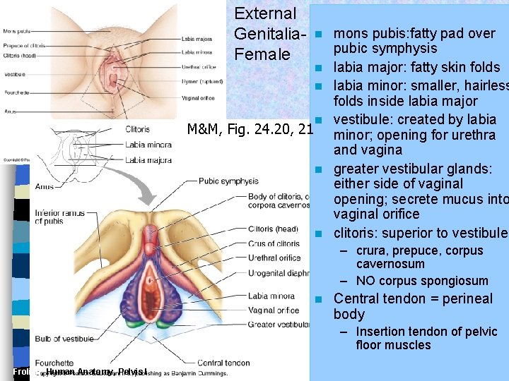
External Genitalia. Female n n n M&M, Fig. 24. 20, 21 n n n mons pubis: fatty pad over pubic symphysis labia major: fatty skin folds labia minor: smaller, hairless folds inside labia major vestibule: created by labia minor; opening for urethra and vagina greater vestibular glands: either side of vaginal opening; secrete mucus into vaginal orifice clitoris: superior to vestibule – crura, prepuce, corpus cavernosum – NO corpus spongiosum n Central tendon = perineal body – Insertion tendon of pelvic floor muscles Frolich, Human Anatomy, Pelvis I
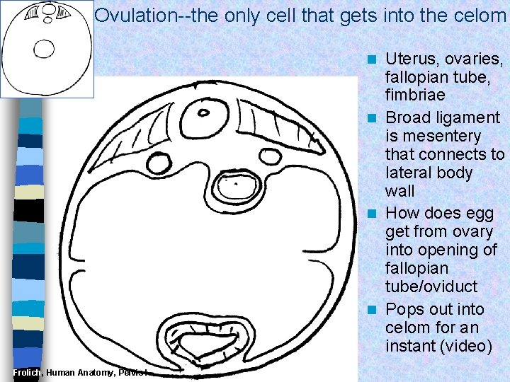
Ovulation--the only cell that gets into the celom Uterus, ovaries, fallopian tube, fimbriae n Broad ligament is mesentery that connects to lateral body wall n How does egg get from ovary into opening of fallopian tube/oviduct n Pops out into celom for an instant (video) n Frolich, Human Anatomy, Pelvis I
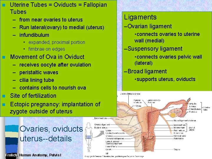
n Uterine Tubes = Oviducts = Fallopian Tubes – from near ovaries to uterus – Run lateral(ovary) to medial (uterus) – infundibulum • expanded, proximal portion • fimbrae on edges n Movement of Ova in Oviduct – – receives oocyte after ovulation peristaltic waves cilia lining tube contains cells to nourish ova Site of fertilization n Ectopic pregnancy: implantation of zygote outside of uterus n Ovaries, oviducts, uterus--details Frolich, Human Anatomy, Pelvis I Ligaments –Ovarian ligament • connects ovaries to uterine wall (medial) –Suspensory ligament • connects ovaries pelvic wall (lateral) –Broad ligament • supports uterus, oviducts
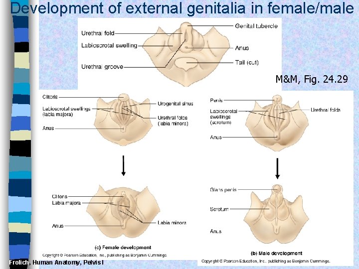
Development of external genitalia in female/male M&M, Fig. 24. 29 Frolich, Human Anatomy, Pelvis I
Development of Reproductive Organs Gonadal ridge: forms in embryo at 5 weeks and gives rise to gonads (intermediate mesoderm with kidneys) n Wolffian ducts: form male duct (vas deferens) n Mullerian ducts: form female duct (uterine tube) n – Both ducts are present in embryo-only one develops! n External genitalia develops from same structures – Labioscrotal swelling: Scrotum = Labia major – Urethral folds: Penile Urethra = Labia minor – Genital tubercle: Penis = Clitoris Frolich, Human Anatomy, Pelvis I
Frolich, Human Anatomy, Pelvis I
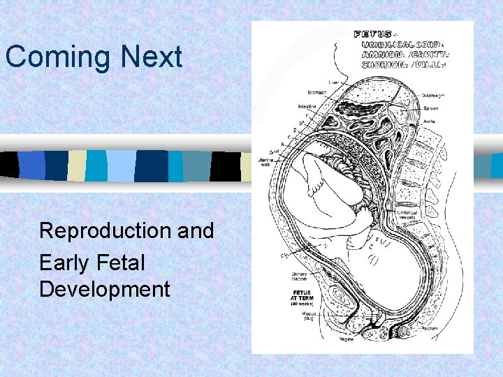
Coming Next Reproduction and Early Fetal Development
- Slides: 19