PELVIS and ACETABULUM FRACTURES Assoc Prof Melih Gven

PELVIS and ACETABULUM FRACTURES Assoc. Prof. Melih Güven Yeditepe University Hospital Department of Orthopaedics and Traumatology

Learning Objectives 1. Should be able to list steps of emergency intervention for pelvic trauma patient 2. Should be able to explain the term of stability of pelvis fractures and also classify the pelvic fractures by stability 3. Should be able to tell the X-rays to be investigated for pelvis fractures and also evaluate X-rays 4. Should be able to list emergency attempts for haemodynamic instability 5. Should be able to list pelvic fracture that should be treated conservatively and also list conservative treatment methods 6. Should be able to define simple and complex acetabulum fractures 7. Should be able to tell the X-rays to be investigated for acetabulum fractures and also evaluate X-rays 8. Should be able to list conservative and surgical treatment options for acetabular fractures

Pelvic injuries 3% of all fractures Can be found in 25% of polytraumatized patients 42% of deaths due to motor vehicle accidents Can be mortal

LOAD TRANSFER

VESSELS AND NERVES PASSING THROUGH THE GREATER SCIATIC NOTCH NERVES Sciatic nerve Superior gluteal nerve Inferior gluteal nerve Internal pudental nerve Posterior femoral cutaneous nerve VESSELS Superior gluteal ınferior gluteal ınternal puden. Tal MUSCLE Piriformis

LIGAMENTS Between lomber vertebrae and ilium Between L 5 transverse process and sacrum Between sacrum and ilium Between sacrum and ischium Between sacrum and cocsyx Between pubic bones Posterior Anterior

PELVIC FRACTURES Anatomy

SACROILIAC LIGAMENTS Anterior sacroiliac Interosseous Posterior sacroiliac

ROTATIONAL STABILIZERS Ligament of symphisis pubis Sacrospinous ligament Anterior sacroiliac ligament Short posterior sacroiliac ligament

VERTICAL STABILIZERS Interosseous sacroiliac ligament Long posterior sacroiliac ligament Iliolumbar ligament Lateral lumbosacral ligament Sacrotuberous ligament

PELVIC FRACTURES Vascular anatomy

PHYSICAL EXAMINATION Leg length discrepancy Haematoma, dermal abrasion on pelvic region Abnormal pelvic internal or external rotational deformity, instability with palpation Pull/push test Crepitation Pain and sensibility

RADIOLOGICAL EVALUATION AP pelvis radiograph is sufficient in 90% of all cases

INLET RADIOGRAPH Posterior displacement Rotational deformity

OUTLET RADIOGRAPH Ø Ø Vertical displacement Sacral fractures

COMPUTERIZED TOMOGRAPHY

OTHER EXAMINATIONS Genitourinary evaluations Vascular evaluations

CLASSIFICATION

TILE CLASSIFICATION TYPE A : Stable A 1 : Avulsion fractures of pelvic ring A 2 : Pelvic ring stable, minimally displaced fractures

TILE CLASSIFICATION TYPE B : Rotational unstable, vertically stable fractures B 1 : AP compression • Stage I: < 2. 5 cm • Stage II: > 2. 5 cm • Stage III: Bilateral B 2 : Lateral compression: ipsilateral B 3 : Lateral compression: contralateral

TILE CLASSIFICATION TYPE C : Both rotational and vertically unstable C 1 : Unilateral C 2 : Bilateral C 3 : Associated acetabulum fracture

YOUNG & BURGESS CLASSIFICATION

BLEEDING DUE TO PELVIC FRACTURES 80% presacral venous plexus 20% major arterial (sup gluteal > int pudendal > obturator > lat sacral…) 4 – 18% of patients die due to bleeding

SURGICAL INDICATIONS HEMODYNAMIC INSTABILITY Systolic P < 100 mm Hg Urine < 30 ml/hour Tachycardia Metabolic acidosis > 4 unites blood transfusion requirement within 4 hours 10 times more mortality rate according to normotensive

SURGICAL INDICATIONS HEMODYNAMIC INSTABILITY

SURGICAL INDICATIONS MECHANIC INSTABILITY LC 2? , LC 3 APC 2, 3 VS

Radiologic instability criteria Posterior or vertical displacement above 1 cm Posterior diastasis instead of impaction Diastasis of symphisis pubis above 2, 5 cm Avulsion fractures: • Spina ischiadica • Lateral border of sacrum • L 5 transverse process

Polytrauma Damage control Pain control Early mobilization

Pelvic Ex-Fix reduces blood loss Stabilization of blood clot Reduction of bleeding bone surfaces Decreasing of pelvic volume Should be applied before laparotomy

İliac Supraacetabular

Arterial hemorrhage • Internal pudendal • Superior gluteal artery • Iliolumbar artery • Lateral sacral artery • Internal iliac artery • Unaccountable blood loss despite stabilization and aggressive ressuscitation
 Gastrointestinal injury Open pelvic fractures Neurologic injury")
COMPLICATIONS HEMORRHAGE Genitourinary tract injury (%16) Gastrointestinal injury Open pelvic fractures Neurologic injury

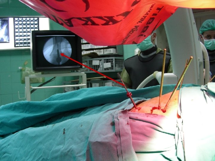
TREATMENT Non-surgical Traction Pelvic clemp Pneumatic dressing Surgical Eksternal fixation Open reduction and internal fixation of SP and SI joints Percutaneous screws for SI joint

TREATMENT

ACETABULUM FRACTURES Letournel classification

Radiologic anatomy Anteroposterior Iliac oblique Obturator oblique

ACETABULUM FRACTURES Treatment with rest or traction in simple fractures Total hip replacement for impaction fractures at the femoral head Open reduction and internal fixation in active young patients
, femoral, superior gluteal Heterotopic ossificaiton Infection %4.")
ACETABULUM FRACTURES COMPLICATIONS Nerve injury; sciatic (%30), femoral, superior gluteal Heterotopic ossificaiton Infection %4. 2 Chondrolysis

Thank you …
- Slides: 40