Pelvic Organ Prolapse POP Dr Ahlam AlKharabsheh Assistant
 Dr. Ahlam Al-Kharabsheh Assistant Professor, OBS & GYN department Mu’tah")
Pelvic Organ Prolapse (POP) Dr. Ahlam Al-Kharabsheh Assistant Professor, OBS & GYN department Mu’tah University

Pelvic Organ Prolapse A prolapse is a protrusion of an organ or structure beyond its normal anatomical site. Pelvic Organ Prolapse (POP) is usually classified according to its location and the organ contained within it.

Prevalence It affects 12 – 30 % of multiparous and 2% of nulliparous women. It is extremely common problem. About 11 % of women will have one kind of the operation for prolapse during their life. * 1 - MULTIPARITY 2 - AGE : AFTER 40 Y
: Urethra+")
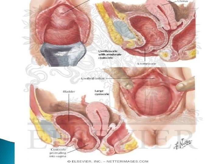
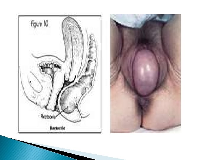
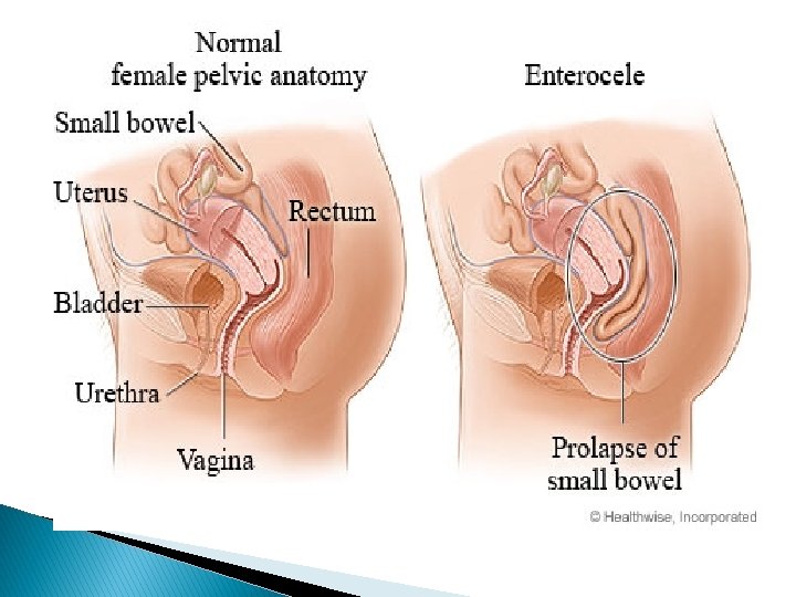
Classification I- Vaginal wall prolapse: A- Anterior vaginal wall prolapse (Antrior compartment prolpase): Urethra+ bladder Ø urethrocele. Ø Cystocele. Ø cystourethrocele. B- Posterior vaginal wall prolapse (Postrior compartment prolapse): rectum Ø Rectocele. Ø Enterocele. Herniation of small bowl C- Apical vaginal wall prolapse: (Apical compartment prolapse) Ø Vault prolapse (after hysterectomy). Uterus _+ cervix

support mechanisms of pelvic organs : First support mechanism Is Mechanical support : Uterine axis ; normal is anteverted anteflexed retroverted >> wide angle between cervix and vagina Second support is : delanceys level of support (three levels) :
 Suspension : Uterus , cervix and upper vagina are suspended from above")
1) Suspension : Uterus , cervix and upper vagina are suspended from above by structures bind them to sacrum ; by uterosacral and cardinal ligaments If defect in these ligaments : prolapse of uterus , cervix , upper vagina In hysterectomy ; we divide these ligaments , so upper vagina is at risk for prolapse (vault prolapse) 2) Attachment : Support middle part of vagina by end pelvic fascia to pelvic wall End pelvic fascia cover levator ani muscle 3) Fusion : Support distal part of vagina by fusion anteriorly with urethra , posteriorly with perineal body and laterally with levator ani muscle
")
- - Muscular : Pelvic diaphragm (levator ani muscle; puborectalis , pubococcygus , iliococcygus) Ligaments and fibrous structures : endopelvic fascia , uterosacral ligament , cardinal ligament , board & round ligaments give minimal support to uterus

Delancy’s level of suppport Antiverted c-v angle Antiflexed c-u angle Cervical vaginal cervical uterus

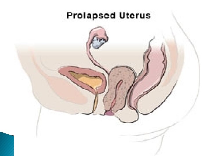
Classification II- Uterine prolapse: Of 3 grades: Grade 1: descent within the vagina. Grade 2: Descent of the cervix outside the introitus but not the body of the uterus. Grade 3: Descent of the whole uterus outside the introitus (Procidentia). Prolapse of all compartment ( anterior , posterior , apical ) III- Combined type.
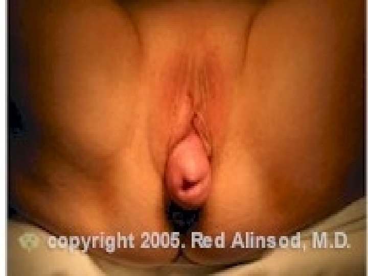

procedentia

Old women : Decrease pubic hair Atrophy of skin Loss of ruge in the vagina Need speculum examination because the cervix is not clear
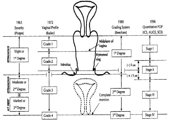
 Grade 0 Grade 1 Grade 2 Grade")
The Baden and Walker Grading System (1968) Grade 0 Grade 1 Grade 2 Grade 3 Grade 4 no prolapse halfway to the hymen prolapse to the introitus prolapse halfway beyond the hymen complete prolapse
 Quantitative Pelvic Organ Prolapse (POP-Q) The topography of vagina is described")
ICS CLASSIFICATION (1996) Quantitative Pelvic Organ Prolapse (POP-Q) The topography of vagina is described using six points (2 on anterior vaginal wall, 2 on the superior vagina, 2 on the posterior vaginal wall). In addition to other 3 points.
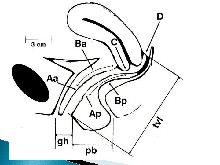

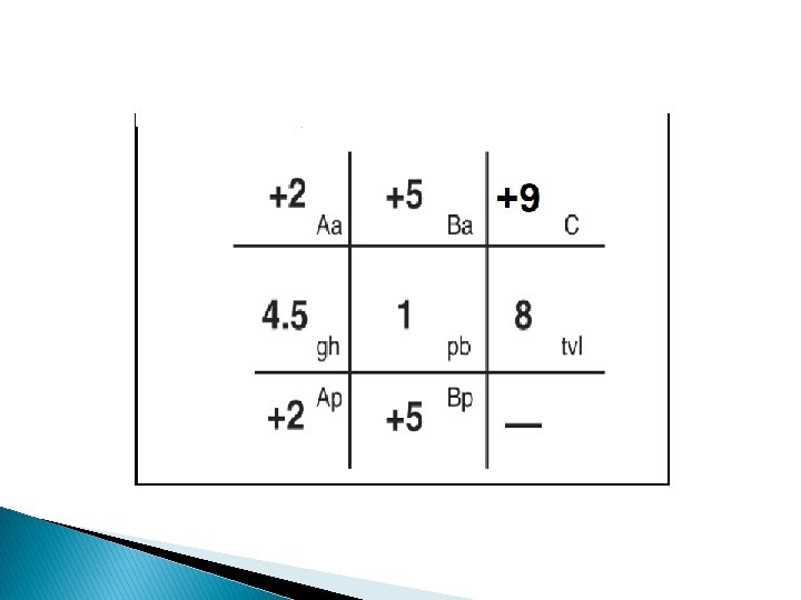
Divide the vagina to 6 points ; 2 points on Anterior Vaginal W all : Aa : 3 cm above hymen Ba : 3 cm above Aa (6 cm above hymen ). 2 points on Posterior Vaginal Wall Ap : 3 cm above hymen Bp : 6 cm above hymen C : cervix or cuff if no cervix (in hysterectomy) D : posterior fornix (douglus pouch) >> disappear in pt with hysterectomy Gh : between midpoint of E. urethral meatus & posterior part of introitus pb : between posterior forshitt & midpoint of External anal sphincter TVL : at rest & reduced
 Quantitative Pelvic Organ Prolapse (POP-Q), Simplified Stage 0: no prolapse Stage")
ICS CLASSIFICATION (1996) Quantitative Pelvic Organ Prolapse (POP-Q), Simplified Stage 0: no prolapse Stage I: the most distal portion of the prolapse is > 1 cm above the level of the hymen Stage II: the most distal portion of the prolapse is 1 cm proximal to or distal to the hymen Stage III: the most distal portion of the prolapse is > 1 cm below the plane of the hymen. Stage IV: complete eversion of the total length of the vagina.
: Prolapse may occur in nulliparous. In")
Etiology of Prolapse 1 - Congenital (genetic factor): Prolapse may occur in nulliparous. In case of collagen disorders , marfin syndrome , … More common in certain races than others. It is familial. 2. 5 risk 2 - Childbirth: Multiparity. ( para 1 : 4 fold , para 2: 8 fold , para 3: 10 fold ) Prolonged labor. Difficult vaginal delivery. (The single major factor for POP) Prolapse not seen in cs

Etiology of Prolapse 3 - After hysterectomy. (Incidence 1 -10% & usually accompanied vault prolapse by enterocele (70%). 4 - Raised intra-abdominal pressure: Chronic cough. Chronic constipation. (Cretinism) Pregnancy, labor and delivery. Large pelvic and abdominal tumor. Ascitis. 5 - Ageing: common in post menopausal women. 6 - Obesity (BMI >25).

Old age , procidentia Prolapse with fibroid and ulcer

Clinical features History: Lump protruding from the vagina either on straining or even at rest. Ø Lower abdominal discomfort and back pain. ( prolapse specific symptoms Ø In anterior compartment prolapse: urinary frequency, Ø urgency, voiding difficulty, urinary tract infections and stress incontinence. Ø Posterior compartment prolapse: incomplete bowel emptying, constipation. Sexual dysfunction. Ø (relater symptoms ) Ø

deep dyspareunia , coital incontinence Splinting : reduce the mass back to the vagina then start voiding Digitation : to facilitate defecation

POPDI-6: Pelvic organ prolapse distress inventory 6 PISQ-12: Pelvic organ prolapse/urinary incontinence sexual function questionnaire ** Procidentia: Bloody vaginal discharge due to ulceration and infection of the most dependent part of prolpase(Decubitus ulcer).

Abdominal examination: to exclude tumors, organomegaly and ascitis. Vaginal examination: Ø On dorsal position, the prolapse could be seen protruding through the introitus. If not, the patient should be asked to push down or cough.

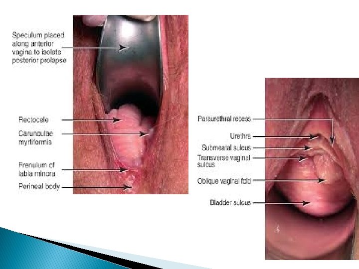
Clinical features Ø Any ulceration should be detected. Ø Bimanual exam to exclude pelvic tumors. Ø By Sim’s speculum and the patient in the left lateral position, the type of prolapse should be identified. Ø By combined rectal and vaginal digital exam, we can differentiate between rectocele and enterocele.

Differential diagnosis Congenital or inclusion vaginal cysts. Urethral diverticulum. Large uterine polyp. Pedunculated fibroid.

Investigations In case of urinary symptoms, GUE, urine culture, cystometry, and cystoscopy may be considered to exclude local causes in the bladder. In major degree of prolonged uterine prolapse, renal function should be studied to exclude renal failure due to kinking of the ureters. Imaging study: MRI.

Treatment The choice of treatment depends on: Ø The patient wish. Ø Age of patient and parity. Ø Preservation of sexual function. The treatment is conservative and/or surgical.

History of POP treatment Kahun gynecological papyrus Hippocratic Succussion

19 th century Pessary 1867

Conservative treatment Attempt should be made to correct obesity, chronic cough and constipation. Ø If decubitus ulcer is found, then local estrogen for 7 days should be used. Ø Pelvic floor muscle exercises. Ø Pessary: Support Pessary: Ring Pessary: A silicon rubber-based ring pessaries are most popular for conservative therapy. Used for all stage prolapse (best for mild P (grade 1, 2), fit with intercourse , easily removed and reinserted by pt Space- Filling Pessary: Donut, Gellhorn. Used in advance stage 3, 4 , not fit with intercourse and should be removed , need doctor to remove and reinsert
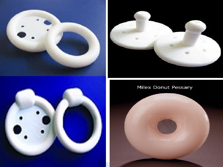

Conservative treatment They are inserted in the vagina, but should be changed at regular intervals. The use of ring pessaries my be complicated by vaginal ulceration and infection. Indications of pessaries: 1. 2. 3. 4. As a therapeutic test. Medically unfit for surgery or refused surgery. During and after pregnancy. While awaiting for surgery.

Surgical treatment: Cystourethrocele: Anterior colporrhaphy operation. Rectocele: Posterior colpo-perinorrhaphy. Enterocele: Posterior colporrhaphy with excision of the peritoneal sac. Uterine Prolapse: Vaginal hysterectomy: in elderly patients and those who completed the family or with other uterine or cervical pathology. Adequate vault support of the utero-sacral ligement or the sacrospinous ligament (SSL fixation) is needed.
 Manchester operation: amputation of the cervix, bringing of the cardinal")
Surgical treatment (uterine sparing) Manchester operation: amputation of the cervix, bringing of the cardinal ligaments and uterosacral ligaments anterior to the lower uterine segment followed by vaginal repair. Sacrohysteropexy: this is an abdominal operation. It involves attachment of a synthetic mesh from the uterocervical junction (isthmus) to the anterior longitudinal ligament of the sacrum. Trans-vaginal mesh (TVM):

Surgical treatment Vault prolapse: Sacrocolpopexy: The vaginal vault is attached to the sacrum by synthetic mesh. Sling operation: The vaginal vault is slinged to the anterior abdominal wall by two strips of anterior rectus sheath. Both operations are carried out by abdominal approach. Vaginal procedures: Sacrospinous ligament fixation (SSLF), Uteroscaral ligament suspension, ileococcygeous suspension, Vaginal mesh kits,

- Slides: 44