PELVIC NERVES Dr Abeera SarfrazDr Muhammad Zubair Nerves

PELVIC NERVES Dr Abeera Sarfraz/Dr Muhammad Zubair

Nerves derived from the lumbar and sacral plexuses and the sympathetic chain form extensive networks across the pelvic walls. These branches supply the pelvis and perineum and also the lower abdomen and the lower limb. Lumbar Plexus The lumbar plexus lies in the posterior abdominal wall. Two branches, the lumbosacral trunk and the obturator nerve, have notable relations to the pelvic walls. Lumbosacral Trunk Part of the anterior ramus of the fourth lumbar nerve emerges from the medial border of the psoas muscle and joins the anterior ramus of the fifth lumbar nerve to form the lumbosacral trunk. This trunk enters the pelvis by passing down in front of the sacroiliac joint and joins the sacral plexus.

OBTURATOR NERVE The obturator nerve is a branch of the lumbar plexus (L 2, 3, and 4). It emerges from the medial border of the psoas muscle in the abdomen and accompanies the lumbosacral trunk down into the pelvis. It crosses the front of the sacroiliac joint and runs forward on the lateral pelvic wall in the angle between the internal and external iliac vessels. On reaching the obturator canal (i. e. , the upper part of the obturator foramen, which is devoid of the obturator membrane), it splits into anterior and posterior divisions that pass through the canal to enter the adductor region of the thigh. Pelvic Branches Sensory branches supply the parietal peritoneum on the lateral wall of the pelvis.
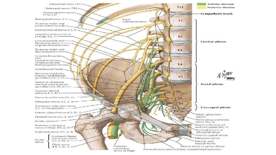

SACRAL PLEXUS The sacral plexus lies on the posterior pelvic wall in front of the piriformis muscle. It is formed from the lumbosacral trunk and the anterior rami of the first, second, third, and fourth sacral nerves. The lumbosacral trunk passes down into the pelvis and joins the sacral nerves as they emerge from the anterior sacral foramina. Relations Anteriorly: the internal iliac vessels and their branches and the rectum. Posteriorly: the piriformis muscle.
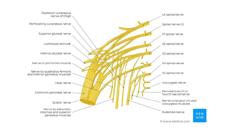

Branches to the lower limb that leave the pelvis through the greater sciatic foramen; 1. Sciatic nerve (L 4 and 5; S 1, 2, and 3) is the largest branch of the plexus and the largest nerve in the body. BRANCHES & DISTRIBUTION 2. Superior gluteal nerve supplies the gluteus medius and minimus and the tensor fasciae latae muscles. 3. Inferior gluteal nerve supplies the gluteus maximus muscle. 4. Nerve to the quadratus femoris muscle also supplies the inferior gemellus muscle. 5. Nerve to the obturator internus muscle also supplies the superior gemellus muscle. 6. Posterior cutaneous nerve of the thigh supplies the skin of the buttock and the back of the thigh.

Branches to the pelvic muscles, pelvic viscera, and perineum: 1. Pudendal nerve (S 2, 3, and 4) leaves the pelvis through the greater sciatic foramen and enters the perineum through the lesser sciatic foramen. 2. Nerve to the piriformis muscle is a small branch to only this muscle. 3. Pelvic splanchnic nerves constitute the sacral part of the parasympathetic system and arise from the second, third, and fourth sacral nerves. They are distributed mainly to the pelvic viscera. 4. Perforating cutaneous nerve supplies the skin of the lower medial part of the buttock

Sympathetic fibers derived from the sympathetic chain and parasympathetic fibers from the sacral nerves intertwine and form expansive plexuses on the pelvic walls and viscera. These autonomic fibers supply the pelvis and perineum and also the hindgut. Pelvic Part of Sympathetic Trunk AUTONOMIC NERVES The pelvic part of the sympathetic trunk is continuous above, behind the common iliac vessels, with the abdominal part. It runs down behind the rectum on the front of the sacrum, medial to the anterior sacral foramina. The sympathetic trunk has four or five segmentally arranged ganglia. Below, the two trunks converge and finally unite in front of the coccyx. Branches Gray rami communicantes to the sacral and coccygeal nerves Fibers that join the hypogastric plexuses

PELVIC SPLANCHNIC NERVES The pelvic splanchnic nerves form the parasympathetic part of the autonomic nervous system in the pelvis. The preganglionic fibers arise from the second, third, and fourth sacral nerves and synapse in ganglia in the inferior hypogastric plexus or in the walls of the viscera. Some of the parasympathetic fibers ascend through the hypogastric plexuses and thence via the aortic plexus to the inferior mesenteric plexus. The fibers are then distributed along branches of the inferior mesenteric artery to supply the large bowel from the left colic flexure to the upper half of the anal canal.

SUPERIOR HYPOGASTRIC PLEXUS The superior hypogastric plexus is situated in front of the promontory of the sacrum. It forms as a continuation of the aortic plexus and from branches of the third and fourth lumbar sympathetic ganglia. It contains sympathetic and sacral parasympathetic nerve fibers and visceral afferent nerve fibers. The superior hypogastric plexus divides inferiorly to form the right and left hypogastric nerves. Inferior Hypogastric Plexuses The inferior hypogastric plexuses lie on each side of the rectum, the base of the bladder, and the vagina. Each plexus is formed from a hypogastric nerve (from the superior hypogastric plexus) and from the pelvic splanchnic nerves. It contains postganglionic sympathetic fibers, preganglionic and postganglionic parasympathetic fibers, and visceral afferent fibers. Branches pass to the pelvic viscera via small subsidiary plexuses

Nerve Pressure from Fetal Head CLINICAL NOTES During the later stages of pregnancy, when the fetal head has descended into the pelvis, the mother often complains of discomfort or aching pain extending down one of the lower limbs. The discomfort, caused by pressure from the fetal head, is often relieved by changing position, such as lying on the side in bed. Nerve Invasion by Malignant Tumors Malignant tumors extending from neighboring viscera can invade the sacral plexus or its branches. A carcinoma of the rectum, for example, can cause severe intractable pain down the lower limbs

Referred Pain from Obturator Nerve The obturator nerve lies on the lateral wall of the pelvis and supplies the parietal peritoneum. An inflamed appendix hanging down into the pelvic cavity could cause irritation of the obturator nerve endings, leading to referred pain down the inner side of the right thigh. Inflammation of the ovaries can produce similar symptoms. Caudal Anesthesia (Analgesia) Anesthetic solutions can be injected into the sacral canal through the sacral hiatus. The solutions then act on the spinal roots of the second, third, fourth and fifth sacral and coccygeal segments of the cord as they emerge from the dura mater. The roots of higher spinal segments can also be blocked by this method. The needle must be confined to the lower part of the sacral canal, because the meninges extend down as far as the lower border of the second sacral vertebra. Caudal anesthesia is used in obstetrics to block pain fibers from the cervix of the uterus and to anesthetize the perineum.

The pelvic fascia is formed of connective tissue and is continuous above with the fascia lining the abdominal walls. Below, the fascia is continuous with the fascia of the perineum. The pelvic fascia can be divided into parietal and visceral layers. PELVIC FASCIA Parietal Pelvic Fascia The parietal pelvic fascia lines the walls of the pelvis and is named according to the muscle it overlies. Where the pelvic diaphragm is deficient anteriorly, the parietal pelvic fascia becomes continuous through the opening with the fascia covering the inferior surface of the pelvic diaphragm. In the perineum, it covers the perineal membrane and forms the fascia deep to that structure.

Visceral Pelvic Fascia The visceral layer of pelvic fascia covers and supports all the pelvic viscera. In certain locations, the fascia thickens and extends from the viscus to the pelvic walls and provides added support. These fascial condensations are termed ligaments and are named according to their attachments (e. g. , the pubovesical and the sacrocervical ligaments). Cervical Fascial Ligaments In the female, the fascial ligaments attached to the uterine cervix are of particular clinical importance because they assist with the support of the uterus and thus prevent uterine prolapse. The visceral pelvic fascia around the uterine cervix and vagina is commonly referred to as the parametrium.
- Slides: 15