Pelvic mass in Prepubertal Girls Dr Zohreh Yousefi


Pelvic mass in Prepubertal Girls Dr. Zohreh Yousefi Professor OF Mashhad University of Medical Sciences Gynecologist Oncologist

Origin of Pelvic mass non gynecologic urinary tract Gasterointestinal Otheres

Wilms' tumor or nephroblastoma cancer of the kidneys that typically occurs in children Most nephroblastomas are unilateral Typical symptoms are: -large abdomen -abdominal pain -Fever -nausea and vomiting -blood in the urine (20% high blood pressure in some cases)

rupture of Wilms' tumor and hemorrhage risk of peritoneal dissemination of the tumor metastasis it is usually to the lung It is highly responsive to treatment 90% of patients surviving at least five years

Neuroblastoma the second most common solid tumour in childhood 8% of the total number of children's cancers Neuroblastoma is a cancer of specialised nerve cells called neural crest cells In some children, the neuroblastoma can occur in nerve tissue along side the spinal cord in the neck , chest, abdomen or pelvis

Signs and symptoms If the tumor is in the abdomen, may be swollen and they may complain of constipation or have difficulty passing urine blood pressure may also be high diagnose neuroblastoma blood, urine, or bone marrow tests x-rays; CT or MRI scans; and MIBG

The treatment of neuroblastoma depends on the age of the child the size position of the tumour biology (including the MYCN status) and whether the neuroblastoma has spread Treatment Surgery Chemotherapy

Appendicitis

Appendicitis is most common in teens and young adults in their early 20 s children younger than 4 years are at the highest risk for a rupture. Up to 80 percent of appendicitis cases in this age group occurred with rupture young children have fewer of the classic symptoms of nausea, vomiting and pain localized in the lower right portion of the abdomen than do teenagers and young adults

ultrasound and CT scan images can be helpful, but are not always conclusive, even if they are available on an emergency basis CT scans in particular expose young children to radiation which should be avoided if possible The only absolute way to diagnose the condition is surgery

Gynecologic causes of Ovary Uterian Cervicovaginal pelvic mass

Müllerian duct abnormality congenital entities that result from nondevelopment, defective vertical or lateral fusion, or resorption failure of the müllerian the majority are asymptomatic when a müllerian duct becomes obstructed may present with an abdominal mass and dysmenorrhea If the patient is not treated in a timely fashion the consequences can be severe, extending even to infertility

ultrasonography initially to delineate any abnormalities in the genital tract US cannot help identify the type of MDA In contrast, MRI is a valuable technique for noninvasive evaluation of the female pelvic anatomy and accurate MDA classification If obstruction is present surgical correction of the MDA may be required
 Transverse US scan (C) with a fluid-debris level (arrow) shows a")
Neonatal hematometrocolpos. (a) Transverse US scan (C) with a fluid-debris level (arrow) shows a huge cystic mass

Duplex uterus with an obstructed hemivagina in a 12 -year-old girl Transverse US scan shows a normal left uterus (arrow) and a dilated right uterus (U).
 and a visible endometrium")
Neonatal uterus Longitudinal US scan shows a prominent cervix (arrows) and a visible endometrium (arrowheads). Some fluid (F) is seen within the vagina

• Wolffian duct Remnant ØThey are mimic tumor of ovary ØThey are small but may enlarge and infarct • Mullerian Dact Remnant ØThese are incidental finding at laparotomy and Ø cause no difficult or symtom

The gynecologic causes of a pelvic mass Neoplastic mass In younger girls Cervico-vaginal rare


The gynecologic causes of a pelvic mass Neoplastic mass In girls younger than 9 years of age, approximately 80% ovarian tumors were found to be malignant Fewer than 2% of ovarian malignancies occur in children and adolescents Non-epithelial tumors predominate

Germ cell tumors ½ -2/3 of ovarian neoplasms in younger than 20 years of age Develop from primordial germ cells Termed as malignant, though do not have high malignant potential

Germ cell tumors Germinoma Teratoma

Typically grow rapidly- 60% seen in first 2 decades of life producing symptoms of distention and abdominal fullness Torsion may occur, producing an acute abdomen obvious malignancy with involvement of opposite ovary ( Dysgerminoma) • all have a tendency to spread to the paraaortic lymph node • isosexual precocity reported

ultrasonographic appearance is very different with heterogeneous and solid components Karyotype Unilateral oophorectomy and lymphadenectomy followed by adjuvant chemotherapy Follow up necessary

Juvenile Granulosa Cell Tumors derive from granulosa cells tend to be of low malignity most of them have mixed components both solid and liquid Present as pelvic mass

Sex cord stromal tumours
")
Secrete estrogen and sometimes prolactin precocious pseudopuberty , galactorrhea measurement of anti-Müllerian hormone (AMH) and inhibin and carletenin

Treatment -unilateral oophorectomy -cystectomy and surgical staging careful examination of the contralateral ovary Postoperative follow-up consists of ultrasonography and tumor marker for -Good prognosis several years

NON NEOPLASTIC MASS Ovarian Cysts more common in the neonatal and adolescent periods ( 3 and 8 years ) decreases of frequency functional cysts in early childhood then increases as puberty

The Various Etiologies of Ovarian Cysts in Prepubertal Girls • Derived degenerated follicular cysts • ovarian gonadotropin stimulation • failure of follicular apoptosis • interaction with other hormonal secretion

Small cysts are more frequent than large cysts Small cysts asymptomatic discovered incidentally on ultrasonography elongated ovarian ligament of the abdominal location of tumor a predisposition to torsion
 acute or sub acute non-specific signs (nausea, vomiting,")
symptomatic cases: ( Abdomino -Pelvic pain) acute or sub acute non-specific signs (nausea, vomiting, urinary disorders) Precocious development may be transient onset breast development increasing formation during ovarian cyst

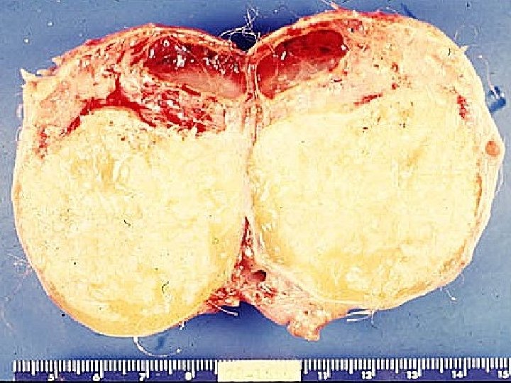
Benign Teratoma Dermoid cysts most frequent neoplasms in childhood mean age of diagnosis being 10 years heterogeneous appearance in ultrasonography a solid cystic component containing ectodermal tissue (skin, hair, dentin) calcifications are also clearly visible on plain radiographs

Treatment Preferable method in dermoid cyst in a young woman shell it out from the ovarian stroma and preserving functioning tissue

Autonomous Cyst Mc. Cune-Albright Syndrome is a sporadic disorder in small girls between 2 and 5 years ( early childhood) recurrent ovarian cysts diffuse anomalies

skin pigmentation skin spots café-au-lait polyostotic fibrous dysplasia Mc. Cune-Albright syndrome characterizes the symptoms bone and cutaneous signs detected several years later confirm the diagnosis by molecular studies ( mutations of Gs proteins )

The best known type of precocious pseudopuberty metrorrhagia and rapid breast development premature thelarche Central precocious puberty (elevated estradiol) Very low LH and FSH levels

Treatment Unilocular cysts conservatively <50 mm may be followed This is a gonado-independent form of puberty usual puberty inhibiting treatments are ineffective Only aromatase inhibitors seem to have a certain efficacy

Management of ovarian cysts syndrome with endocrine hormonal investigations are necessary ultrasound-guided percutaneous aspiration Recurrence or surgically by laparaoscopy in the individual case.


Diagnostic Arguments of Ovarian Cysts in Prepubertal Girls Limitation of pelvic capacity in prepubertal child • pelvic mass very quickly becomes abdominal location Examination abdominal palpation bimanual rectoabdominal investigate the condition of the contralateral ovary

Transrectal ultrasonography For girl who had not intercourse No alternative imaging modality has demonstrated sufficient superiority to USG to justify its routine use

Ultrasonographic signs of malignancy Adnexal pelvic mass with area of complexity -Irregular border -solid patterns within the mass -Dense multiple septae color Doppler hypervascularized tumor

Transvaginal Ultrasonography Ø Ultrasonographic signs of malignancy Adnexal pelvic mass with area of complexity Irregular border solid patterns within the mass Dense multiple septae

Tumor Markers a useful diagnostic aid in difficult to analyze by ultrasonography and in surveillance after tumor removal αFP in endodermal sinus tumor and embryonic carcinomas and immature teratomas. (β-HCG) in choriocarcinomas and dysgerminoma CA-125 levels either in peripheral blood or in the cyst fluid after aspiration LDH in dysgerminomas

Management • clinical signs • sonographic appearance • volume of the mass • finally its persistence Unilocular cysts are virtually always benign Will regress in 3 to 6 months Close observation is recommended surgical therapy for a functional ovarian mass can result adhesions and adversely affect future fertility

surgical treatment is required • complicated cyst • • hemorrhage • • ovarian torsion • • Solid masses • larger than approximately 8 cm

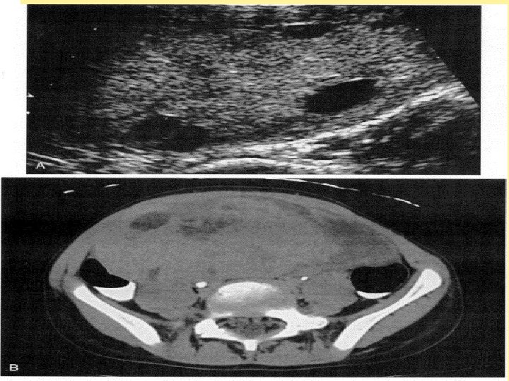
Torsion of a normal ovary in a 10 -year-old girl with severe acute pelvic pain Transverse US scan shows a markedly enlarged right ovary with peripheral follicles (arrows)
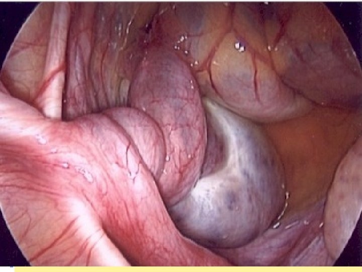
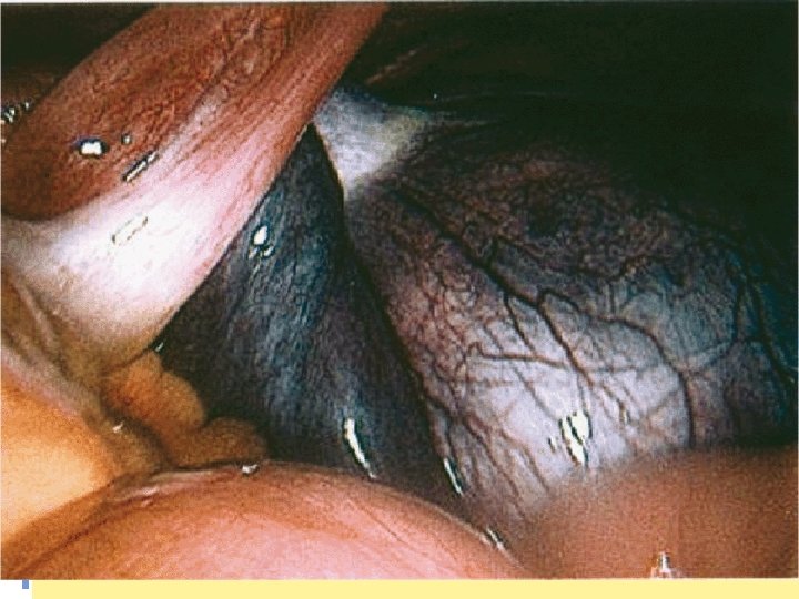

Management of adnexal torsion Ø Detorsion! Only procedure which should be performed Estimation of the degree of necrosis during surgery → inaccurate Color, size, and edema → not reflect the true damage to ovarian tissue Ischemic-hemorrhagic, black bluish appearance result of venous and lymphatic stasis rather than gangrene Any additional procedure should be avoided Ovarian cystectomy of the black-bluish ischemic should be avoided handling of the edematous friable and ischemic adnexa is risky additional damage to the ovary a high percentage of functional cysts

Management of adnexal torsion Suspicous adnexal torsion ü Emergency detorsion, only! Adnexectomy avoided Ovarian function is preserved in 88 -100% of cases Edema associated torsion Interval cystectomy Recurrence Rare Repeat torsion → ovarian fixation

Thank you for your attention !
- Slides: 56