Pelvic girdle Dr Kroly Altdorfer pelvis major pelvis


")


 SAGITTAL DIAMETER of amplitudo pelvis ANATOMICAL CONJUGATE Pelvic")


- Slides: 22
Pelvic girdle Dr. Károly Altdorfer
pelvis major pelvis minor - GREATER PELVIS - LESSER PELVIS SEPARATED BY LINEA TERMINALIS
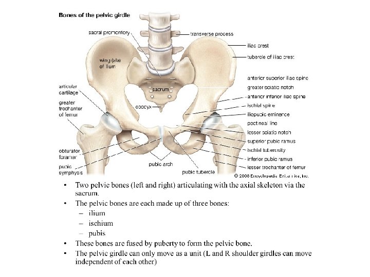
BONES
INCLINATIO PELVIS Male: 60° female: 65° Vesalius Leonardo
Connections LUMBOSACRAL synchondrosis, syndesmosis intervertebral disc anterior and posterior longitudinal ligg. flava interspinal and supraspinal ligg. lig. iliolumbale (stabilizes the joint) ZYGAPOPHYSIAL JOINT Synovial joint between the 5 th lumbar vertebra and the correspondig articular process of the sacrum SACROCOCCYGEAL SYNCHONDROSIS
SACROILIAC JOINTS Pubic symphysis
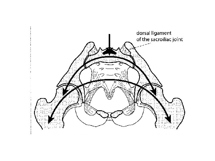
1. Sacroiliac joints: hip bones + sacrum auricular surfaces of sacrum and ilium hyalin cart. superficialy, fibrous cart. deeply! Capsule: tight, dense, limited articular cavity Ligaments: - ventral sacroiliac ligaments (weak) - dorsal sacroiliac ligaments (strong) - interosseal sacroliliac ligaments (strong) 2. Pubic symphysis - symphyseal surface of pubis and an interpubic disc - synchondrosis, fibrocartilage Not a real joint, however, it may contain a „cavity” within the disc. Ligaments: superior pubic lig. (inf. !) arcuate pubic lig.
SACROILIAC JOINTS Sacroiliac ligg. dorsal interosseal Hyalin cart. ventral Fibrous cart. Pubic symphysis
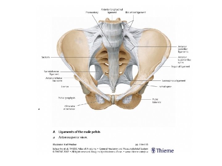
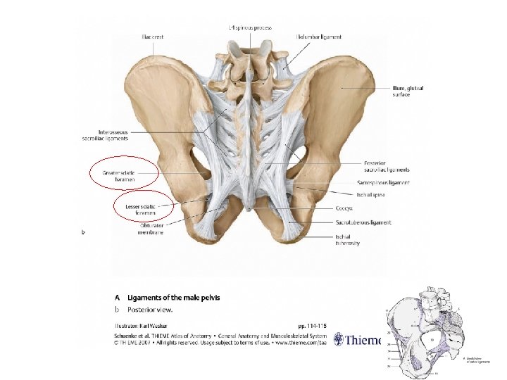
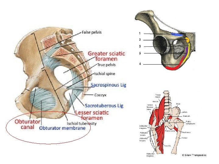
Ligaments of the pelvis - sacrospinous ligament - sacrotuberal ligament - obturator membrane - inguinal ligament (-- aponeurosis of the external oblique abdominis m. !)
Inguinal ligament, subinguinal hiatus Inner hip muscles: -psoas major -iliacus (-psoas minor)
Greater sciatic foramen. divided by piriformis m. forms suprapiriform and infrapiriform hiatuses
MALE AND FEMALE PELVIS differences INCLINATIO PELVIS Male: 60° female: 65°
DIAMETERS MALES FEMALES TRANSVERSE DIAMETER OBLIQUE DIAMETER SAGITTAL DIAMETERS
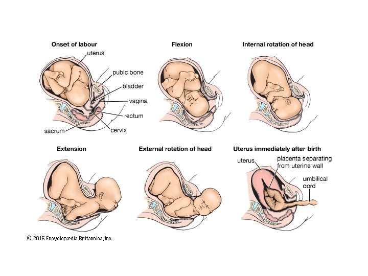
DIAMETERS FEMALE PELVIS DIAGONAL CONJUGATE (DIAMETER) SAGITTAL DIAMETER of amplitudo pelvis ANATOMICAL CONJUGATE Pelvic inlet OBSTETRIC CONJUGATE Pelvic outlet AXIS PELVIS
Folio 9 v from the Epitome of Vesalius, Basel, 1543.
Images: • Réthelyi-Szentágothai: Functional anatomy • Dr. János Hanics