
PEDIATRIC UROLOGICAL EMERGENCIES Traumatic Rapid assessment and management





- Slides: 46
PEDIATRIC UROLOGICAL EMERGENCIES Traumatic
• Rapid assessment and management of urological emergencies are essential to preservation of urological health • Children with acute abdominal pain sholud be evaluated immediately • Failure to recognize true urologic emergencies may result in renal failure, organ damage, or loss of sexual function
Classification Traumatic • Renal trauma • Ureteral injury • Bladder trauma • Urethral injury • Penile trauma • Testicular trauma Non traumatic • Congenital malformation • Hematuria • Renal colic • Urinary retention • Obstructive pyelonephritis • Acute scrotum • Paraphimosis
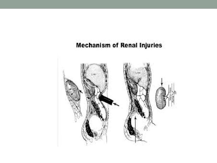
Renal trauma • Accounts for greater than 60% of the pediatric genitourinary injuries • 80 -85% blunt trauma • Penetrating trauma – associated abdominal visceral injuries • Mechanism of trauma : motor vehicle collision, fall from height, direct blow to the torso, sport injury, . . . • Pediatric kidney is more susceptive to trauma (more pliable thoracic cage, weaker abdominal musculature, less perineal fat, sits in a lower position) • Rarely occurs in isolation (often associated with liver, spleen, closed head, orhopedic injuries)
Renal injury – clinical manifestation : • Pain localized to one flank area or over the abdomen • Gross or microscopic hematuria !Renal injury can be present even without hematuria. The degree of hematuria does not correlate with degree of injury! • Ecchymosis in the flank or upper quadrants of the abdomen • Palpable mass (retroperitoneal hematoma or urinary extravasation) • Abdominal tenderness • Fractured ribs
Grading (The American Association for the Surgery of Trauma grading system)
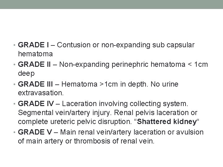
• GRADE I – Contusion or non-expanding sub capsular hematoma • GRADE II – Non-expanding perinephric hematoma < 1 cm deep • GRADE III – Hematoma >1 cm in depth. No urine extravasation. • GRADE IV – Laceration involving collecting system. Segmental vein/artery injury. Renal pelvis laceration or complete ureteric pelvic disruption. “Shattered kidney“ • GRADE V – Main renal vein/artery laceration or avulsion of main artery or thrombosis of renal vein.
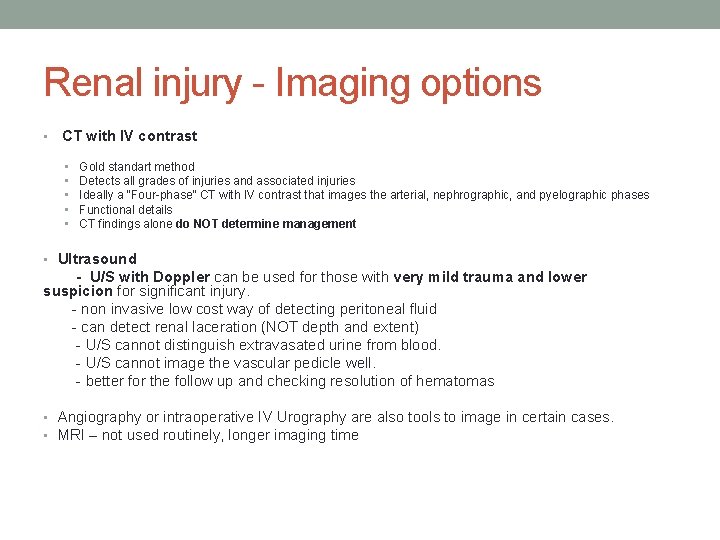
Renal injury - Imaging options • CT with IV contrast • • • Gold standart method Detects all grades of injuries and associated injuries Ideally a “Four-phase” CT with IV contrast that images the arterial, nephrographic, and pyelographic phases Functional details CT findings alone do NOT determine management • Ultrasound - U/S with Doppler can be used for those with very mild trauma and lower suspicion for significant injury. - non invasive low cost way of detecting peritoneal fluid - can detect renal laceration (NOT depth and extent) - U/S cannot distinguish extravasated urine from blood. - U/S cannot image the vascular pedicle well. - better for the follow up and checking resolution of hematomas • Angiography or intraoperative IV Urography are also tools to image in certain cases. • MRI – not used routinely, longer imaging time
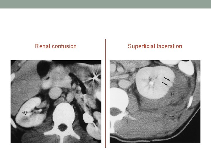
Renal contusion Superficial laceration
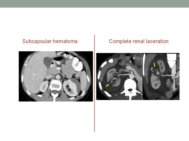
Subcapsular hematoma Complete renal laceration
Management • Overall, renal trauma in children is managed conservatively, even with higher grade injuries • These higher grade injuries require a careful, tailored approach to each individual • Surgery is recommended for hemodynamically unstable patient and with severe intraabdominal PENETRATING injuries. • Surgery or interventional radiology may be required for : massive urinary extravasation, extensive (>20%) nonviable tissue, Arterial injury, Incomplete staging
Complications • Urine extravasation - Most common complication of Stage IV injuries • Urinoma • May be acute, or present weeks-months later. • Pain, fever, ileus, and palpable mass may be signs • Perinephric abscess - Rare complication • Secondary hemorrhage • Delayed bleeding occurs in 13 -25% of Grade III-V injuries • Usually seen in first 2 -3 weeks after trauma • AV fistula formation • Rare and exclusively from stab wounds • Pseudoaneurysm • Impaired renal function • Hypertension - Still controversial
Ureteral injury • External trauma (20%) – associated with renal and bladder injuries, penetrating, blunt trauma (deceleration injury) • Surgical injury (80%) – iatrogenic, pelvic surgical procedure, endoscopic manipulation
Ureteral injury – clinical features • peritonitis, pain, fever, paralytic ileus • Hematuria – poor sensitivity • hydronephrosis
Ureteral injury - Investigation • IVP, CT Ureteral dilatation, deviation Delayed or no visualisation of renal unit Urinary/contrast extravasation
Ureteral injury - reconstruction • Reimplantation of proximal end of ureter on bladder • Primary ureterostomy • Transureterostomy • Ileal interposition flap • Permanent cutaneous flap
Bladder trauma • Causes : road traffic accidents, blow, kick or fall, stabs, gunshot injuries, diathermy, iatrogenic injuries • Classic triad : suprapubic pain, difficulty passage of urne, hematuria • Classification : intraperitoneal/extraperitoneal
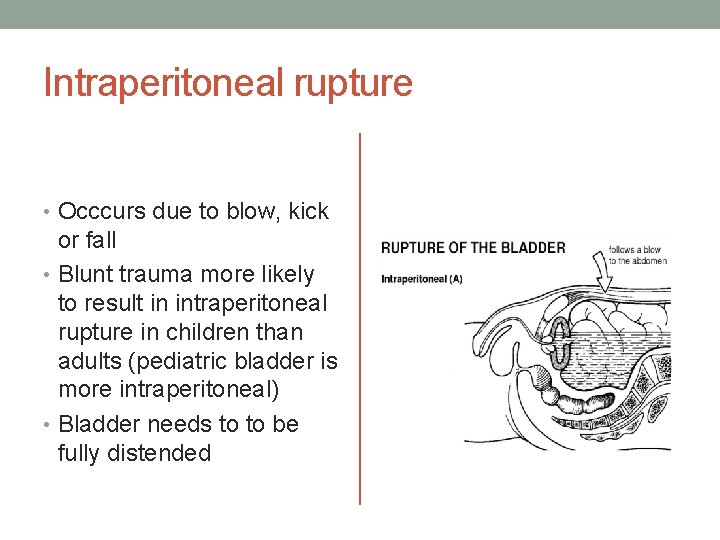
Intraperitoneal rupture • Occcurs due to blow, kick or fall • Blunt trauma more likely to result in intraperitoneal rupture in children than adults (pediatric bladder is more intraperitoneal) • Bladder needs to to be fully distended
Intraperitoneal rupture - clinical features • Sudden pain in suprapubic region • Shock and syncope • Diffuse abdominal pain • Abdominal distension • Lately : peritonitis, guarding rigidity, rebound tenderness
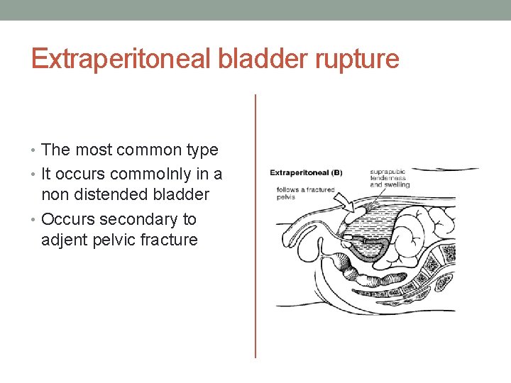
Extraperitoneal bladder rupture • The most common type • It occurs commolnly in a non distended bladder • Occurs secondary to adjent pelvic fracture
Extraperitoneal injury – clinical features • Blood and urine in the extraperitoneal space • Suprapubic tendernees and pain • Scrotal swelling • Inability to micturate • Associated with other injuries
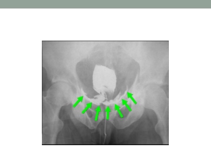
Investigation • Plain x-ray (ground glass appearance, fractured pelvis) • Peritoneal tap (conformed presence of urine) • Cystography (leak from the bladder) • U/S • CT scan
Treatment • Emergency laparotomy • Catheter drainage • Bladder is needed to be sutured in two layers • Peritoneal and estraperitoneal wash, prevesical scape and peritoneal cavity are drained • Adequate specific antibiotics (to prevent sepsis)
Complications of bladder injury • UTI (cystitis, pyelonephritis) • Peritonitis, pelvic abscess • Vesicovaginal or retrovesical fistula • Paralytic ileus • Haemorrhage
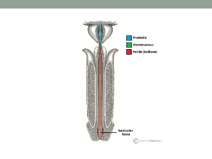
Urethral injury • Rare (short and mobile urethra, protected by the pubic bone) • Urtethral injury account for 3, 4% of the children with traumatic injuries • Mainly occur with pelvic fractures • Straddle injuries (blow to the perineum) • In males the urethra is divided into the anterior and posterior part (urogenital diaphragm) • The posterior urethra consists of the membranous and the prostatic urethra • The anterior part is formed by the bulbar and penile urethra
Classification of urethral injury • Anterior /Posterior • Depending on circumference of the urethral wall : Complete/incomplete • Depending on the thickness of the urethra : total/partial • Goldman classification – based on the results of retrograde urethrography
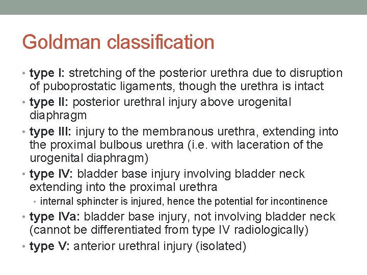
Goldman classification • type I: stretching of the posterior urethra due to disruption of puboprostatic ligaments, though the urethra is intact • type II: posterior urethral injury above urogenital diaphragm • type III: injury to the membranous urethra, extending into the proximal bulbous urethra (i. e. with laceration of the urogenital diaphragm) • type IV: bladder base injury involving bladder neck extending into the proximal urethra • internal sphincter is injured, hence the potential for incontinence • type IVa: bladder base injury, not involving bladder neck (cannot be differentiated from type IV radiologically) • type V: anterior urethral injury (isolated)
Goldman classification
Anterior urethral injuries • blunt trauma, straddle-type injuries, • predominantly in boys, mostly the bulbous urethra is compressed between the object and symphysis • Clinical features : 1. blood in external meatus 2. perineal, scrotal and labial haematoma 3. retention of urine
Therapy • Patient should be told not to try to micture (extravasation of urine!) • Gentle attempt of urethral catheterization. If able to pass, it is left in place • If catheter fails to pass, suprapubic cystostomy is done • antibiotics • reconstruction - bulbous urethra is exposed throught perineal midline incision • Perineal urethrostomy – if is not possible to suture the tear
Posterior urethral injuries • linked with pelvis fractures (road traffic accidents, falls from height), during instrumentation (catheterisation) • boys have predisposition for complete posterior urethral rupture (intraabdominal location of the bladder, more cranially placed prostate) • Clinical features : blood in external meatus failure in passing urine extravasation of urine „floating prostate“
Therapy • The extent and depth of the traumatic posterior urethral distraction defect, with or without involvement of the external sphincter complex, is frequently difficult to predict before surgical intervention • Transpubic urethroplasty • Bulboprostatic anastomosis from perineal approach
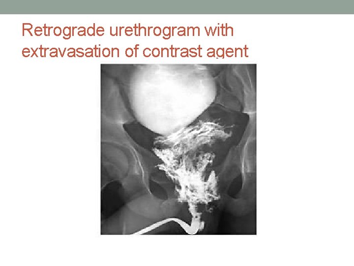
Diagnosis • Clinical assesment : presence of pelvic fracture, straddle • • • injuries, penetrating trauma close to urethra, blood on meatus, hematuria, hematoma – typical butterfly configurationof the hematoma on perineum, scrotal enlargement or labial swelling CT – complex analysis of the entire involved bone structure and soft tissue organs MRI - evaluating posterior urethral injuries and estimating the length of the urethral obliteration as well as the degree of fibrosis of the external sphincter complex Retrograde uretrography - gold standard method, able to reveal foreign bodies Anterograde cystourethrogram - in posterior urethral ijnuries USG - with insufflation of saline solution distending the lumen Cystoscopy – therapeutic transurethral splinting
Retrograde urethrogram with extravasation of contrast agent
• The management of the urethral trauma remains controversial largely because of the limited expertise of most pediatric urologists. • The aim of therapy should be to minimize remote damages such as urethrocutaneous fistula, periurethral diverticula, urethral stricture, incontinence and impotence.
Penile injuries • Penile injuries in children can be due to several causes: traffic accidents, iatrogenic trauma, animal bite, electrocution, zipper injuries and hair strangulation • trivial or more severe • Functional and psychosocial consequences • Fracture of penis – disruption of the tunica albuginea with ruptute of corpus cavernosus
Testicular injury Mechanism : direct blow, straddle injury, penetrating injury • Haematocele • Haematoma • Testicular rupture, laceration • Traumatis epididymitis • Testicular dislocation/torsion Clinical features : pain, bruising, swelling, haematuria
Hematoma • Usually focal, may be multiple • US : Difficult to identify, may appear isoechois or may have a diffusely heterogenous echotexture • Color Dopler imaging helps differentiate hematomas from tumors • Large testicular hematoma may need drainage
Hematocele
Hematocele
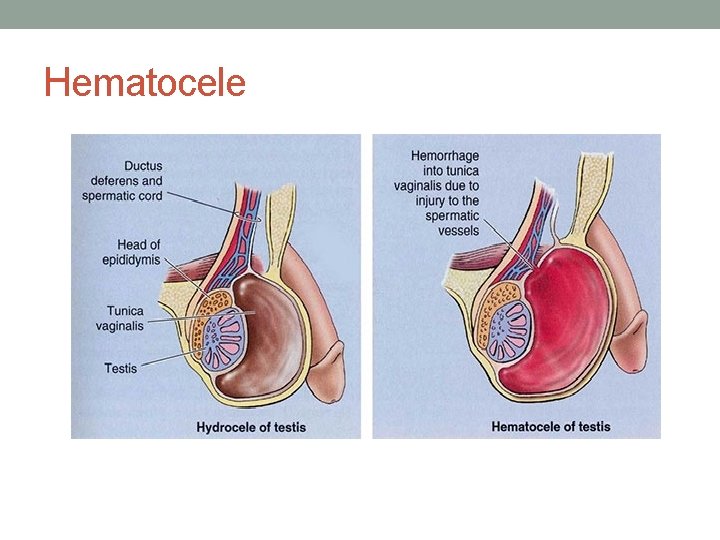
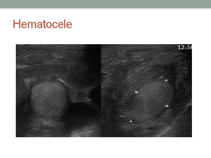
Hematocele • Blood within tunica vaginalis – collection that separate the visceral and parietal layers • Most common finding in the scrotum after blunt injury • May represent severe testicular injury • Acute onset of a large hematocele may result in reduced blood flow to the testes (extrinsic pressure on the vessels) • Investigation : USG (blood is more echogenis than fluid) • Treatment : surgical exploration, drain, repair of rupture
Testicular rupture • More than 80% of ruptured testes can be salvaged, if surgical repair is performed within 72 hours of injury
THANK YOU!