Pediatric Radiology presented by Dr ALIYA SHEMISA Pediatric

Pediatric Radiology presented by : Dr. ALIYA SHEMISA.

Pediatric chest Techniques Disease Radiological findings

Chest techniques 1. Plain chest X-ray CXR & Fluoroscopy. 2. Computed Tomography Scan CT Scan. 3. High resolution CT HRCT 4. Magnetic resonance images MRI 5. Angiography ( conventional, CT, MRI) 6. Ultrasonographic scan USS

The normal neonatal NN chest X-ray has the following features: - Thymus : may be prominent. - Heart shadow : is quite prominent & globular in outline - Normal cardiothoracic ratio is up to 65%. -Diaphragms : normally lie at the level of the 6 th rib anteriorly.

Normal Neonatal chest x-ray

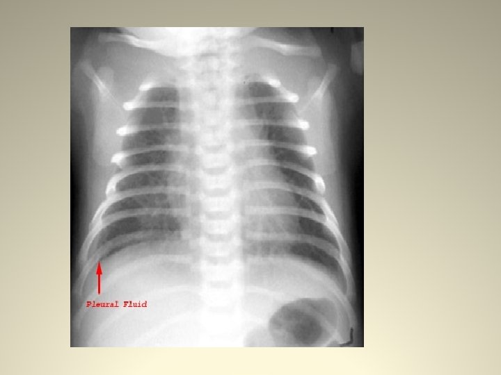
Finally, it should be remembered that acutely ill neonates may have various tubes and vascular catheter visible on CXR and it is important to be checked & to be correctly positioned as : - Endotracheal tube: tip is 5 cm above carina. - Nasogastric tube: ends few 10 cm distal to GE junction • Below Lt. Hemidiaphragm. - Umbilical artery catheter: tip in lower thoracic aorta T 6 -T 10 away from renal artery origins. - Umbilical vein catheter: tip at T 8 -T 9 lower right atrium

Complications - malposition of nasogastric tube into the trachea and bronchus can lead to pneumonia and pulmonary laceration. vascular catheters can cause perforation of the vessel and thrombosis

Acutely ill NN chest X-ray

Respiratory Neonatal Distress Disease
: surfactant")
Causes of Respiratory NN distress disease: Medical causes : 1. Respiratory Distress Syndrome(RDS): surfactant deficiency 2. Transient Tachypnea of newborn 3. Meconium aspiration syndrome 4. Neonatal pneumonia Surgical causes : 1. Congenital diaphragmatic hernia 2. Congenital Cystic Adenomatoid Malformation 3. Congenital lobar emphysema 4. Sequestration

Respiratory Distress Syndrome RDS - Also known as surfactant- deficiency disorders. - Is a relatively common condition resulting from insufficient production of surfactant lead to increase in the surface tension of alveoli causing alveolar collapse. - Usually present in the 1 st few hours of life in premature baby with symptoms like: tachypnea, expiratory grunting, nasal flaring & the baby may or may not cyanosed.

CXR findings: - bilateral symmetrical reticulo-granular densities with air bronchogram. - lung whiteout in severe cases. Treatment: - Surfactant administration - Antibiotic coverage - Chest intubation in severe cases.

Transient Tachypnea of NN TTN - is delayed clearance of intra-uterine pulmonary fluids - post C-section or prolonged vaginal delivery - wet lung with small amount of pleural effusion - has rapid recovery after 2 -3 days. - treated by Antibiotics & intubation is usually not required

Meconium aspiration syndrome - Meconium is staining of amniotic fluid - because of its thick consistency, it is aspirated in tracheo-bronchial tree causing obstruction & asymmetrical lung atelectasia, (i. e it acts as Foreign body). - usually treated by intubation & Antibiotic coverage.

Neonatal Pneumonia -caused by streptococcus bacterial infection - CXR may mimic Meconium aspiration syndrome or similar to RDS as patches of Reticulo-granular densities. - treated by Antibiotics.

Complications of respiratory distress disease 1. Acute complications : - pulmonary interstitial emphysema & O 2 toxicity: from treatment -pneumothorax : post intubation - pulmonary hemorrhage 2. Chronic complications : - bronchopulmonary dysplasia - recurrent chest infection - subglottic stenosis post intubation

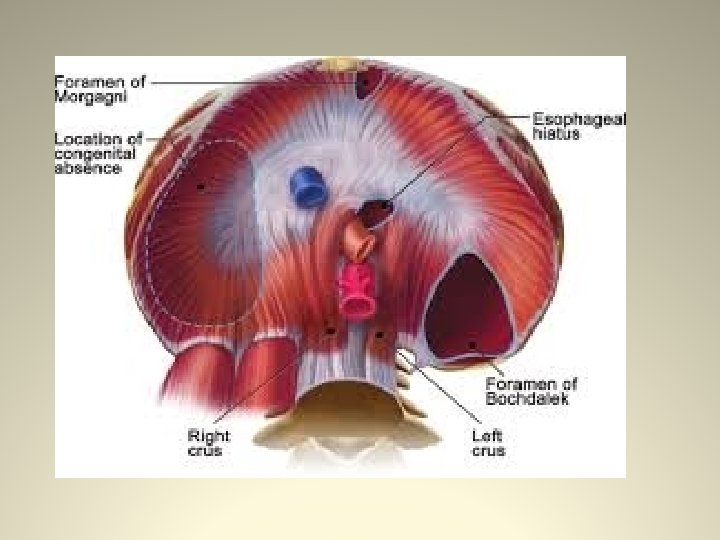
Congenital diaphragmatic hernia CDH - is a defect in the diaphragm that result in herniation of abdominal contents into the thoracic cavity & has 2 types Bochdalek hernia : more common in Lt. side (75%) Morgagni hernia : common on Rt. Anterior & central - both need surgical intervention.

Congenital cystic adenomatoid malformation CCAM - congenital large pulmonary cyst >2 cm surrounded by multiple smaller cysts. - if there is significant mass effect in the chest, CCAM is surgically removed. - CXR : cystic lesion of air density & may contain air-fluid level or may appear as solid lesion. - CT scan chest is modality of choice

Congenital lobar emphysema CLE - overexpansion of one or more lobes - commonly effect Lt. upper & Rt. Middle lobe - CXR : hyperlucent affected lobes - CT scan is modality of choice for diagnosis - treated by surgical resection

Pulmonary sequestration - is a lung tissue that is not connected to tracheobronchial tree. - has systemic blood supply from Aorta mainly - venous drainage either from IVC or pulm. Veins - has 2 types : intralobar & extralobar which commonly present in NN & can cause RDS - CT, MRI or Angiography are modality of choice for diagnosis.

The differential diagnosis of neonatal respiratory distress sign & symptoms; . -When assessing a neonatal chest X-ray (CXR) the clinical setting should always be borne in mind: *In distress premature infant with respiratory distress, hyaline membrane disease will be the most likely diagnosis. * in distressed full term neonate following cesarean; Retained lung fluid TTS will be the most common diagnosis. * in full term delivery with meconium-stained liquor; Meconium aspiration syndrome is suggested diagnosis.

- Slides: 25