Pediatric Nursing Genitourinary Disorders Lecture 15 Acute Post


- Slides: 33
Pediatric Nursing Genitourinary Disorders Lecture 15
Acute Post infections Glomerulonephritis n n Is an inflammation of the glomeruli of the kidneys. It is most often a response of a group A beta-hemolytic streptococcal infection of the skin or pharynx. It can be caused by other organisms such as Staphylococcus, Pnuemococcus. Or it can be due to immunologic abnormalities, effects of drugs or toxins, systematic diseases & viruses. It occur more often in children from 2 -12 years of age.
Pathophysiology n n Antibody-antigen complexes become logged in the glomerular. Leading to inflammation & obstruction. The glomerular membranes are thickened & capillaries in the glomeruli are obstructed by the damaged tissue cells, leading to a decreased GFR. Vascular permeability increases, allowing protein, RBCs to be excreted. Sodium & water are retained, expanding the intravascular & interstitial compartments. Low levels of proteins in serum will result in shifting of fluid from intravascular to intracellular compartment & resulting in the characteristic findings of edema.
Clinical manifestations n n n Many children are asymptomatic, in other children the onset abrupt with flank or midabdominal pain, irritability, malaise & fever. Hematurea is present in all cases, Mild preorbital edema occurs early along with dependent edema of the feet & ankles Edema may progress to pulmonary effusion (dyspnea & crackles), or ascites. Acute hypertension may cause an encephalopathy that includes headache, nausea, vomiting, irritability, lethargy, & seizures. Oligurea may or may not be present.
Diagnostic tests n n n n BUN, Cr are elevated. Serum protein is decreased due to mild or moderate proteinurea. WBC, ESR are elevated. Serum lipid levels are increased in about 40% of cases. (due to increase production of lipoprotein). An elevated antistreptolysin O (ASO) titer, reflects an the presence of antibody from a recent pharyngeal streptococcal respiratory infections. The anti-DNase B titer is used for detecting antibodies in recent skin infection. urine analyses reviles hematurea, proteinurea. Anemia is common in the acute phase due to diluted serum, as well as hemoglobin & hematocrit levels.
Clinical therapy. n n n Treatment is supportive & to relive symptoms. Bed rest is a key component of treatment plan in the acute phase. Edema & mild to moderate hypertension should be treated with sodium restriction (diet) & diuretics such as Fursemide. A course of antibiotic is given to ensure treatment of the original infectious agent. Fluid requirement are determined by monitoring of urinary output, wt, B/P & serum electrolytes. Low salt high protein diet.
Nursing interventions n 1. 2. 3. 4. n 1. 2. 3. Monitor fluid status: by monitoring V/Ss, electrolyte status, intake & output. Monitoring levels of ascites by measuring abdominal girth. Monitor urine specific gravity. Educate parents, family & visitors about fluid restriction as a therapy to prevent over intake. Prevent infections: Monitor signs of infection such as fever, increased malaise, elevated WBC. Instruct family about hygiene & hand washing. Limit visitors & screen for infection.
n 1. 2. 3. n Prevent skin breakdown: Turn child frequently or at least every 2 hours. Pad the bony prominence or susibtable area with sheepskin. Make sure the child’s bed is free from crumbs or sharp toys or wrinkles of the sheets. Meet nutritional needs: Low salt diet, high protein diet. Encourage parents to bring the child favored food from home to over come the anorexia. Allow the child to eat with other family members. Provide emotional support
Nephrotic Syndrome n n n It is a clinical state characterized by edema, massive proteinurea, hypoalbomemia, hypoproteinemia, hyperlipidimia, & altered immunity. It can be congenital: autosomal recessive disorder (defect in the gene CNF on chromosome No. 19. ) Or can be primary due to other diseases in kidneys such as glomerulonephritis. 90% of cases have one of the primary nephrotic syndrome types which is the minimal change nephrotic syndrome (MCNS). MCNS: drives its name from normal or minimally changed appearance of glomeruli on light microscopic evaluation.
Pathophysiology n n n The mechanism of increase the membrane permeability is usually due to the releasing of the permeability factors from abnormal circulating T cells. And the loss of the negative charge of the glomerular capillary wall. This will lead to , proteinurea, hypoalbuminemia, edema, loss of immunoglobulin resulting in altered immunity. loss of antithrombin III by urine along with reduced levels of factors IX, XI< & XII leading to hypercoagulability
Clinical Manifestations n n n n Edema develops gradually over several weeks. Other signs include snug fit of clothing & shoes. Pallor, hypertension, irritability, anorexia, hematurea, decreased urine output. The urine is frothy or foamy. In some cases, plural effusion occur & progress to respiratory distress. Massive edema resulting in a dramatic weight gain, abdominal pain, with or without vomiting, depending on amount of sodium ingestion. Pale, shiny skin with prominent veins, & brittle (weak) hair.
Diagnostic Tests n n n n Urine analysis. BUN, sodium. Serum albumin: hypoalbuminemia (<25 g/l). Cholesterol Serum electrolytes. Protein > 40 mg/m 2/ hour proteinurea. Microscopic hematurea is present. renal ultrasound.
Clinical Therapy n n n n Hospitalization in sever cases. Treatment focuses in decreasing proteinurea, relieving edema, improving nutrition preventing infections. A corticosteroid (Prednisone) is used to decrease proteinurea. Antihypertensive, diuretics & antibodies are given. Intravenous albumen in sever hypoalbominea. Low salt diet is recommended Relapse occur frequently, mostly in the form of respiratory infections.
Nursing Interventions n 1. 2. 3. 4. Administered medications: Monitor side effects of corticosteroid such as moon face, increased appetite, increased hair growth, abdominal distension, mood swings, hypertension, nausea, & hyperglycemia. Evaluating fasting blood sugar is needed during therapy. If child on diuretics watch for shock signs (hypotension). If child on IV albumin, watch for overload signs (crackles)
n 1. 2. 3. 4. n 1. 2. Prevent infections: Child is at risk for infection due to corticosteroid therapy & loss of immunoglobulins. Educate parents about importance of hygiene & hand washing. Monitor both V/Ss & WBCs. Decreased visitors (child’s social contact) during immunosuppression treatment. Prevent skin breakdown: Turn the child frequently. Keep skin dray & clean.
n 1. 2. 3. Meet nutritional needs: Present meal in attractive ways and in small portion, to increase child appetite. Fluid is not restricted unless sever edema. Promote rest: Educate families to provide child with quiet play such as drawing, watching videos. Monitor signs of fatigue after activity, such as irritability, mood swings. Limiting visitors in the acute phase is important, use telephone as an alternative.
Renal Failure n n 1. 2. 3. Renal failure may be acute or chronic, occurs when the kidney can not excrete wastes & concentrate urine. Acute renal failure occurs over days or weeks & may be reversible. Chronic renal failure occur over months or years, & permanent dysfunction. Both types is characterized by: Azotemia: accumulation of nitrogenous wastes in the blood. Uremia: excess of urea & other waste products in blood. Oligurea: urine out put is < 0. 5 -1 ml/kg/hr.
Acute renal failure It caused by acute glomerulonephritis, poisoning, sepsis, hypovolemia, obstructive uropathy, complications of cardiac diseases or surgery. n The causes is classified into prerenal, postrenal & actual kidney damage: 1 - Prerenal factors: result of decreased perfusion to a normal kidney in association with systematic condition, cased by hypovolemia, septic shock, cardiac failure. 2 - Primary kidney damage: (intrinsic factors), results from infections, ingestion of drugs or poisons. 3 - Postrenal factors: results from obstruction of both kidneys such as in posterior urethral valves. n
Chronic renal failure n n Is a progressive, irreversible reduction of the kidney function. It results from developmental abnormalities of the kidneys or obstructed urine flow & reflux, heredity disease, infections. The progressive dysfunction of nephrons results in end stage renal disease (ESRD). Which is characterized by minimal (<10% of normal) or no renal functioning, anemia, abnormal blood values, metabolic acidosis, fluid & electrolytes imbalance; the child needs dialysis. Elevated B/P is due to water & sodium retention.
Continue… n n Hypocalcaemia occur due to irresponsive parathyroid gland in response to low calcium levels & vitamin D. increasing the risk of fractures. Kidney become unable to produce activated vitamin D & to excrete phosphorus causing increased phosphorus levels. Growth retardation due to disturbances of calcium, phosphorus & vitamin D; decreased caloric intake; & metabolic acidosis. Decreased production of erythropoietin leading to decrease production of RBCs in bone marrow & anemia.
Clinical manifestations Type Clinical manifestations Acute Dark urine or gross hematurea, headache, edema, hypertension, lethargy, nausea, vomiting, oligurea. Mass in flank area, if cyst or tumor, or obstructive lesion is present. Fatigue, malaise, poor appetite, nausea, vomiting, failure to thrive, headache, decreased mental alertness, or ability to concentrate, secondary enuresis, chronic anemia, hypertension, edema, . Fractures with minimal trauma, rickets, bone deformities. Chronic
Diagnostic tests n 1. 2. 3. 4. n 1. 2. 3. Urine analysis: p. H: acidic urine. Specific gravity: high in prerenal, low in intrinsic & normal in postrenal. Protein: positive. hematurea Blood chemistry: potassium, phosphorus, Cr, & BUN are elevated. calcium & p. H are low. Sodium: normal, high or low: according to amount of water in body
Clinical therapy: ARF n n n Treat the underlying cause. Fluid replacement in the acute stage by isotonic solution (RL). Diuretic therapy is needed in patients with pulmonary edema as well as dialysis if poorly responded to diuretics. Fluid requirements must be calculated to meet (Zero Water Balance) intake should be same output. Low potassium diet, in case of sever hyperkalemia give calcium gluconate to exchange sodium for potassium. antibiotic is prescribed for infection.
n n n Give sodium bicarbonate to correct the metabolic acidosis by exchange hydrogen for potassium. Give calcium gluconate to correct hypocalcaemia. Give nitroglycerin to control elevated blood pressure. In CRF: diet need to be low in potassium, sodium & phosphorus. With high calcium. Dialysis is needed. Kidney transplant.
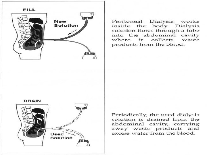
Dialysis n n n It can be peritoneal where a sterile solution containing menials & sugar is administered into the peritoneal cavity where it left for a specific period of time then irrigated carrying wastes. Fluid drained is clear not cloudy. If cloudy it is indication of peritoneal infection Patient needs fewer dietary restriction.
n n Hemodialysis is the second type, where blood is drained from artery into the dialzer to be cleared from waste and electrolytes then return into body through vein. Patients needs fluid & food restrictions
Nursing Interventions n 1. 2. n 1. Prevent complications: Ensure compliance with treatment by monitoring V/S, intake & output, serum electrolytes, level of consciousness. Maintain fluid balance: Estimate child status by monitoring of weight, intake & output, B/P three to four times a day. The aim of maintaining fluid balance is to achieve sodium balance & decrease body weight by 0. 5 -1% a day. Administer medication: Monitor adjusted doses of medication (increase interval or decrease dose).
n A. B. C. D. While performing daily peritoneal dialysis & catheter exit site care with the mother of a child with chronic renal failure, which of the following would be an important step to stress to the mother? Applying an occlusive dressing after cleaning the site. Changing the dressing when the peritoneal space is dry. Examining the site for signs of infection while cleaning the area. Pulling on the catheter to hold taut while cleaning the skin.
n A. B. C. D. The mother of a toddler with nephrotic syndrome asks the nurse what can be done about the child’s swollen eyes. Which of the following would the nurse suggest? Applying cool compresses. Elevating the head of the child’s bed. Applying eye drops every 8 hours. Limiting the child’s television watching.
n A. B. C. D. When developing the discharge teaching plan for a child with chronic renal failure & the family, the nurse would emphasize restriction of which of the following nutrients? Ascorbic acid. Calcium. Magnesium. Phosphorus.
n A. B. C. D. When developing the plan of care for school-aged child with acute poststerptoccal glomerulonephritis who has a fluid restriction of 1, 000 ml/day; which of the following fluids would the nurse consider as most appropriate for the client’s condition & effective for preventing excessive thirst? Diet cola. Ice chips. Lemonade. Tap water.
n A. B. C. D. The nurse determines that interventions for decreasing fluid retention have been effective when the child which nephrotic syndrome demonstrates evidence of which of the following? Decreased abdominal girth. Increased caloric intake. Increased respiratory rate. Decreased heart rate.