Pediatric Nephrology Review Anke Banks Anke banksahs ca

Pediatric Nephrology Review Anke Banks Anke. banks@ahs. ca Jan. 23, 2020

Outline • Approach to the abnormal urinalysis o Hematuria • Macro • Micro o Proteinuria • Hypertension • Weird stuff o Barrter’s o Gitelmans o Maybe RTAs

Approach to the Abnormal Urinalysis

Case 1 • 5 year old boy o 1 week history of swelling around his eyes o One episode of brown coloured urine o History of sore throat and ear ache about 2 weeks ago • The nursing assessment states: o o Temp 36. 8 C HR 110 RR 20 BP 95/60

Hematuria

Gross vs microscopic Gross • Can see with the naked eye • Does not necessarily reflect a large degree of blood loss o As little as 1 m. L of blood per liter of urine can induce a visible colour change
")
Gross vs Microscopic • ≥ 5 red blood cells per high powered field (hpf) in a spun urine sediment

General Approach to Gross Hematuria 1. Is it blood or just red urine? 2. Are there red blood cells in the urine or just a hemepositive urinalysis? 3. Where is it coming from (glomerular vs. nonglomerular)? 4. No really, where is it coming from?

General Approach to Gross Hematuria 1. Is it blood or just red urine? 2. Are there red blood cells in the urine or just a hemepositive urinalysis? 3. Where is it coming from (glomerular vs. nonglomerular)? 4. No really, where is it coming from?

1. Is it blood or just red urine? • Urine can be red for many reasons other than blood • How can you answer the above question? • Need to do a urinalysis to be sure blood is present

1. Is it blood or just red urine? Red urine Hemepositive urinalysis Hemenegative urinalysis

Urinalysis - 1 • Urinalysis: Appearance: clear Colour: red Specific gravity: 1. 020 (reference range: 1. 003 -1. 030) p. H: 7. 0 (reference range 5. 0 -8. 0) Leukocyte esterase: negative Nitrite: negative Protein: negative (reference range: neg-0. 15) Glucose: normal Ketones: negative Blood: negative

Urinalysis - 1 • Urinalysis: Appearance: clear Colour: red Specific gravity: 1. 020 (reference range: 1. 003 -1. 030) p. H: 7. 0 (reference range 5. 0 -8. 0) Leukocyte esterase: negative Nitrite: negative Protein: negative (reference range: neg-0. 15) Glucose: normal Ketones: negative Blood: negative

Causes of red urine Heme negative on U/A • Meds • Food dyes o Doxorubicin o Beets o Chloroquine o Blackberries o Ibuprofen o Food colouring o Iron sorbitol • Metabolites o Nitrofurantoin o Bile pigments o Rifampin o Melanin o Deferoxamine o Urates o Phenolphthalien o Porphyrin o Phenazopyridine o Methemoglobin

General Approach 1. Is it blood or just red urine? 2. Are there red blood cells in the urine or just a hemepositive urinalysis? 3. Where is it coming from (glomerular vs. nonglomerular)? 4. No really, where is it coming from?

General Approach 1. Is it blood or just red urine? 2. Are there red blood cells in the urine or just a heme -positive urinalysis? 3. Where is it coming from (glomerular vs. nonglomerular)? 4. No really, where is it coming from?

Urinalysis - 2 • Urinalysis: Appearance: clear Colour: red Specific gravity: 1. 020 (reference range: 1. 003 -1. 030) p. H: 7. 0 (reference range 5. 0 -8. 0) Leukocyte esterase: negative Nitrite: negative Protein: negative (reference range: neg-0. 15) Glucose: normal Ketones: negative Blood: 250 (++++) Ery/u. L • Urine Microscopy: RBCs: 0 -2 (reference range: 0 -3 RBCs/hpf) WBC: 0 -2 (reference range: 0 -8 WBCs/hpf)

Urinalysis - 2 • Urinalysis: Appearance: clear Colour: red Specific gravity: 1. 020 (reference range: 1. 003 -1. 030) p. H: 7. 0 (reference range 5. 0 -8. 0) Leukocyte esterase: negative Nitrite: negative Protein: negative (reference range: neg-0. 15) Glucose: normal Ketones: negative Blood: 250 (++++) Ery/u. L • Urine Microscopy: RBCs: 0 -2 (reference range: 0 -5 RBCs/hpf) WBC: 0 -2 (reference range: 0 -8 WBCs/hpf)

Question What can cause a urinalysis to be “positive” for blood when no red blood cells are seen on microscopy? A. B. C. D. E. Free hemoglobin Myoglobin High urine calcium A and B only All of the above

2. Are there RBC’s in the urine? • How does the urine dipstick work? • Hydrogen peroxide. o This catalyzes a reaction between hemoglobin (or myoglobin) and a dye (chromogen tetramethylbenzidine)

2. Are there RBC’s in the urine? Red urine Heme-positive urinalysis Red blood cells present Hemenegative urinalysis Red blood cells absent Free hemoglobin Myoglobin Hemolysis Rhabdomyolysis • éLDH • ébilirubin • écreatine kinase • é myoglobin
 o Cells")
2. Are there RBC’s in the urine? • Urine centrifugation (sometimes reported) o Cells are spun down • Sediment red or brown o Blood! • Supernatant red or brown o Not blood!

General Approach 1. Is it blood or just red urine? 2. Are there red blood cells in the urine or just a hemepositive urinalysis? 3. Where is it coming from (glomerular vs. nonglomerular)? 4. No really, where is it coming from?

Case 1 revisited • 5 year old boy o 1 week history of swelling around his eyes o One episode of brown coloured urine o History of sore throat and ear ache about 2 weeks ago • The nursing assessment states: o Temp 36. 8 C, HR 110, RR 20, BP 95/60 • U/A

• • Case 1 revisited Urinalysis: Appearance: clear Colour: red Specific gravity: 1. 020 (reference range: 1. 003 -1. 030) p. H: 7. 0 (reference range 5. 0 -8. 0) Leukocyte esterase: negative Nitrite: negative Protein: 2 g/L (reference range: neg-0. 15) Glucose: normal Ketones: negative Blood: 250 (++++) Ery/u. L Urine Microscopy: RBCs: >100(reference range: 0 -3 RBCs/hpf) WBC: 20 -50 (reference range: 0 -8 WBCs/hpf) RBC casts present Hyaline casts: 5 -10 (reference range: 0 -8 WBCs/hpf)

• • Case 1 revisited Urinalysis: Appearance: clear Colour: red Specific gravity: 1. 020 (reference range: 1. 003 -1. 030) p. H: 7. 0 (reference range 5. 0 -8. 0) Leukocyte esterase: negative Nitrite: negative Protein: 2 g/L (reference range: neg-0. 15) Glucose: normal Ketones: negative Blood: 250 (++++) Ery/u. L Urine Microscopy: RBCs: >100 (reference range: 0 -3 RBCs/hpf) WBC: 20 -50 (reference range: 0 -8 WBCs/hpf) RBC casts present Hyaline casts: 5 -10 (reference range: 0 -8 WBCs/hpf)

3. Where is it coming from? • Divide into glomerular and non-glomerular • What are some of the distinguishing features of these two types of hematuria?

3. Where is it coming from? Glomerular Urine appearance Urinalysis Microscopy Non-glomerular

3. Where is it coming from? Glomerular Urine appearance • Urinalysis • Microscopy • • • Non-glomerular Tea or cola coloured Clots not present Protein >2+ • Dysmorphic RBCs RBC casts • Bright red or pink blood May see clots • Protein <2+ • Normal RBC morphology No casts •

Non-glomerular blood https: //www. everydayhealth. com/urine/what-causes-blood-urine-hematuria/

Non-glomerular blood

Glomerular bleeding

Glomerular bleeding

Glomerular bleeding

Glomerular bleeding

• • • Case 1 revisited Urinalysis: Appearance: clear Colour: red Specific gravity: 1. 020 (reference range: 1. 003 -1. 030) p. H: 7. 0 (reference range 5. 0 -8. 0) Leukocyte esterase: negative Nitrite: negative Protein: 2 g/L (reference range: neg-0. 15) Glucose: normal Ketones: negative Blood: 250 (++++) Ery/u. L Urine Microscopy: RBCs: >100 (reference range: 0 -3 RBCs/hpf) WBC: 20 -50 (reference range: 0 -8 WBCs/hpf) RBC casts present Hyaline casts: 5 -10 (reference range: 0 -8 WBCs/hpf) IMPRESSION: likely glomerular source

General Approach 1. Is it blood or just red urine? 2. Are there red blood cells in the urine or just a hemepositive urinalysis? 3. Where is it coming from (glomerular vs. nonglomerular)? 4. No really, where is it coming from?

Etiology of gross hematuria • Most common causes of gross hematuria in children • Depends on the setting • How about in those presenting to ER? o UTI o Irritation of the meatus or perineum o Trauma Ingelfinger JR, et al, Pediatrics. 1977; 59(4): 557 Patel HP, Bissler JJ, Pediatr Clin North Am. 2001; 48: 1519

Etiology • Less common causes of gross hematuria o Acute nephritis • Post-infectious glomerulonephritis • Ig. A nephropathy o Nephrolithiasis o Coagulopathy o Sickle cell disease o Drug-induced hemorrhagic cystitis o Malignancy (Wilms or rarely transitional cell carcinoma) o Hypercalciuria o Post-micturition urethral bleeding o No cause Ingelfinger JR, et al, Pediatrics. 1977; 59(4): 557 Patel HP, Bissler JJ, Pediatr Clin North Am. 2001; 48: 1519

Differential for hematuria Extra. Glomerular Isolated renal disease • PIGN – Postinfectious • Ig. A nephropathy • Alport Syndrome • Thin BM disease • MPGN/FSGS • Anti-GBM disease Upper urinary tract Systemic disease • HSP nephritis • Hemolytic uremic syndrome • SLE nephritis • ANCA vasculitis Tubulointerstitial Vascular Hemoglobinopathy Lower urinary tract Crystalluria Anatomic • Inflammation • Infection • Urolithiasis • Trauma • Coagulopathy • Heavy exercise • Munchausen/ • Munch by proxy

Differential for hematuria Extra. Glomerular Isolated renal disease • PIGN – Postinfectious • Ig. A nephropathy • Alport Syndrome • Thin BM disease • MPGN/FSGS • Anti-GBM disease Upper urinary tract Systemic disease • HSP nephritis • Hemolytic uremic syndrome • SLE nephritis • ANCA vasculitis Tubulointerstitial Vascular Hemoglobinopathy Lower urinary tract Crystalluria Anatomic • Inflammation • Infection • Urolithiasis • Trauma • Coagulopathy • Heavy exercise • Munchausen/ • Munch by proxy

Differential for hematuria Upper urinary tract Tubulointerstitial • Pyelonephritis • Acute interstitial nephritis • Acute tubular necrosis Hemoglobinopathy Vascular • Arterial/venous • Sickle cell disease/trait thrombosis • Malformations • Aneurysm • Nutcracker syndrome Anatomic • Cystic kidney disease • Tumor • Trauma Crystalluria • Calcium • Oxalate • Uric acid

Bolenz, C; Schröppel, B; Eisenhardt, A; Schmitz-Dräger, B J; Grimm, M The Investigation of Hematuria Dtsch Arztebl Int 2018; 115(48): 801 -7; DOI: 10. 3238/arztebl. 2018. 0801

Gross hematuria - history Usually there are clues! • Recent heavy exercise or trauma • Incontinence, dysuria, frequency, or urgency • Loin to groin (or sometimes just abdo) pain • Flank pain without radiation but with fever, dysuria, and frequency • Timing of hematuria o o o • • Colour of urine History of illness o • Recent vs concurrent Past medical history of pre-disposing conditions o • Initial Continuous Terminal Sickle cell, coagulopathies Medication history o o Cyclophosphamide NSAIDs eosinophilic cystitis (nonsteroidal anti-inflammatory drug or antihistamines) [7 -9], or interstitial nephritis Ingelfinger JR, et al, Pediatrics. 1977; 59(4): 557 Up. To. Date, Accessed 2020

Gross hematuria - PE More clues! • BP – high vs low? • Edema • Rash • Purpura • Genitalia erosions • Abdo mass Ingelfinger JR, et al, Pediatrics. 1977; 59(4): 557 Up. To. Date, Accessed 2020

Next steps

Copyrights apply

Case 1 revisited • 5 year old boy o 1 week history of swelling around his eyes o One episode of brown coloured urine o History of sore throat and ear ache about 2 weeks ago • The nursing assessment states: o Temp 36. 8 C, HR 110, RR 20, BP 95/60 • U/A – likely glomerular source

Glomerular Isolated Renal Disease • • • PIGN – Post-infectious Ig. A nephropathy Alport Syndrome Thin BM disease MPGN/FSGS Anti-GBM disease Systemic disease • • Henoch Schonlein purpura nephritis Hemolytic uremic syndrome Systemic lupus erythematous nephritis ANCA vasculitis • Granulomatosis with polyangiitis (GPA) • Microscopic polyangiitis

Case 1 revisited • What do you want to know on history? • What are you looking for on physical exam?

Key history for glomerular hematuria • HPI: o Recent illness o Concurrent illness • ROS o HEENT: hearing loss, o Resp: Cough, hemoptysis o GI: abdo pain, diarrhea (? bloody? ) o MSK: arthralgia o Derm: rash, purpura
 o Recurrent “UTIs”")
Key history for glomerular hematuria • PMHx: o Hematuria (previous episodes) o Recurrent “UTIs” that are culture negative • Family Hx o Hematuria o Hearing loss o Prominent history of renal failure in males

• • Key PE for glomerular hematuria Measure BP Edema (or recent weight gain) HEENT: oral ulcers, retinal changes Resp: wheeze, poor air entry Abdo – abdo discomfort MSK: edema, heat, pain Derm – purpura, rash

Case 1 revisited • 5 year old boy o 1 week history of swelling around his eyes o 1 episode of brown coloured urine o History of sore throat and ear ache about 2 weeks ago • The nursing assessment states: o Temp 36. 8 C, HR 110, RR 20, BP 95/60 • U/A – appears glomerular • Additional hx: o No other episodes of hematuria, no family members with hematuria, ROS negative. • PE: • Mild peri-orbital edema otherwise normal exam

Glomerulonephritis • Inflammation of the nephrons • Key features of GNs? • PHAROH o Proteinuria – usually non-nephrotic o Hematuria o Azotemia o RBC casts o Oliguria o Hypertension

Nephritic clinical presentation in children • Acute GN o Post-infectious GN o HSP nephritis o Chronic GNs: Ig. A, MPGN, lupus • Rapidly progressive GN o Primary GN (isolated renal disease): Ig. A, MPGN o Secondary GN: ANCA-associated, lupus, PIGN, HSP • Recurrent macroscopic hematuria o Ig. A, Alport • Chronic glomerulonephritis o Primary GN: MPGN, Ig. A, anti-GBM disease o Secondary GN: lupus nephritis, granulomatosis with polyangiitis Uptodate, GN in children, accessed Sept. 2016)

Investigations for glomerular hematuria 1. For everyone o U/A, Urine microscopy • Glomerular vs non glomerular

Investigations for glomerular hematuria 2. Symptomatic gross hematuria o S/Sx of glomerular disease • Standard labs: CBC, lytes, Cr, urea, albumin • Special tests: C 3, C 4, ASOT, ANA • Based on hx: ANCAs, Hep B/C screen, anti-GBM • Immunoglobulins? ? ? • Imaging: ultrasound

Investigations for glomerular hematuria 3. Asymptomatic gross hematuria o o Urine: U/A, Urine Ca/Cr, U Cx Standard labs: creatinine, C 3 Imaging: US (r/o calculus) Family testing – for hematuria (possible TBM or Alports disease)

Case 1 revisited • 5 year old boy o 1 week history of swelling around his eyes o One episode of brown coloured urine o History of sore throat and ear ache about 2 weeks ago • The nursing assessment states: o Temp 36. 8 C, HR 110, RR 20, BP 95/60 • U/A – likely glomerular source • Labs o o Creatinine and urea - slightly increased Lytes normal C 3 is low, C 4 is normal Throat swab grows Group A strep

Post-infectious glomerulonephritis • AKA post-streptococcal GN • Epidemiology o The most common cause of acute GN in children o Age: usually > 3 years (most commonly 5 -12 y) o Sporadic or epidemic of group A streptococcal infection Eison et al. , Ped Neph (2011) 26: 165 Uptodate, PSGN, accessed Sept. 2016)

Post-infectious glomerulonephritis • Pathophysiology o Certain group A strep strains are nephritogenic o Infectious agents (staph or strep)produce nephritogenic antigens that may be deposited in the glomerulus o The humoral response activates the alternative complement pathway o Complement activation leads to • Generation of chemotactic and chemokinetic peptides (C 3 a, C 5 a, C 3 b) that attract WBC to the site of injury • Formation of membrane attack complex (C 5 b-C 9) which may cause direct injury to glomerular cells Eison et al. , Ped Neph (2011) 26: 165 Uptodate, PSGN, accessed Sept. 2016)

Post-infectious glomerulonephritis • Clinical presentation o 3 phases of disease 1. Latent o o o From time of strep infection to acute phase Throat infection: 1 -3 weeks Skin infection: 3 -6 weeks o Edema • From fluid and sodium retention Hematuria • Tea-colored Hypertension • From salt and water retention 2. Acute o o 3. Recovery
 26: 165")
Post-infectious glomerulonephritis 2 weeks Eison et al, Ped Neph (2011) 26: 165

Post-infectious glomerulonephritis • Labs Renal function – ↑creatinine U/A – hematuria ± RBC casts, proteinuria, pyuria Urine protein to creatinine ratio Complement • Decrease in C 3 in 90% of people • Should return to normal 4 -8 weeks from nephritis presentation o Throat or skin cx o Serology • Anti-streptolysin O (ASO) • Streptozyme (measures 5 different streptococcal antibodies) o Positive o o

Post-infectious glomerulonephritis • Management o Typically supportive o Treat hypertension • Loop diuretics • Sodium and water restriction • Occasionally calcium channel blocker o Rarely – dialysis o Bx: course deviates from typical PIGN o Throat swab • Follow-up o C 3 should recover o Hematuria – resolves within 3 -6 months o Proteinuria – decrease slowly (can still see elevation up to 10 years post-illness)

Microscopic Hematuria

Case 2 • 14 yo male, previously well, presents to the emergency room • 3 d hx URTI • 2 d hx of puffiness to his eyes

• • Case 2 Urinalysis: Appearance: clear Colour: yellow Specific gravity: 1. 020 (reference range: 1. 003 -1. 030) p. H: 7. 0 (reference range 5. 0 -8. 0) Leukocyte esterase: negative Nitrite: negative Protein: 0. 3 g/L (reference range: neg-0. 15) Glucose: normal Ketones: negative Blood: 250 (++++) Ery/u. L Urine Microscopy: RBCs: >30 (reference range: 0 -3 RBCs/hpf) WBC: 0 -2 (reference range: 0 -8 WBCs/hpf)

Microscopic hematuria 1. Is it persistent? 2. Where is it coming from (glomerular vs. nonglomerular)? 3. No really, where is it coming from?

Microscopic hematuria 1. Is it persistent? 2. Where is it coming from (glomerular vs. nonglomerular)? 3. No really, where is it coming from?

1. Is it persistent? • If patient is Asx with isolated micro hematuria o Repeat U/A weekly x 2 • Persistent hematuria o Positive repeat test after 6 mo

1. Is it persistent? • Most common causes of persistent microscopic hematuria (not necessarily isolated) o Glomerulopathies – Ig. A, Alport, TBM, PIGN o Hypercalciuria o Nutcracker syndrome

Microscopic hematuria 1. Is it persistent? 2. Where is it coming from (glomerular vs. nonglomerular)? 3. No really, where is it coming from?

2. Where is it coming from? • Is it isolated or is there accompanying proteinuria or other findings? • Isolated o Can keep following with U/As q 3 -6 mo o Consider additional tests if still present at 1 y • Urine Calcium to Creatinine ratio (R/O hypercalciuria) • Test parents (thin basement membrane or hereditary nephritis) • Consider US (R/O nutcracker)

2. Where is it coming from? • Is it isolated or is there accompanying proteinuria or other findings? • Proteinuria present • Quantify proteinuria • Measure Cr • Special tests (C 3 etc) as noted in macro hematuria o If sx are also present, use these to further direct investigations as in macro hematuria

• • • Case 2 Urinalysis: Appearance: clear Colour: yellow Specific gravity: 1. 020 (reference range: 1. 003 -1. 030) p. H: 7. 0 (reference range 5. 0 -8. 0) Leukocyte esterase: negative Nitrite: negative Protein: 3 g/L (reference range: neg-0. 15) Glucose: normal Ketones: negative Blood: 250 (++++) Ery/u. L Urine Microscopy: RBCs: >30 (reference range: 0 -3 RBCs/hpf) WBC: 0 -2 (reference range: 0 -8 WBCs/hpf) IMPRESSION: likely glomerular source

Microscopic hematuria 1. Is it persistent? 2. Where is it coming from (glomerular vs. nonglomerular)? 3. No really, where is it coming from?

Back to the case… • Urinary hx o Denies dysuria, urgency, frequency, supra-pubic pain • Recent URTI over last 2 d • Denies all else

Differential for hematuria Extra. Glomerular Isolated renal disease • PIGN – Postinfectious • Ig. A nephropathy • Alport Syndrome • Thin BM disease • MPGN/FSGS • Anti-GBM disease Upper urinary tract Systemic disease • HSP nephritis • Hemolytic uremic syndrome • SLE nephritis • ANCA vasculitis Tubulointerstitial Vascular Hemoglobinopathy Lower urinary tract Crystalluria Anatomic • Inflammation • Infection • Urolithiasis • Trauma • Coagulopathy • Heavy exercise • Munchausen/ • Munch by proxy

Back to the case… • PMHx: o Hematuria happened once before a couple of months ago when he had an URTI (forgot to tell anyone) • Meds: none • Allx: none, IUTD • Soc. Hx: denies ingestions • Fam. Hx: o No one with hematuria, hearing loss, kidney stones

Back to the case… • PE o Normotensive o Mild periorbital edema, normal exam

Back to the case… • What do you want to order?

Differential for hematuria Extra. Glomerular Isolated renal disease • PIGN – Postinfectious • Ig. A nephropathy • Alport Syndrome • Thin BM disease • MPGN/FSGS • Anti-GBM disease Upper urinary tract Systemic disease • HSP nephritis • Hemolytic uremic syndrome • SLE nephritis • ANCA vasculitis Tubulointerstitial Vascular Hemoglobinopathy Lower urinary tract Crystalluria Anatomic • Inflammation • Infection • Urolithiasis • Trauma • Coagulopathy • Heavy exercise • Munchausen/ • Munch by proxy

Back to the case… • Ucx: negative • Urine protein: creatinine: 306 mg/mmol (normal <25, nephrotic range >250) • Urine Ca: creatinine: 0. 4 (normal <0. 6 mmol/mmol)

Back to the case… • • • CBC – slightly elevated WBC and neuts Lytes – N Albumin 29 (low) Cr 84, urea 4. 9 (normal) ESR 66 and CRP 117

Back to the case… • Special tests o C 3, C 4, ASOT, ANCAs, anti-GBM – normal o Immunoglobulins - normal

Back to the case… • US kidneys o Enlarged kidneys, increased parenchymal echogenicity o No hydronephrosis or hydroureter o Normal doppler exam

Back to the case… • Biopsy o Acute tubular injury o Ig. A nephropathy (Ig. A mesangial staining)

Ig. A Nephropathy • 2: 1 Males : Females • Most prevalent GN worldwide o Japan, France, Italy and Australia: 18 -40% of all GNs o US, UK - 2 -10% • Peak incidence in 2 nd and 3 rd decade of life

Ig. A Nephropathy • Typical presentation o Macroscopic hematuria following an URTI by 1 -2 days • Other presentations o 30 -40% • Asx microscopic hematuria ± proteinuria o <10% • Nephrotic syndrome • Mixed nephrotic/nephritic • Rapidly progressive glomerulonephritis o Acute nephritis (hematuria, proteinuria, renal insufficiency, HTN)

Ig. A Nephropathy • Labs: usually normal o Normal complement o 20 -50% will have elevated serum Ig. A • Diagnosis o Can only be made with renal biopsy • See mesangial Ig. A deposits

Ig. A Nephropathy • Prognosis o 20% - ESRD in 10 y o 20% - ESRD in 25 y o 60% - no progression • Worse prognosis o o Older age Hypertension Proteinuria (>1 g in 24 h urine) Persistent microscopic hematuria • Better prognosis o Recurrent macroscopic hematuria

Ig. A Nephropathy • Treatment o Controversial o Some RCTs show benefits from prednisone • Decrease proteinuria • Slow progression of renal failure o RAS blockade (ACEI or ARB) o Probably little benefit from fish oils • Overall: o Consider steroids if nephrotic syndrome or renal insufficiency
")
A few others… Thin Basement Membrane Nephropathy Hereditary Nephritis (aka Alport Syndrome)
 • Pathology o Mutation in gene encoding one of")
Hereditary nephritis (aka Alport syndrome) • Pathology o Mutation in gene encoding one of the 6 alpha chains that comprise type 4 collagen (COL 4) • Genetics o 80% of mutations are X-linked COL 4 A 5 • Encodes alpha 5 chain of type IV collagen • Females have variable penetrance (depends on type of mutation and degrees of lyonization of the X chromo) o Other forms include • AR – 15% (early onset renal failure in males and females) • AD – 5% (renal failure of varying severity)
 • Clinical o Kidneys • Persistent microscopic hematuria •")
Hereditary nephritis (aka Alport syndrome) • Clinical o Kidneys • Persistent microscopic hematuria • Proteinuria (late childhood/earlyadolescence) • Renal failure (50% by age 25 y) • Interstitial nephritis • Macroscopic hematuria (with illness in some pts) • Hypertension (usually in teens) o ENT • SNHL in 50% by age 15 y o Ophtho • Lenticonus of anterior lens capsule • Retinopathy • Diagnosis o - biopsy

Thin Basement Membrane Nephropathy • AKA Benign Familial Hematuria • Pathology: o Defects in COL 4 A 3 or COL 4 A 4 • Epidemiology o 5 -9% of general population (i. e. common!) • Clinical Presentations o Microscopic hematuria • persistent or intermittent • Asymptomatic (usually incidentally discovered) o Macroscopic hematuria • May occur with URTI o Usually normal BP and no proteinuria

Thin Basement Membrane Nephropathy • Diagnosis o Usually clinical based on features of isolated micro hematuria with benign course o Definitive = renal biopsy • Course o Usually benign o Some – slowly progressive renal failure via glomerular injury • Increased risk with o famhx of renal failure o Proteinuria

Isolated and persistent glomerular hematuria Feature TBM disease Ig. A Alports Gross hematuria Uncommon (<10%) Episodic, gross hematuria is common May have episodic gross hematuria with illness Family hx of hematuria Yes (AD inheritance, 50% of first degree relatives will have) Rare (case reports) Yes (X-linked inheritance) Family hx of renal failure No Rare Yes Other features Fam Hx of deafness, ocular defects (especially x-linked disease)

Summary - hematuria • Approach to pediatric gross hematuria 1. 2. 3. 4. Is it blood or just red urine? Are there red blood cells in the urine or just a heme-positive urinalysis? Where is it coming from (glomerular vs. non-glomerular)? No really, where is it coming from? • Approach to pediatric microscopic hematuria 1. 2. 3. Is it persistent? Where is it coming from (glomerular vs. non-glomerular)? No really, where is it coming from? • PIGN o o o Common Presents after skin or throat strep infection with hematuria, hypertension, edema, low C 3 and positive streptozyme Need to measure a follow-up C 3 at 6 weeks post-illness to ensure that this is PIGN and not another etiology such as MPGN or shunt nephritis

Proteinuria

Case 3 • 3 year old male, previously well. • Mom notes that he had a cold 2 weeks ago and was prescribed amoxil. • Developed swelling around his eyes about 5 days after starting the amoxil. The antibiotic was stopped because of this “allergic reaction” and an antihistamine was prescribed • Swelling around eyes has not improved and now the child has edema to his abdomen • Nursing assessment: HR 80, RR 18, BP: 90/60
")
Periorbital edema Infokid. org (http: //www. infokid. org. uk/nephroticsyndromefrequentlyrelapsing)

Pitting edema Scrotal edema http: //www. medicalzone. net/differentialdiagnosis-of-bilateral-pitting-ankleedema. html http: //www. myhealth. gov. my/en/nephrot ic-syndrome-2/

• • • Case 3 Triage nurse sent a urine while the child was waiting. Urinalysis: Appearance: clear Colour: yellow Specific gravity: 1. 015 (reference range: 1. 003 -1. 030) p. H: 7. 0 (reference range 5. 0 -8. 0) Leukocyte esterase: negative Nitrite: negative Protein: >3 (++++) g/L (reference range: neg-0. 15) Glucose: normal Ketones: negative Blood: negative Ery/u. L Urine Microscopy: RBCs: 0 -2 (reference range: 0 -3 RBCs/hpf) WBC: 0 -2 (reference range: 0 -8 WBCs/hpf) Hyaline casts: 5 -10 (reference range: 0 -8 WBCs/hpf)

Outline • • Measuring proteinuria Normal protein excretion Abnormal protein excretion Approach

Measuring Proteinuria www. nephcure. org

Measuring proteinuria • Urinalysis o Detects albumin concentration o Reported as • g/L (neg to >3 g/L) • + (neg to 4+) www. vetbook. org

Measuring proteinuria • Problems with a U/A o False positives • Alkaline urine • Radiocontrast • Antiseptic (chlorhexidine) • Gross hematuria • Dipstick left in urine too long/delay in reading o May underestimate protein amount in dilute urine

Problem with the u/a Protein: 2 g/L Protein: trace

Measuring proteinuria • Quantitative measures o Protein to creatinine ratio o 24 hour urine protein collection www. therapak. com

Normal protein excretion

Normal protein excretion • Urine protein to creatinine ratio: o Child greater than 2 y: < 20 mg/mmol o 6 -24 month old: < 50 mg/mmol • 24 hour urine protein: o <100 mg/m 2/d or <4 mg/m 2/h

Normal protein excretion • Where is it coming from? o Tubular epithelium o Plasma protein

Normal protein excretion • Why is there not more protein in the urine? o Composition of the glomerular capillary wall o Reabsorption of the LMW proteins by the proximal tubule

Abnormal protein excretion

Abnormal protein excretion • Abnormal o Protein to creatinine ratio: >20 mg/mmol - 200 mg/mmol o 24 urine protein: >4 mg/m 2/h - 40 mg/m 2/h • Nephrotic o Protein to creatinine ratio: > 200 mg/mmol o 24 urine protein: > 40 mg/m 2/h

Proteinuria Transient • Fever • Exercise • Seizure • Hypovolemia Orthostatic Persistent Glomerular 1 o • Minimal Change • Congenital nephrotic syndrome • Focal and segmental flomerulosclerosis • Ig. A • MPGN • Membranous • Alports Tubular 2 o 1 o • Acute PIGN • Diabetes • Lupus • Cystinosis • Dents nephritis disease • Wilsons disease • Lowes disease 2 o • ATN • Heavy metal poisoning • Tubulointerstitial disease • Obstructive uropathy

Reasons for increased protein • Glomerular proteinuria o Increased filtration of macromolecules across glomerular caps o Causes: • Glomerular disease • Fever • Intensive exercise • Orthostatic

Reasons for increased protein • Tubular proteinuria o Decreased reabsorption of LMW proteins in the proximal tubule o Often associated with other proximal tubular defects o LMW proteins are not detected on dipstick (dipstick measures albumin)

Reasons for increased protein • Overflow proteinuria o Increased production of LMW proteins in the body leads to increased presence in the urine o Not seen in kids

• • • Back to case 3 What is your approach to this child with this U/A: Urinalysis: Appearance: clear Colour: yellow Specific gravity: 1. 015 (reference range: 1. 003 -1. 030) p. H: 7. 0 (reference range 5. 0 -8. 0) Leukocyte esterase: negative Nitrite: negative Protein: >3 (++++) g/L (reference range: neg-0. 15) Glucose: normal Ketones: negative Blood: negative Ery/u. L Urine Microscopy: RBCs: 0 -2 (reference range: 0 -3 RBCs/hpf) WBC: 0 -2 (reference range: 0 -8 WBCs/hpf) Hyaline casts: 5 -10 (reference range: 0 -8 WBCs/hpf)

Approach to Asx child with mild proteinuria 1. Child with minimal protein on dip (1+ or 0. 3 g/L) o Repeat U/A o Do a protein to creatinine ratio on 1 st morning void • Why?

Orthostatic proteinuria • AKA: postural proteinuria • Definition: o Increased protein excretion in the upright position o Returns to normal when the patient is supine • Epidemiology: o Accounts for the majority of persistent proteinuria in children (60% of cases) and adolescents (75% of cases) o Prevalence: 6% • Pathogenesis: o Not well understood

Orthostatic proteinuria • Presentation: o Incidental diagnosis • Labs: o First morning void: normal protein to creatinine ratio (<20 mg/mmol) o Upright sample (later in day): elevated o Or 24 h split sample (day – does not contain 1 st am void; night-all urine from time of sleep and includes 1 st am void) • Outcomes: o No adverse effects on renal function • Management o No follow-up required

Approach to Asx child with mild proteinuria 2. Repeat U/A shows blood or pyuria or protein to creatinine ratio >20 mg/mmol • Proceed with further evaluation • Hx: Hepatitis B/C, drugs, family hx • PE: hypertension • Investigations: Renal function, lytes, cholesterol, albumin o May also do C 3, C 4, ANA, HIV, Hepatitis B/C o Kidney US

Approach to Asx child with mild proteinuria 2. Repeat U/A shows blood or pyuria or protein to creatinine ratio >20 mg/mmol • If all further evaluation is negative o Repeat urinalysis x 2 and refer to Nephro if still abnormal • If further eval shows something funny o Refer to Nephro

Back to case 3 • • Repeat U/A: same Urine protein to creatinine ratio: 1200 mg/mmol What is going on here? What do you want to do next?

Approach to child with nephrotic range proteinuria 3. U/A shows blood or pyuria or protein to creatinine ratio >200 mg/mmol • DDx of nephrotic range proteinuria in children: o Minimal change disease o o o Congenital nephrotic syndrome Membranoproliferative glomerulonephritis Focal and segmental glomerulosclerosis Ig. A nephropathy Membranous glomerulonephropathy

Approach to child with nephrotic range proteinuria 3. U/A shows blood or pyuria or protein to creatinine ratio >200 mg/mmol • DDx of nephrotic range proteinuria in children: • Further evaluation of nephrotic range proteinuria should include o BLOOD: Electrolytes, creatinine, urea, cholesterol, albumin and C 3 o OTHER: depend on s/sx accompanying proteinuria but could include • ANA, Hepatitis B/C serology, HIV o RENAL BIOPSY: selective – consider if greater than 12 y old

Question Which of the following features are NOT associated with minimal change disease? A. B. C. D. E. Absence of hypertension Absence of hematuria Age 10 -15 years Normal renal function All of the above are features of minimal change disease
 o o Nephrotic range proteinuria Hypoalbuminemia")
Minimal Change Disease • Clinical features (Protein LEAC) o o Nephrotic range proteinuria Hypoalbuminemia (Albumin < 30 g/L) Edema Hyperlipidemia • Pathogenesis o Proteinuria • Increased filtration of albumin across the glomerulus • Loss of anionic charge without any structural change noted on light microscopy • Electron microscopy: epithelial podocyte effacement

Minimal Change Disease

Minimal Change Disease • Epidemiology o Most common form of childhood nephrotic syndrome (>90% of cases between 1 and 10 years, 50% of cases after 10 years of age) • Clinical presentation o Usually a trigger • URTI, insect bite, stressor, vaccination o Edema o May have s/sx of decreased effective circulating volume

Minimal Change Disease • Clinical presentation o Key features on presentation that differentiate MCD from other forms of NS • Age < 6 years of age • Absence of hypertension • Absence of microscopic hematuria • Normal complement levels • Normal renal function

Minimal Change Disease • Investigations o Renal biopsy: • If presentation is typical – a renal biopsy is not necessary and can proceed with treatment • However, if child does not respond to therapy in a typical way, need to consider biopsy

Minimal Change Disease • Treatment o Prednisone • 2 mg/kg/day for 6 weeks then 1. 5 mg/kg/day on alternating days for 6 weeks • 80% of children will respond (protein to creatinine ratio returns to normal)

Minimal Change Disease Outcomes o Relapse: (80 -90% of patients will experience one or more relapse) • Treat with prednisone 2 mg/kg/d until u/a is neg or trace x 3 days then 1. 5 mg/kg/d eod for 4 weeks o Frequent relapse: (4 or more relapses per year) • Stay on low-dose steroid • Try second line agents: cyclophosphamide, cyclosporine, tacrolimus, MMF, rituximab o Steroid-dependent: (relapse on tapering dose of steroid) • Stay on low-dose steroid • Try second line agents: cyclophosphamide, cyclosporine, tacrolimus, MMF, rituximab o Steroid-resistant (no response after 8 weeks or pred) • Renal biopsy • ACE inhibitors • BOTTOM LINE: those that respond to steroids have better outcomes

Summary - proteinuria • Approach to pediatric proteinuria o Repeat urinalysis to determine if the protein was transient o Do a quantitative analysis (urine protein to creatinine ratio) on a first morning void – rule out orthostatic proteinuria o Based on quantitative amount of proteinuria, proceed with either • Repeat urinalysis and urine protein to creatinine ratio on 2 occasions • Further labs • Referral to pediatric nephrologist • Minimal change disease o Most common cause of nephrotic range proteinuria in children o Usually steroid responsive o If presents with typical features and is steroid responsive, does not require biopsy

Hypertension

Hypertension • Either systolic and/or diastolic BP ≥ 95 th percentile measured upon three or more occasions • Stage 1: SBP and/or DBP between the 95 th percentile and 5 mm. Hg above the 99 th percentile • Stage 2: SBP and/or DBP ≥ 99 th percentile plus 5 mm. Hg.

Urgency vs Emergency • A severe symptomatic elevation in BP with evidence of acute target organ damage • Brain (seizures, increased intracranial pressure) • Kidneys (renal insufficiency) • Eyes (papilledema, retinal hemorrhages, exudates) • Heart (heart failure)

Urgency vs Emergency Urgency • A severe elevation in BP without sx or evidence of acute target organ damage • Brain (seizures, increased intracranial pressure) • Kidneys (renal insufficiency) • Eyes (papilledema, retinal hemorrhages, exudates) • Heart (heart failure)

Treatment for Emergencies • Lower BP promptly by up to 25% in the first 8 h • Use labetalol IV infusion or bolus • Other choices include hydralazine, esmolol

Treatment for urgencies • Urgent evaluation • If urgency arises from o An acute process (e. g. GN) with rapid change in MAP, • consider prompt IV Rx o A chronic process (CKD) where BP has increased gradually over time • consider oral Rx with BP lowering over several days

Treatment for urgencies • Oral meds • UTD says nifedipine is no longer used • Clonidine

Now for the rare stuff that the Royal College loves most

Barrter and Gitelman Syndrome

Bartter Syndrome

Bartter Syndrome • Looks like chronic furosemide use • Why? o Bartter’s genetic defect results in decreased activity in one of several electrolyte transporters in the loop of Henle’s thick ascending limb o Furosemide inhibits salt absorption in the loop of Henle’s thick ascending limb • So what should you see?

Bartter Syndrome • Volume problems o o o Impaired Na. Cl absorption Polyuria and polydipsia Volume depletion Activation of RAAS Lowish blood pressure

Bartter Syndrome • Serum electrolyte problems o Hypokalemia o Metabolic alkalosis o Normal or mildly decreased Mg • Urine lytes o Normal to increased urine Ca

Bartter Syndrome • Other o Growth and mental retardation o Depending on the lesion, can present in prenatal/infancy period or later in life • Genetics o AR • Prevalence o 1/1, 000

Gittelman Syndrome

Gitelman Syndrome • Looks like chronic thiazide use • Why? o Gittelman’s genetic defect results in decreased activity in one of several electrolyte transporters in the distal tubule o Thiazides inhibit salt absorption in the distal tubule

Gitelman Syndrome • Volume status o Mild volume depletion o Activation of RAAS o Normal blood pressure (usually)

Gitelman Syndrome • Serum lytes o Hypokalemia o Metabolic alkalosis o Hypomagnesemia • Urine lytes o Hypocalciuria

Gitelman Syndrome • Other o Usually diagnosed later in life • Genetics o AR • Prevalence o 1/40, 000

• Genetics Similarities o AR • Volume status o Impaired Na. Cl reabsorption leading to • Volume contraction • Hyperreninemia (from volume contraction) • Hyperaldosteronism (from high renin) o No hypertension • Metabolic abnormalities o Hypokalemia o Metabolic alkalosis

Comparison chart Bartter Gitelman Drug Furosemide Thiazide Volume Mild-mod depletion Mild depletion BP Low Normal Serum lytes Low potassium, met alk N to low Mg Low potassium, met alk Low Mg Urine N to high Ca Low Ca Presents Infancy to early years Adult Prevalence Very rare (1/1, 000) Rar-ish (1/40, 000)

RTA
![Renal handling of bicarbonate • Assuming o GFR of 180 L/day and [HCO 3]](http://slidetodoc.com/presentation_image_h2/815bd3dc004231cd2622080ec085fa96/image-163.jpg "Renal handling of bicarbonate • Assuming o GFR of 180 L/day and [HCO 3]")
Renal handling of bicarbonate • Assuming o GFR of 180 L/day and [HCO 3] 24 m. Eq/L o Filtered load HCO 3 is 4. 3 Eq/day Koeppen MB. 2009. Adv Physiol Educ 33(4): 275 -81

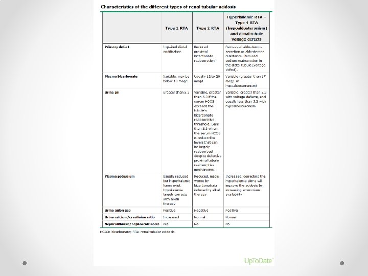
Overall problem in RTAs • Too little base o Reduction in proximal bicarbonate reabsorptive capacity o Bicarbonate is “wasted” in the urine o Wasting continues until serum bicarbonate has Proximal fallen so low RTA such that all the bicarbonate that is filtered through the glomerulus and into the • Too much acid o Impaired distal acid secretion o Progressive retention of hydrogen ions leads to decrease in Distalthan RTA 5. 5) serum HCO 3 and abnormally high urine p. H (greater

Proximal RTAs Isolated Inherited Transient Meds Carbonic anhydrase inhibitors -topiromate - acetazolamide Fanconi’s Acquired Genetic Renal disease Meds 1. Tubulointerstitial nephritis 2. Tx 3. Membranous nephopathy 1. Anti-cancer - Ifosfamide 2. Abx - Aminoglycosides 3. Anti-retrovirals - Cidofovir Heavy Metals Lead Cadmium Mercury 1. Neonates - Mitochondrial - Galactosemia - Tyrosinemia 2. Infants - Cystinosis - Lowes 3. Childood - Dent’s - Cystiniosis - Wilson’s

Proximal RTAs • Major features o Degree of acidosis is self-limited • HCO 3 usually 14 -20 (compare to <10 in distal RTA) o Hypokalemia • Decreased proximal sodium reabsorption means increased distal sodium reabsorption (when sodium goes in, potassium is secreted thus potassium falls) o When HCO 3 is given o No longer steady state. o Will have HCO 3 loss – thus urine p. H will increase o Will have increased distal delivery of Na (with HCO 3) and water. Thus more Na reabsorbed and resulting worsening HYPOKALEMIA

Distal RTAs • Major features o Inability to excrete daily acid load • HCO 3 usually <10 • Urine p. H is 5. 5 or higher (i. e. not acidic when you would expect it to be with acidemia) o Hypokalemia • Mutation in distal proton pump means no H secreted into lumen • Sodium moving into principal cell creates negative charge • K moves out down electroneg gradient o When HCO 3 is given • HCO 3 reaches distal tubule and improves p. H thus less K out • Improved HYPOKALEMIA

Distal RTAs Genetic Acquired Meds Amphotericin Ifosfamide Lithium Toluene Autoimmue disease Sjogrens SLE RA AI hepatitis Renal disease H-ATPase defect -2 forms – both AR - Present in infancy with poor growth and severe met acid - Obstructive uropathy - MSK - Renal transplant Hypercalciuric conditions rejection - Hyper. PTHism - Vit D intox Cl-HCO 3 exchanger defect

Urine anion gap • Difference between the positive charges and negative charges in urine o UAG = Urine (Na + K –Cl) • Usual state: more Na and K in excreted than Clo POSITIVE charge • With diarrhea: loss of bicarbonate salts in the stool (Na. HCO 3 and KHCO 3) o Decreased Na and K in urine but Cl unchanged o NEGATIVE charge • Metabolic acidosis: want to excrete H+ o H+ is excreted as ammonium (NH 3+H →NH 4+) o Ammonium travels with Clo Thus should see NEGATIVE charge • In patients with met acidosis from impaired H excretion o Will have same as usual state without increase H excretion o POSITIVE charge
- Slides: 170