
Pediatric Nephrology Pearls KRISTA RAY APN FNPBC SENIOR
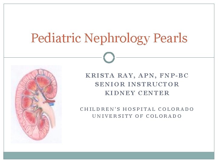
Pediatric Nephrology Pearls KRISTA RAY, APN, FNP-BC SENIOR INSTRUCTOR KIDNEY CENTER CHILDREN’S HOSPITAL COLORADO UNIVERSITY OF COLORADO
Objectives � Consider differential diagnoses associated with hematuria and proteinuria. � Identify effective screening tools and diagnostic studies to identify children with kidney disease. � Describe the long term health consequences of kidney disease in a pediatric patient.
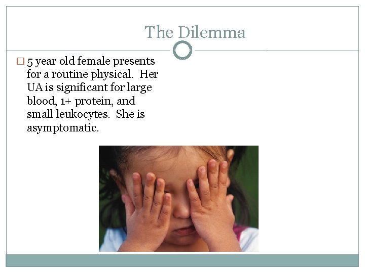
The Dilemma � 5 year old female presents for a routine physical. Her UA is significant for large blood, 1+ protein, and small leukocytes. She is asymptomatic.
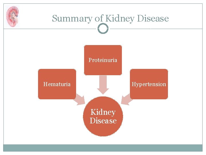
Summary of Kidney Disease Proteinuria Hematuria Hypertension Kidney Disease
The Needle in the Haystack � 12 -74 cases/million � 5 -10% proteinuria � 3 -4% hematuria � <0. 1% diagnosed by random screening actually have kidney disease

AAP guidelines for UA Screening � Avoid UA if patient asymptomatic or previous was normal � Need to perform >800 UAs to diagnose 1 case of CKD = ~$3000/case � So why do we still do them? ? ?
Complications of CKD �CVD (#1 cause of death) Dyslipidemia Obesity Inflammatory toxins Platelet dysfunction Carotid stenosis �HTN (70%) >50 -90 th%tile x 3 readings
Complications of CKD �Anemia <11. 0 g/dl �Growth Failure (multi factorial) food averse �Electrolyte imbalances �Endocrine abnormalities Glucose metabolism disturbed Delayed puberty Resistance to growth hormone Thyroid disorders �Bone Mineral Disorders
More effective Alternative to UA: BP Screening � Annual age 3 years � Earlier with significant risk factors or PMH of congenital renal anomaly � Manual more accurate! � Appropriate cuff size critical!
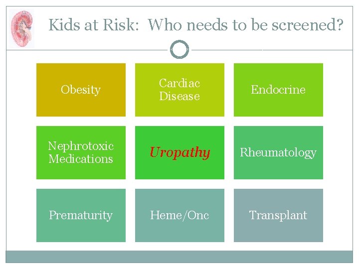
Kids at Risk: Who needs to be screened? Obesity Cardiac Disease Endocrine Nephrotoxic Medications Uropathy Rheumatology Prematurity Heme/Onc Transplant
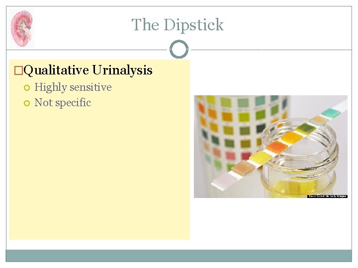
The Dipstick �Qualitative Urinalysis Highly sensitive Not specific
Specimen Collection � Timing is everything! � 1 st AM Specimen � Clean catch midstream � Read results at time indicated

Proteinuria � Earliest indicator of kidney injury � Negative and Trace “Normal” � 1+ or > needs follow up � >2+ may be nephrotic range
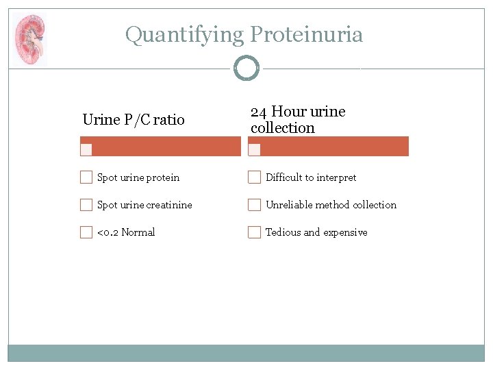
Quantifying Proteinuria Urine P/C ratio 24 Hour urine collection Spot urine protein Difficult to interpret Spot urine creatinine Unreliable method collection <0. 2 Normal Tedious and expensive
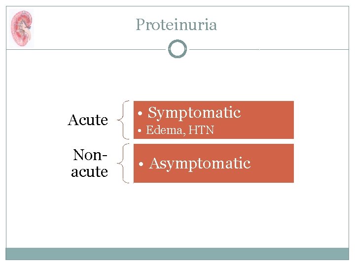
Proteinuria Acute • Symptomatic Nonacute • Asymptomatic • Edema, HTN
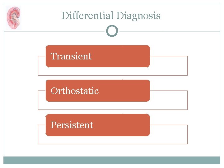
Differential Diagnosis Transient Orthostatic Persistent
Transient Proteinuria � Benign � Physiologic stressor � No work up/referral necessary

Orthostatic Proteinuria � Benign � Present in any specimen other than � 1 st AM
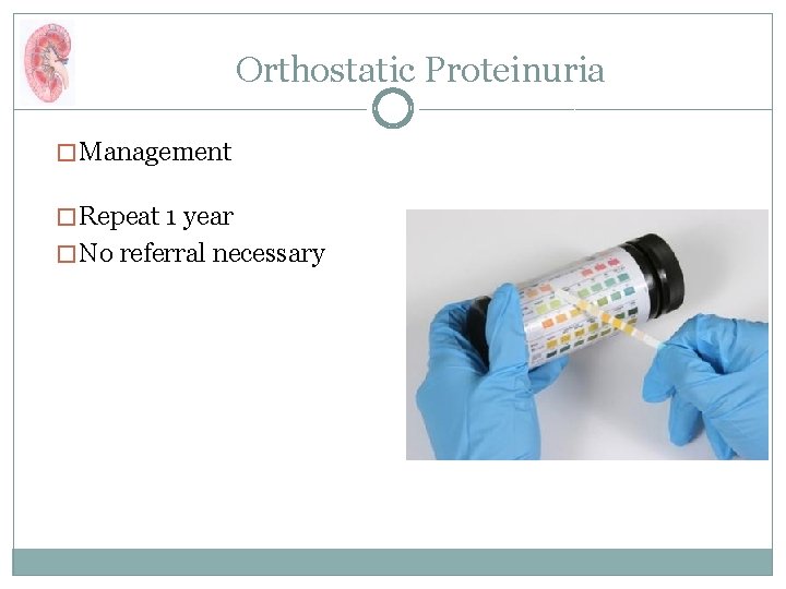
Orthostatic Proteinuria � Management � Repeat 1 year � No referral necessary
Persistent Proteinuria �Renal injury or scarring �>1+ or UPCR >0. 2 Nephrotic (>2. 0) v non nephrotic range Acute Chronic
Nephritis Protein HTN Edema Blood Nephritis
Evaluation: Asymptomatic Proteinuria � Assess for risk factors � Urinary or systemic symptoms? � Refer to nephrology
Diagnostic Testing Labs Imaging • BUN, Cr, Albumin, UPCR • CBC, C 3 • Renal Ultrasound
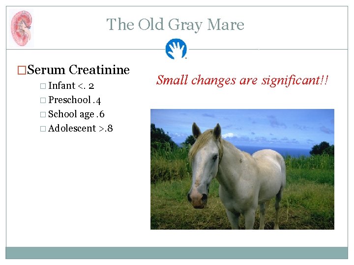
The Old Gray Mare �Serum Creatinine � Infant <. 2 � Preschool. 4 � School age. 6 � Adolescent >. 8 Small changes are significant!!
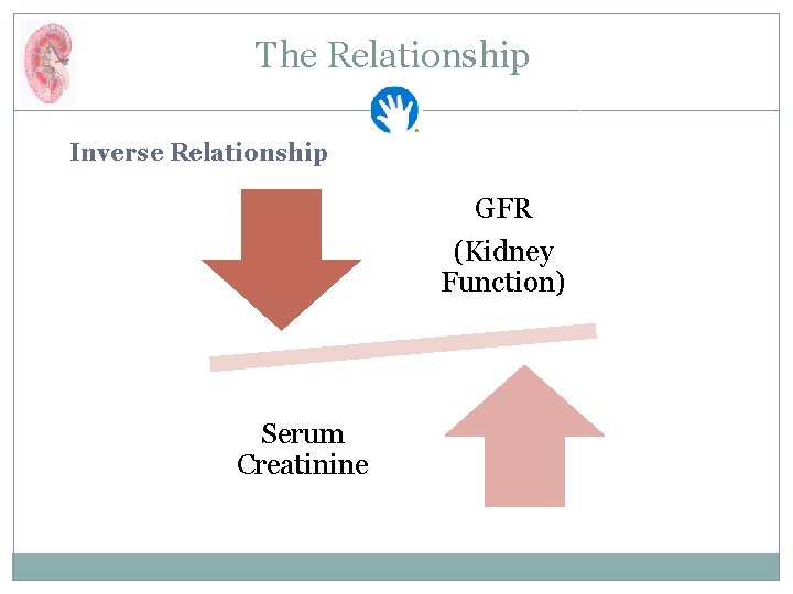
The Relationship Inverse Relationship GFR (Kidney Function) Serum Creatinine
#1 Rule of Hematuria � Microscopic � Gross
Practice Pearl � Dipstick + for blood with negative microscopic UA �What is this? �What about “orange” urine?
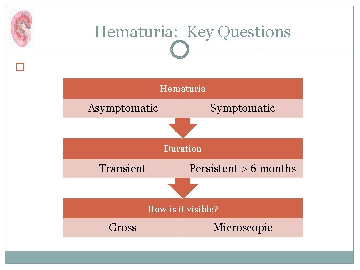
Hematuria: Key Questions � Hematuria Asymptomatic Symptomatic Duration Transient Persistent > 6 months How is it visible? Gross Microscopic
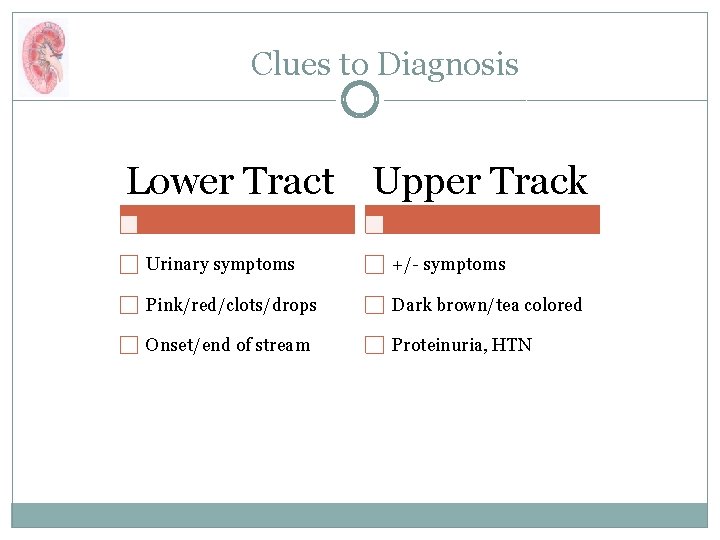
Clues to Diagnosis Lower Tract Upper Track Urinary symptoms +/- symptoms Pink/red/clots/drops Dark brown/tea colored Onset/end of stream Proteinuria, HTN
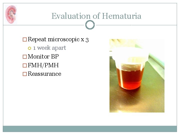
Evaluation of Hematuria � Repeat microscopic x 3 1 week apart � Monitor BP � FMH/PMH � Reassurance
Management of Hematuria � Refer to nephrology > 6 months OR Any time associated with:
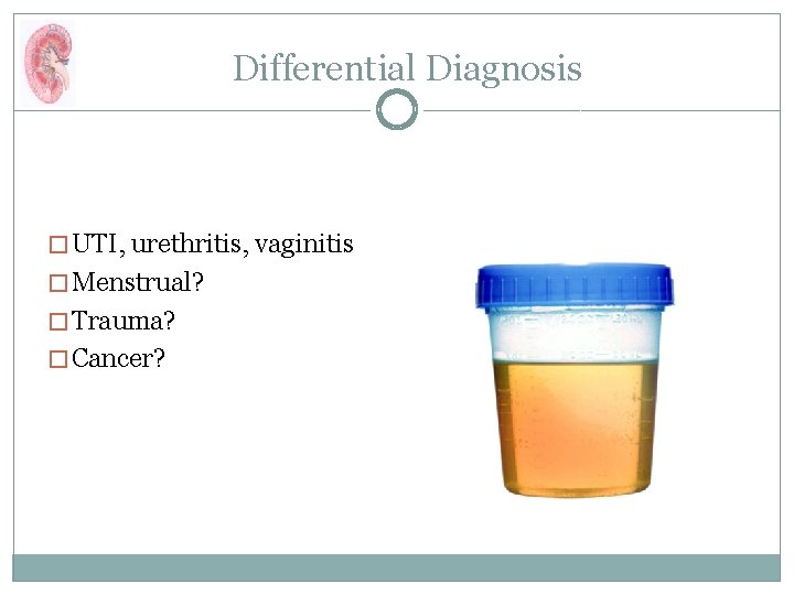
Differential Diagnosis � UTI, urethritis, vaginitis � Menstrual? � Trauma? � Cancer?
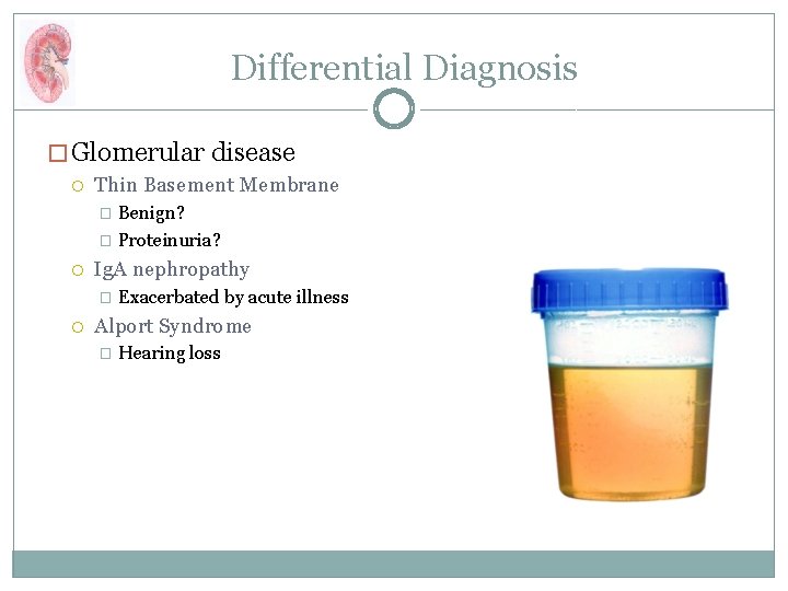
Differential Diagnosis � Glomerular disease Thin Basement Membrane Benign? � Proteinuria? � Ig. A nephropathy � Exacerbated by acute illness Alport Syndrome � Hearing loss
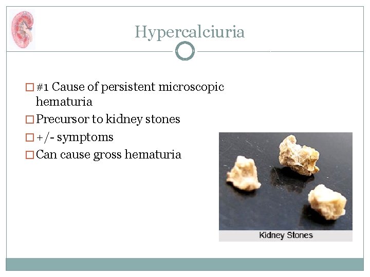
Hypercalciuria � #1 Cause of persistent microscopic hematuria � Precursor to kidney stones � +/- symptoms � Can cause gross hematuria
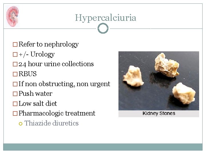
Hypercalciuria � Refer to nephrology � +/- Urology � 24 hour urine collections � RBUS � If non obstructing, non urgent � Push water � Low salt diet � Pharmacologic treatment Thiazide diuretics

School/Sports Participation � Frequent use of restroom � Carry water bottle during class � No restrictions Not even solitary kidney
What Can YOU Do? Avoid or limit nephrotoxic meds � GFR Renal Contrast Protocol � GFR <60 ml/min/1. 73 m 2 < 89 ml/min/1. 73 m 2 Renal Dosing Monitor labs post med changes Surgical clearance/pre op labs Monitor for HTN Reinforce kidney follow up
Just the Facts, Ma’am � Stop “random” testing � 1 st Am UA � Confirm with microscopic � Identify kids at high risk � Routinely screen BP >95 th%tile �Refer early
The End Thank you!
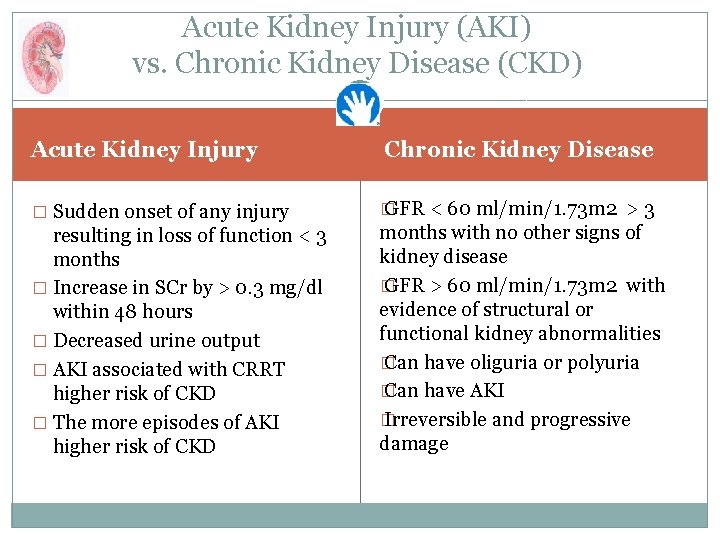
Acute Kidney Injury (AKI) vs. Chronic Kidney Disease (CKD) Acute Kidney Injury � Sudden onset of any injury resulting in loss of function < 3 months � Increase in SCr by > 0. 3 mg/dl within 48 hours � Decreased urine output � AKI associated with CRRT higher risk of CKD � The more episodes of AKI higher risk of CKD Chronic Kidney Disease � GFR < 60 ml/min/1. 73 m 2 > 3 months with no other signs of kidney disease � GFR > 60 ml/min/1. 73 m 2 with evidence of structural or functional kidney abnormalities � Can have oliguria or polyuria � Can have AKI � Irreversible and progressive damage
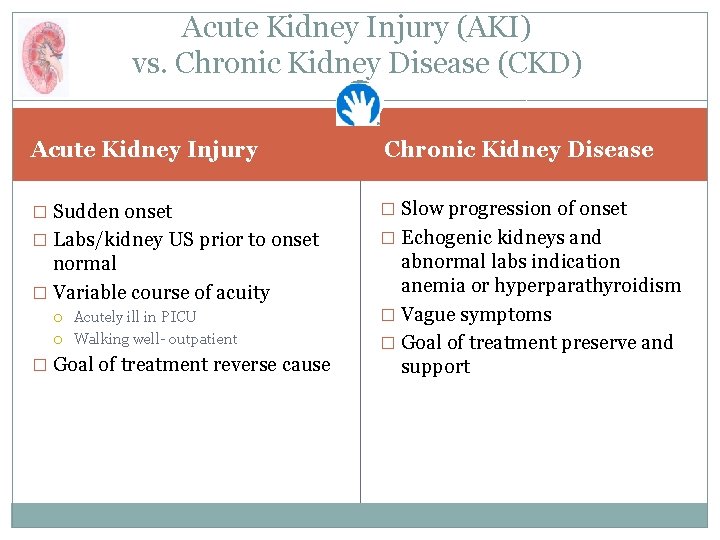
Acute Kidney Injury (AKI) vs. Chronic Kidney Disease (CKD) Acute Kidney Injury Chronic Kidney Disease � Sudden onset � Slow progression of onset � Labs/kidney US prior to onset � Echogenic kidneys and normal � Variable course of acuity Acutely ill in PICU Walking well- outpatient � Goal of treatment reverse cause abnormal labs indication anemia or hyperparathyroidism � Vague symptoms � Goal of treatment preserve and support
- Slides: 41