
PEDIATRIC NEPHOLOGY MODULE 6 th Year Medical school

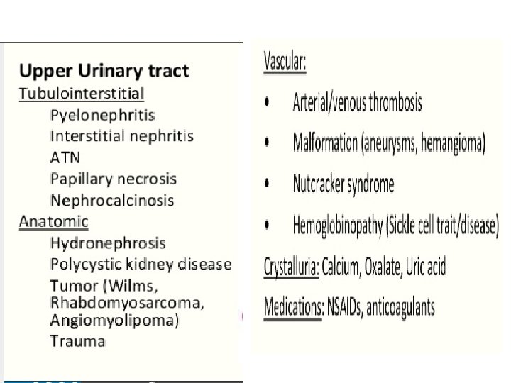




- Slides: 116
PEDIATRIC NEPHOLOGY MODULE 6 th Year Medical school Dr. Reham Almardini
Outline • • • Introduction Hematuria Proteinuria Tubulopathy HUS Acute Kidney Injury CKD RRT Urology- UTI
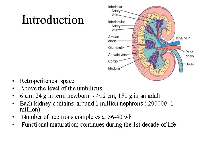
Introduction • • Retroperitoneal space Above the level of the umbilicus 6 cm, 24 g in term newborn - ≥ 12 cm, 150 g in an adult Each kidney contains around 1 million nephrons ( 200000 - 1 million) • Number of nephrons completes at 36 -40 wk • Functional maturation; continues during the 1 st decade of life
Nephrons Number • New nephrons cannot be formed after birth • diseases results in loss of nephrons lead to renal insufficiency • Low birthweight & Prematurity or others • Risk factor for: HT, CKD • Low nephron number: hyperfiltration and eventual sclerosis of “overworked” nephron units
Functions of the kidneys • Filtration and excretion of fluid &waste products • Bp. control • Acid base balance • Production of Erythropoietin and renin • Activation of vitamin D
Hematuria
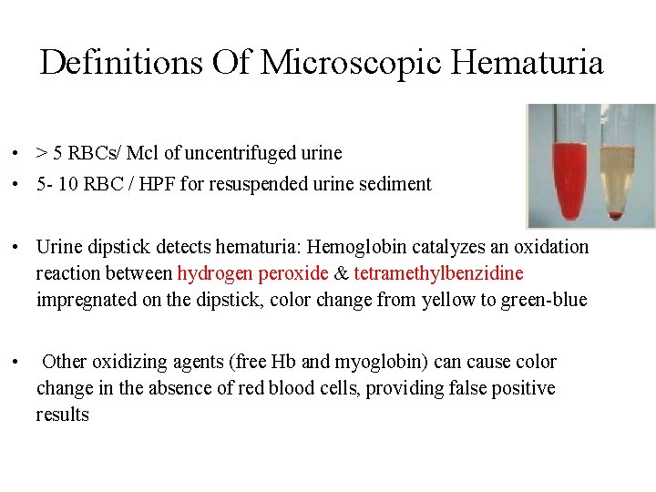
Definitions Of Microscopic Hematuria • > 5 RBCs/ Mcl of uncentrifuged urine • 5 - 10 RBC / HPF for resuspended urine sediment • Urine dipstick detects hematuria: Hemoglobin catalyzes an oxidation reaction between hydrogen peroxide & tetramethylbenzidine impregnated on the dipstick, color change from yellow to green-blue • Other oxidizing agents (free Hb and myoglobin) can cause color change in the absence of red blood cells, providing false positive results
• False positive: Hemoglobin, myoglobin, oxidizing agent as hypochlorite, UTI- microbial peroxidases • False negative; reducing substances as Ascorbic acids, high specific gravity
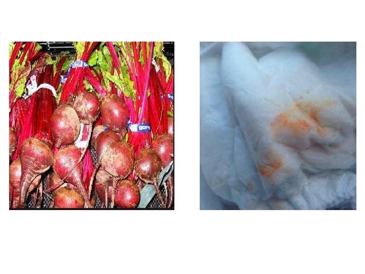
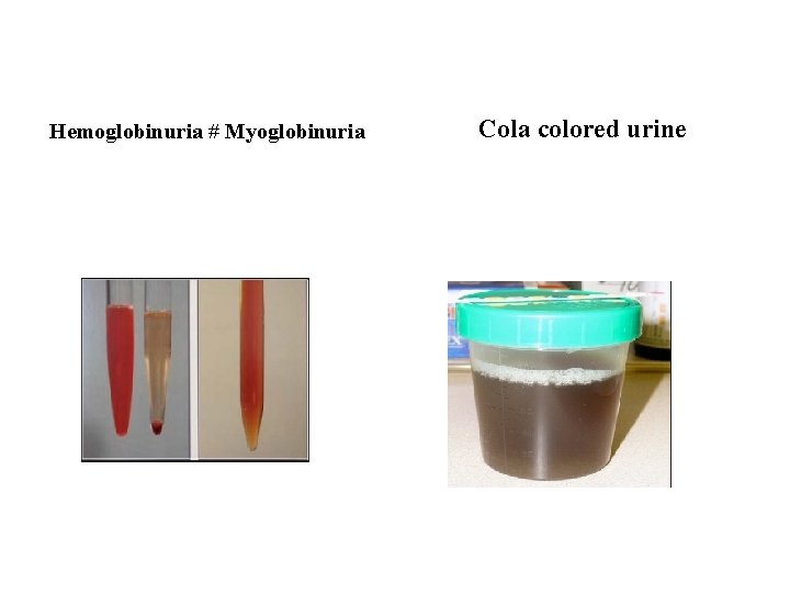
Hemoglobinuria # Myoglobinuria Cola colored urine
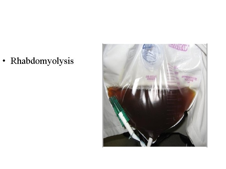
• Rhabdomyolysis
Incidence of Hematuria • Ma. H in a general pediatric population is 0. 13% • Persistent Mi. H occurs in 0. 5%-2% of children, varying with definitions of ‘persistent’ and ‘hematuria’ • If present on more than 3 occasions over 3 weeks, it is termed ‘persistent’ and requires further investigation
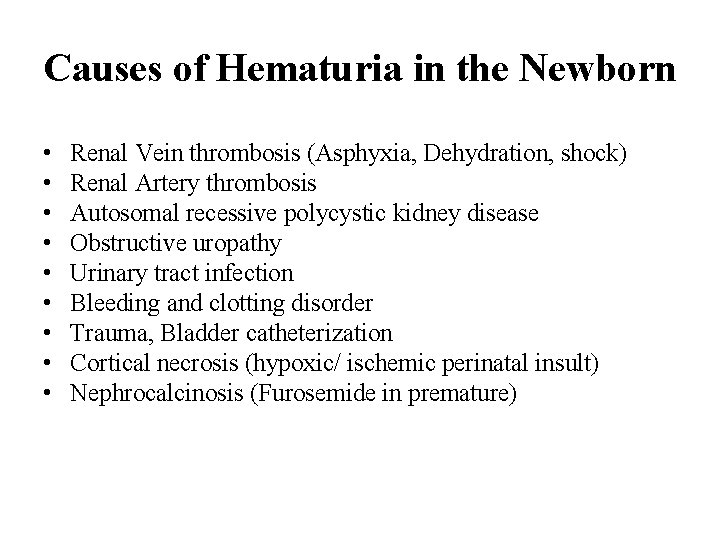
Causes of Hematuria in the Newborn • • • Renal Vein thrombosis (Asphyxia, Dehydration, shock) Renal Artery thrombosis Autosomal recessive polycystic kidney disease Obstructive uropathy Urinary tract infection Bleeding and clotting disorder Trauma, Bladder catheterization Cortical necrosis (hypoxic/ ischemic perinatal insult) Nephrocalcinosis (Furosemide in premature)
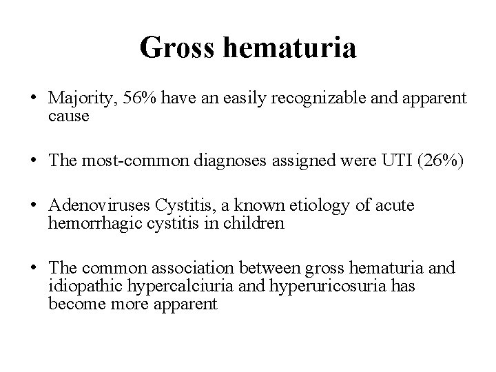
Gross hematuria • Majority, 56% have an easily recognizable and apparent cause • The most-common diagnoses assigned were UTI (26%) • Adenoviruses Cystitis, a known etiology of acute hemorrhagic cystitis in children • The common association between gross hematuria and idiopathic hypercalciuria and hyperuricosuria has become more apparent
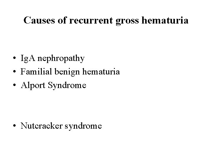
Causes of recurrent gross hematuria • Ig. A nephropathy • Familial benign hematuria • Alport Syndrome • Nutcracker syndrome
• In all children with hematuria or proteinuria a clinical evaluation should include blood pressure measurement compared to a reference of normal values for the child’s gender and height
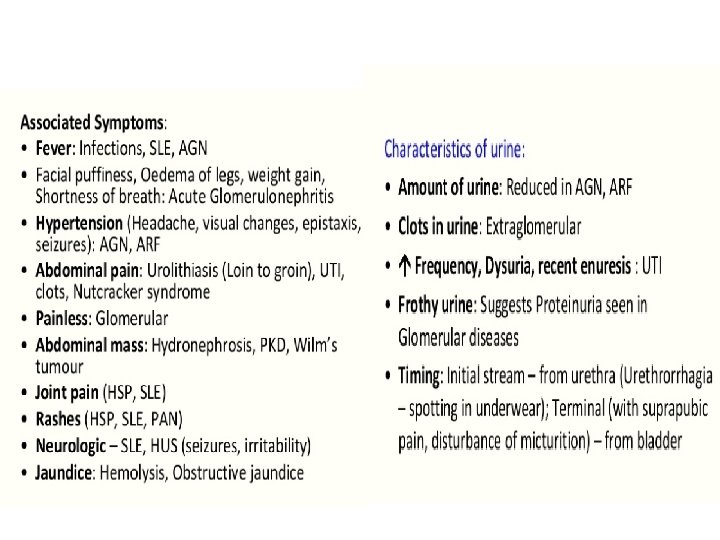
Features suggestive of renal disease requiring urgent investigation • • • Fluid overload, Edema, Ascites, Oligoanuria Hypertension significant proteinuria Systemic symptoms: joint pain or swelling, Vasculitis rash previous episodes and significant past medical history such as recent upper respiratory tract infection • Growth impairment • Hearing abnormalities • The presence of a family history of hematuria, renal disease or hearing impairment suggests a familial cause
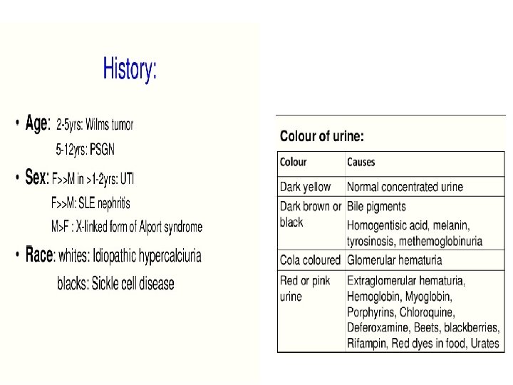
Glomerular • A brownish-red or ‘colacolored’ • Signs Of GN; HT, fluid overload Absence does not exclude GN • Dysmorphic RBC • Glomerular bleeding alone will not cause a drop in the serum haemoglobin. • Anaemia; haemolysis, CRF or heavy LUT bleeding Lower Urinary Tract • Red or burgundy • Contain blood clots • Early stream suggests a urethral focus • Terminal stream suggests a bladder focus (e. g. cystitis, calculus, schistosomiasis). • Pain is more suggestive of a LUT source
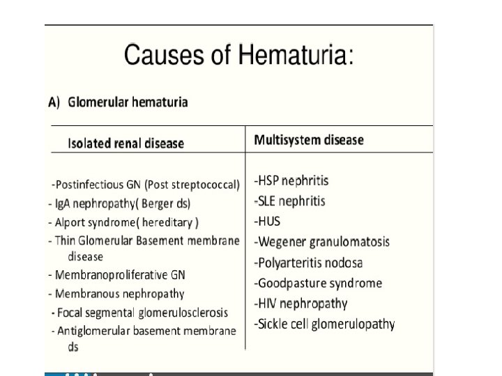
Glomerular Causes • PIGN: Common cause of childhood glomerulonephritis; usually managed in a general pediatric inpatient setting • A personal or family history of Mi. H and intermittent gross hematuriacoinciding or within days of intercurrent infections is characteristic of Ig. A nephropathy • Ig. A nephropathy may present with Mi. H, proteinuria or signs of GN. A raised serum Ig. A is sensitive but not specific • Henoch Schonlein Purpura (HSP) Nephritis is preceded by a history of abdominal pain, bloody stools, vasculitic rash and arthritis
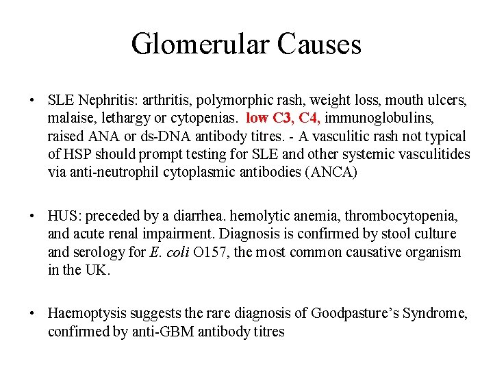
Glomerular Causes • SLE Nephritis: arthritis, polymorphic rash, weight loss, mouth ulcers, malaise, lethargy or cytopenias. low C 3, C 4, immunoglobulins, raised ANA or ds-DNA antibody titres. - A vasculitic rash not typical of HSP should prompt testing for SLE and other systemic vasculitides via anti-neutrophil cytoplasmic antibodies (ANCA) • HUS: preceded by a diarrhea. hemolytic anemia, thrombocytopenia, and acute renal impairment. Diagnosis is confirmed by stool culture and serology for E. coli O 157, the most common causative organism in the UK. • Haemoptysis suggests the rare diagnosis of Goodpasture’s Syndrome, confirmed by anti-GBM antibody titres
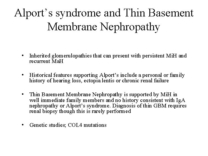
Alport’s syndrome and Thin Basement Membrane Nephropathy • Inherited glomerulopathies that can present with persistent Mi. H and recurrent Ma. H • Historical features supporting Alport’s include a personal or family history of hearing loss, ectopia lentis or chronic renal failure • Thin Basement Membrane Nephropathy is supported by Mi. H in well immediate family members and no history consistent with Ig. A nephropathy or Alport’s syndrome. Diagnosis of thin GBM requires renal biopsy though this is rarely performed • Genetic studies; COL 4 mutations
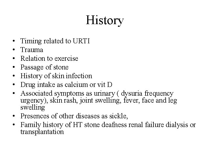
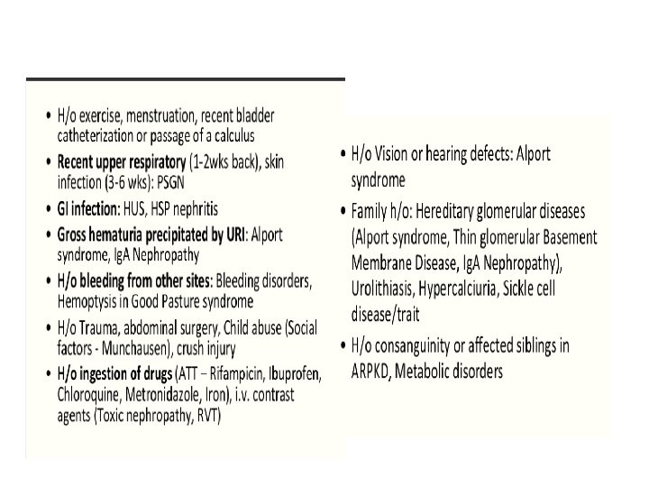
History • • Timing related to URTI Trauma Relation to exercise Passage of stone History of skin infection Drug intake as calcium or vit D Associated symptoms as urinary ( dysuria frequency urgency), skin rash, joint swelling, fever, face and leg swelling • Presences of other diseases as sickle, • Family history of HT stone deafness renal failure dialysis or transplantation
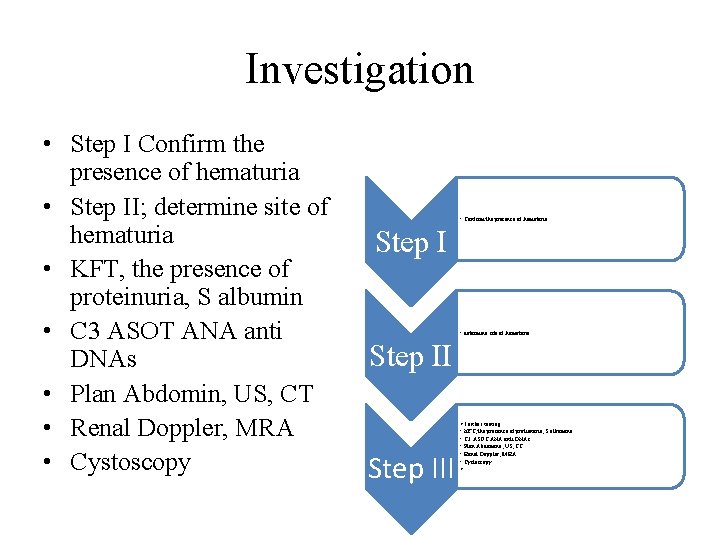
Investigation • Step I Confirm the presence of hematuria • Step II; determine site of hematuria • KFT, the presence of proteinuria, S albumin • C 3 ASOT ANA anti DNAs • Plan Abdomin, US, CT • Renal Doppler, MRA • Cystoscopy • Confirm the presence of hematuria Step I • determine site of hematuria Step III • Further testing • KFT, the presence of protienuria, S albumine • C 3 ASOT ANA anti DNAs • Plan Abdomine, US, CT • Renal Doppler, MRA • Cystoscopy •
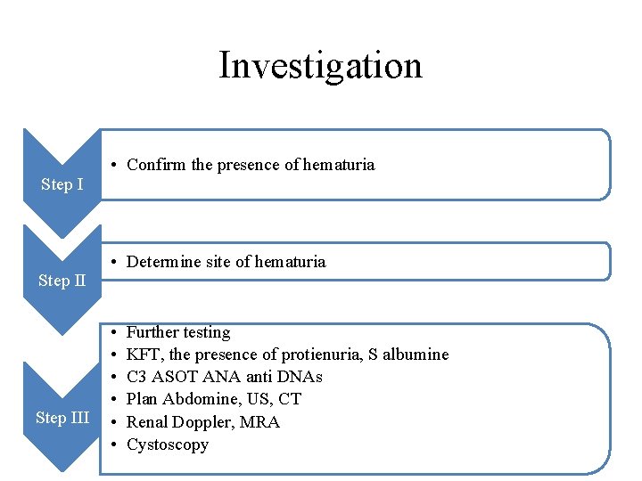
Investigation Step III • Confirm the presence of hematuria • Determine site of hematuria • • • Further testing KFT, the presence of protienuria, S albumine C 3 ASOT ANA anti DNAs Plan Abdomine, US, CT Renal Doppler, MRA Cystoscopy
• If the microscopic hematuria persists unchanged for more than 1– 2 years further evaluation is recommended
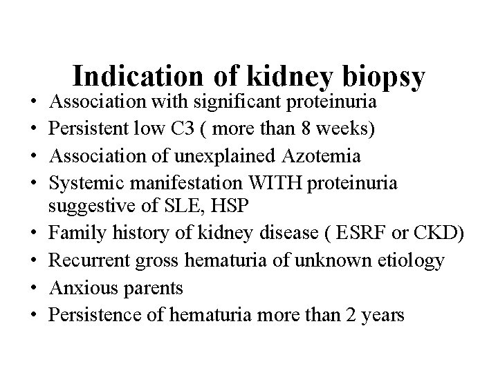
• • Indication of kidney biopsy Association with significant proteinuria Persistent low C 3 ( more than 8 weeks) Association of unexplained Azotemia Systemic manifestation WITH proteinuria suggestive of SLE, HSP Family history of kidney disease ( ESRF or CKD) Recurrent gross hematuria of unknown etiology Anxious parents Persistence of hematuria more than 2 years
Questions
Hemolytic-Uremic Syndrom
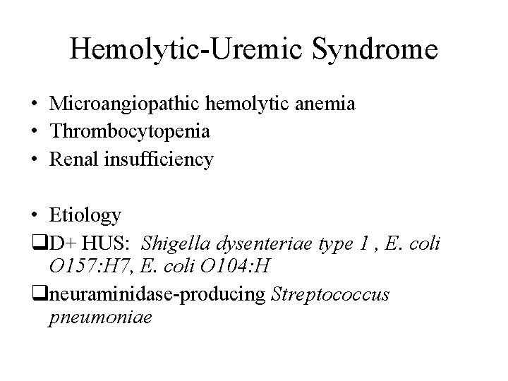
Hemolytic-Uremic Syndrome • Microangiopathic hemolytic anemia • Thrombocytopenia • Renal insufficiency • Etiology q. D+ HUS: Shigella dysenteriae type 1 , E. coli O 157: H 7, E. coli O 104: H qneuraminidase-producing Streptococcus pneumoniae
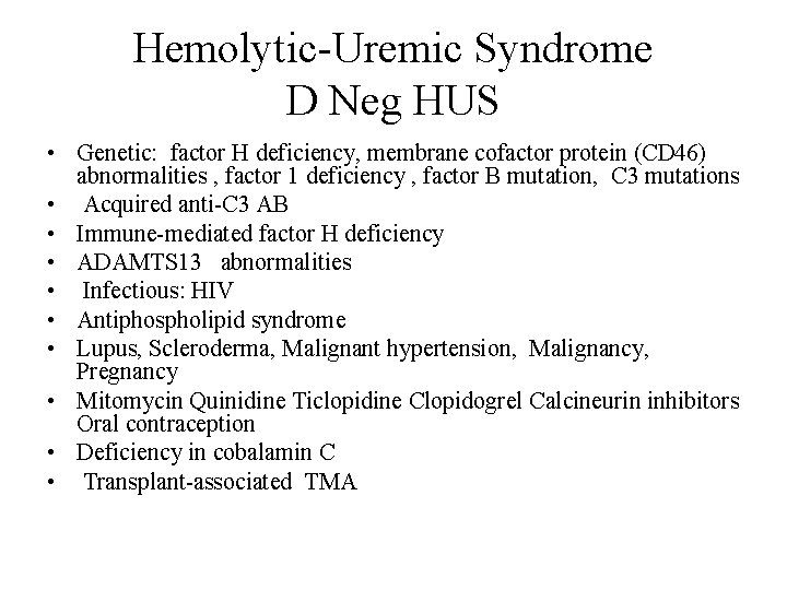
Hemolytic-Uremic Syndrome D Neg HUS • Genetic: factor H deficiency, membrane cofactor protein (CD 46) abnormalities , factor 1 deficiency , factor B mutation, C 3 mutations • Acquired anti-C 3 AB • Immune-mediated factor H deficiency • ADAMTS 13 abnormalities • Infectious: HIV • Antiphospholipid syndrome • Lupus, Scleroderma, Malignant hypertension, Malignancy, Pregnancy • Mitomycin Quinidine Ticlopidine Clopidogrel Calcineurin inhibitors Oral contraception • Deficiency in cobalamin C • Transplant-associated TMA
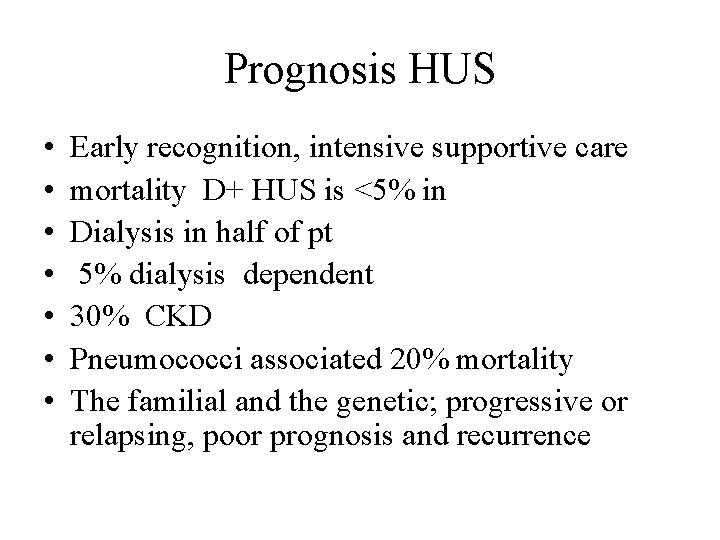
Prognosis HUS • • Early recognition, intensive supportive care mortality D+ HUS is <5% in Dialysis in half of pt 5% dialysis dependent 30% CKD Pneumococci associated 20% mortality The familial and the genetic; progressive or relapsing, poor prognosis and recurrence
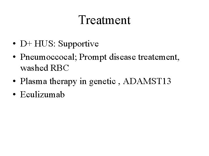
Treatment • D+ HUS: Supportive • Pneumoccocal; Prompt disease treatement, washed RBC • Plasma therapy in genetic , ADAMST 13 • Eculizumab
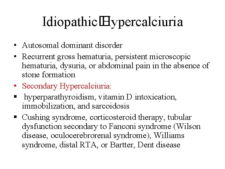
Idiopathic� Hypercalciuria • Autosomal dominant disorder • Recurrent gross hematuria, persistent microscopic hematuria, dysuria, or abdominal pain in the absence of stone formation • Secondary Hypercalciuria: § hyperparathyroidism, vitamin D intoxication, immobilization, and sarcoidosis § Cushing syndrome, corticosteroid therapy, tubular dysfunction secondary to Fanconi syndrome (Wilson disease, oculocerebrorenal syndrome), Williams syndrome, distal RTA, or Bartter, Dent disease
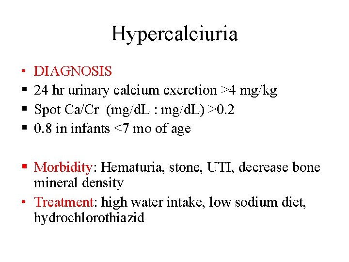
Hypercalciuria • § § § DIAGNOSIS 24 hr urinary calcium excretion >4 mg/kg Spot Ca/Cr (mg/d. L : mg/d. L) >0. 2 0. 8 in infants <7 mo of age § Morbidity: Hematuria, stone, UTI, decrease bone mineral density • Treatment: high water intake, low sodium diet, hydrochlorothiazid
Proteinuria
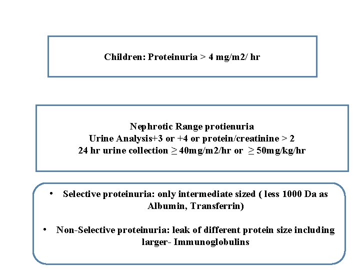
Children: Proteinuria > 4 mg/m 2/ hr Nephrotic Range protienuria Urine Analysis+3 or +4 or protein/creatinine > 2 24 hr urine collection ≥ 40 mg/m 2/hr or ≥ 50 mg/kg/hr • Selective proteinuria: only intermediate sized ( less 1000 Da as Albumin, Transferrin) • Non-Selective proteinuria: leak of different protein size including larger- Immunoglobulins
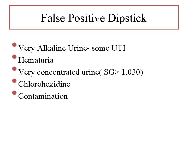
False Positive Dipstick • Very Alkaline Urine- some UTI • Hematuria • Very concentrated urine( SG> 1. 030) • Chlorohexidine • Contamination
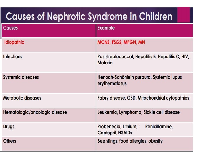
Causes of proteinuria • Non Pathological causes - Orthostatic - Febrile - Exercise induced Pathological Causes - Tubular (Allergic interstitial nephritis, ATN) - Glomerular ( NS, GN) •
Nephrotic Syndrome
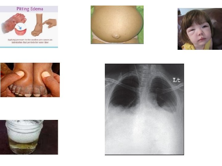
Clinical presentation • Edema, periorbital, morning • Antecedent infection, URTI • Decreased Urine output - frothy or foamy • Abdominal pain • Headache with neurological signs or irritability • • • should raise the suspicion for cerebral venous sinus thrombosis Hematuria (Gross in < 5%, microscopic in 20%) Hypertension (5%) 2 to 10 yrs of age with male predominance(2: 1)
Differential Diagnosis of Nephrotic Syndrome • • • Protein- loosing enteropathy Hepatic failure Protein energy malnutrition Acute and chronic GN Urticaria & Angioedema
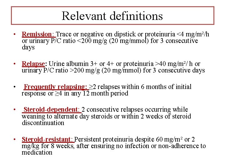
Relevant definitions • Remission: Trace or negative on dipstick or proteinuria <4 mg/m²/h or urinary P/C ratio <200 mg/g (20 mg/mmol) for 3 consecutive days • Relapse: Urine albumin 3+ or 4+ or proteinuria >40 mg/m²/ h or urinary P/C ratio >200 mg/g (20 mg/mmol) for 3 consecutive days • Frequently relapsing: ≥ 2 relapses within 6 months of initial response or ≥ 4 in any 12 month period • Steroid-dependent: 2 consecutive relapses occurring while weaning to alternate day steroids or within 2 weeks of steroid discontinuation • Steroid-resistant: Persistent proteinuria despite 60 mg/m² or 2 mg/kg for 8 weeks, after ensuring no infection or non-adherence to medication
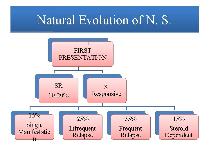
Natural Evolution of N. S. FIRST PRESENTATION SR 10 -20% 15% Single Manifestatio n S. Responsive 25% Infrequent Relapse 35% Frequent Relapse 15% Steroid Dependent
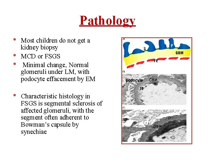
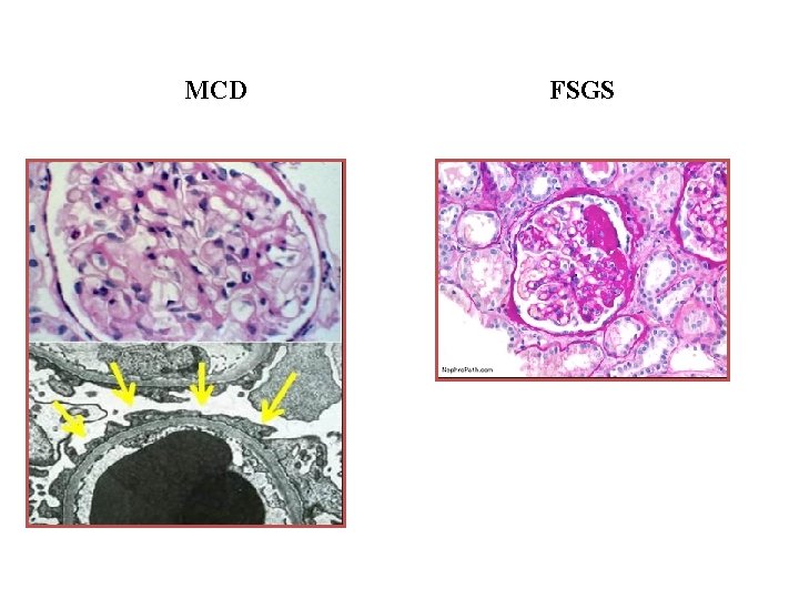
Pathology • • Most children do not get a kidney biopsy MCD or FSGS Minimal change, Normal glomeruli under LM, with podocyte effacement by EM Characteristic histology in FSGS is segmental sclerosis of affected glomeruli, with the segment often adherent to Bowman’s capsule by synechiae
MCD FSGS
Second line investigations in NS • Complement C 3, C 4 • Antinuclear antibody (ANA) • Anti-double- stranded DNA (anti-ds. DNA) and • • ANCA Hepatitis B and C (HBs. Ag, anti HCV) Anti HIV antibodies Genetic mutation analysis Renal biopsy
Indications for Renal biopsy At onset • Age 10 yrs • Gross hematuria, persistent microscopic hematuria • Low C 3 • Sustained hypertension • Renal failure not attributable to hypovolemia • Suspected secondary cause of • Proteinuria despite 4 weeks of daily steroids • Before initiating treatment with tacrolimus
Incidence • • 1∙ 15 to 16∙ 9 / 100 000 children Highest in south Asian compared to European as reported in UK, South Africa, and Canada • SRNS: 2∙ 1 to 27∙ 3% and varies by country of origin • African–American children are more to have biopsy-proven FSGS (42– 72%) & highest proportion of progression to ESRD as compared to European Americans • In south Asian children, FSGS is reported less commonly and ranges from 15 - 39%
Nephrotic syndrome Pathogenesis • • Immune-mediated Systemic circulating factors (eg, su. PAR) Podocyte related factors (eg, ANGPTL 4) Genetic variants
Immune Mediated: dysregulation of T lymphocytes Ø Supportive evidence • Efficacy of immunosuppressive agents in NS • Spontaneous NS remission following infection with measles • Resolution of NS following chemotherapy for Hodgkin’s and other T-cell lymphomas • Lastly, development of NS after allergic reactions to various stings and poisons
Systemic circulating factors • Serum from patients with FSGS induced proteinuria or increased permeability of glomeruli to albumin • There was also successful treatment of recurrent FSGS after kidney transplantation with immuno- adsorption • Maternal transmission of FSGS confirmed a circulating factor • There are numerous factors proposed including: Heparanase, Haemopexin, Angiopoietin-like 4 (ANGPTL 4), Cardiotrophin-like cytokine-1 and, more recently, Soluble Urokinase Plasminogen activator receptor (su. PAR)
Genetics • Genetic risk and SSNS A genetic locus on chromosome 6 p and single nucleotide polymorphisms in HLA-DQA 1 & DQB 1 were associated with SSNS using an exome Array
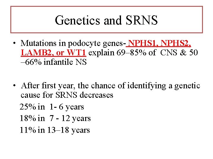
Genetics and SRNS • Mutations in podocyte genes- NPHS 1, NPHS 2, LAMB 2, or WT 1 explain 69– 85% of CNS & 50 – 66% infantile NS • After first year, the chance of identifying a genetic cause for SRNS decreases 25% in 1 - 6 years 18% in 7 - 12 years 11% in 13– 18 years
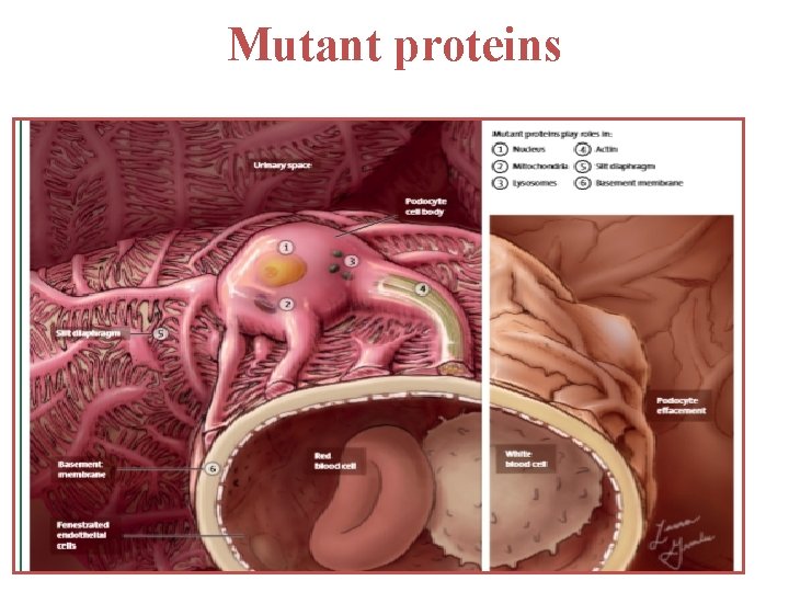
Mutant proteins
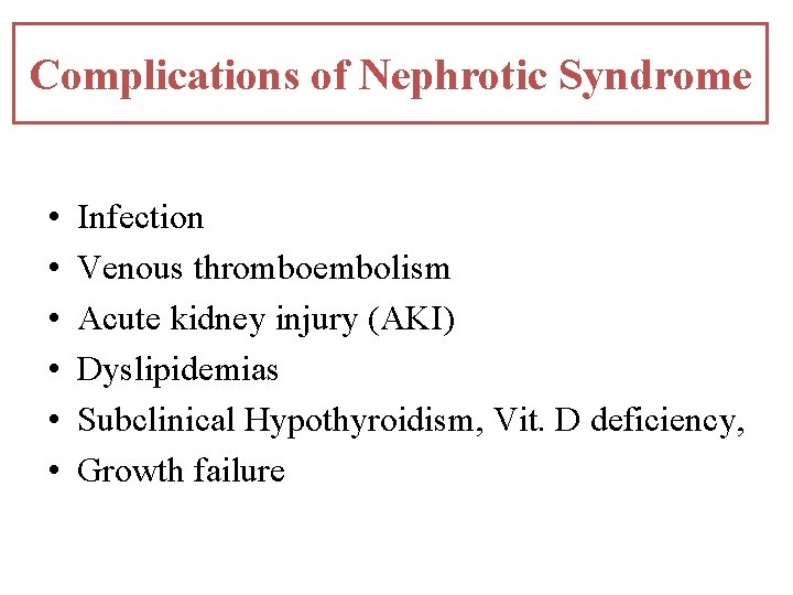
Complications of Nephrotic Syndrome • • • Infection Venous thromboembolism Acute kidney injury (AKI) Dyslipidemias Subclinical Hypothyroidism, Vit. D deficiency, Growth failure
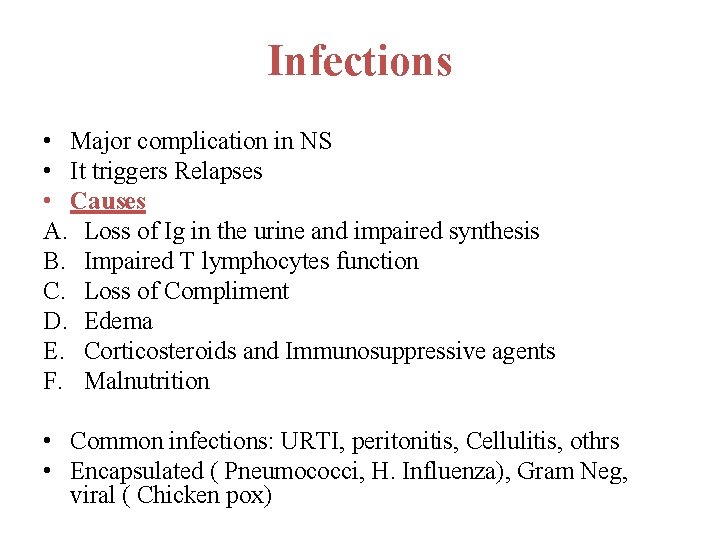
Infections • Major complication in NS • It triggers Relapses • Causes A. Loss of Ig in the urine and impaired synthesis B. Impaired T lymphocytes function C. Loss of Compliment D. Edema E. Corticosteroids and Immunosuppressive agents F. Malnutrition • Common infections: URTI, peritonitis, Cellulitis, othrs • Encapsulated ( Pneumococci, H. Influenza), Gram Neg, viral ( Chicken pox)
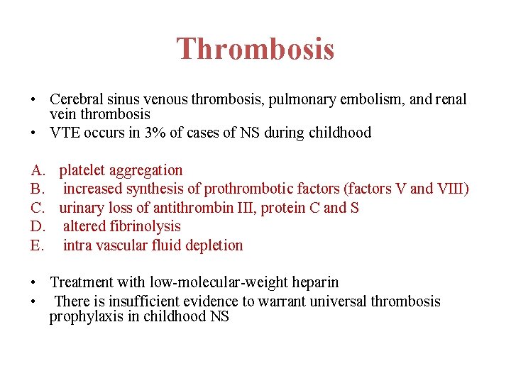
Thrombosis • Cerebral sinus venous thrombosis, pulmonary embolism, and renal vein thrombosis • VTE occurs in 3% of cases of NS during childhood A. platelet aggregation B. increased synthesis of prothrombotic factors (factors V and VIII) C. urinary loss of antithrombin III, protein C and S D. altered fibrinolysis E. intra vascular fluid depletion • Treatment with low-molecular-weight heparin • There is insufficient evidence to warrant universal thrombosis prophylaxis in childhood NS
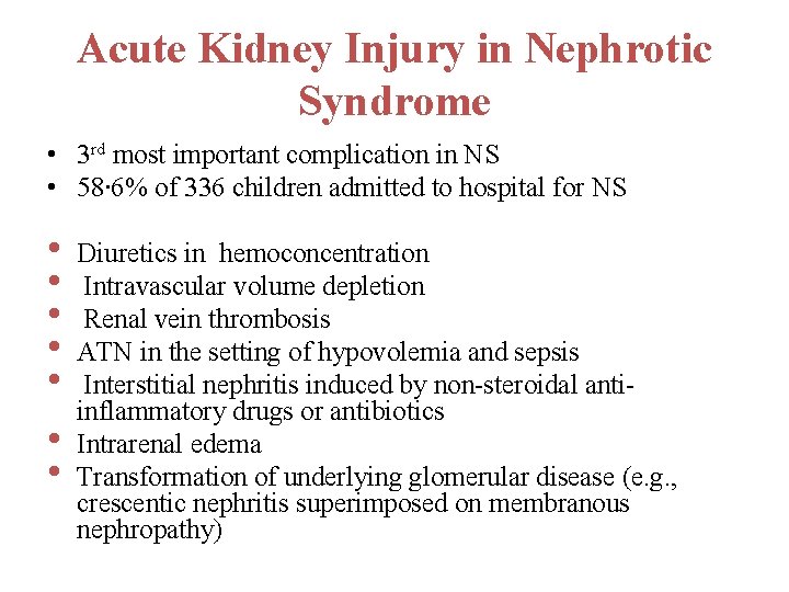
Acute Kidney Injury in Nephrotic Syndrome • 3 rd most important complication in NS • 58∙ 6% of 336 children admitted to hospital for NS • • Diuretics in hemoconcentration Intravascular volume depletion Renal vein thrombosis ATN in the setting of hypovolemia and sepsis Interstitial nephritis induced by non-steroidal antiinflammatory drugs or antibiotics Intrarenal edema Transformation of underlying glomerular disease (e. g. , crescentic nephritis superimposed on membranous nephropathy)
Dyslipidemia • The use of lipid lowering agents for the dyslipidaemia in NS is not advised, unless there is substantial persistent proteinuria with extremely high levels of hypertriglyceridaemia • If statins are initiated, it is only recommended for children over the age of 10 years with monitoring of liver function and creatinine kinase prior to initiating therapy and after 4 weeks
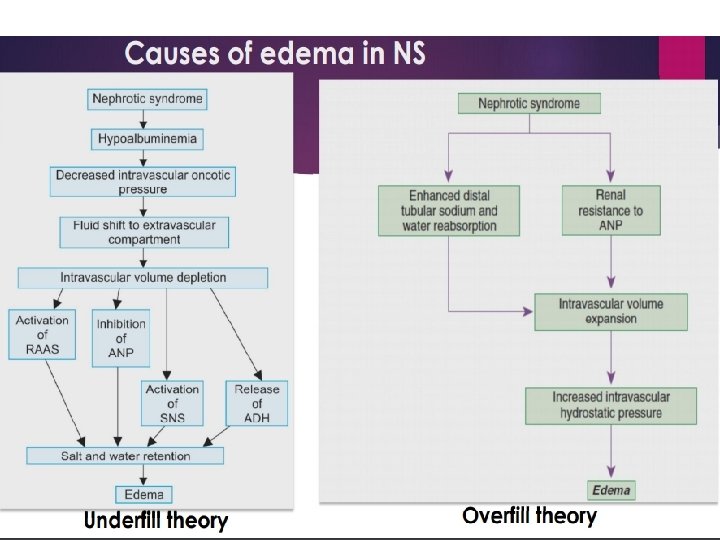
Management Edema in nephrotic syndrome • Salt and fluid restriction with addition of a loop diuretic for severe or symptomatic edema • Albumin infusion, in combination with a loop diuretic • Administration of albumin in children with signs of hypovolemia, as the resultant expansion in intravascular fluid could precipitate pulmonary edema
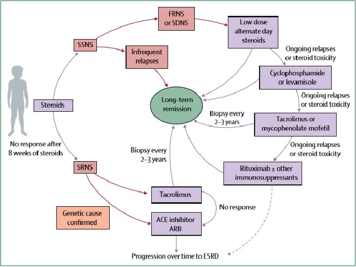
Frequent relapsing -FRNS • Low-dose, alternate-day steroid , typically at the lowest dose possible, or just above the steroid dose associated with the latest relapse
• Several trials suggest that relapses might be reduced if prednisolone is administered daily for 5– 7 days at the onset of upper respiratory tract infection in FRNS or SDNS
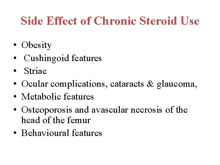
Side Effect of Chronic Steroid Use • • • Obesity Cushingoid features Striae Ocular complications, cataracts & glaucoma, Metabolic features Osteoporosis and avascular necrosis of the head of the femur • Behavioural features
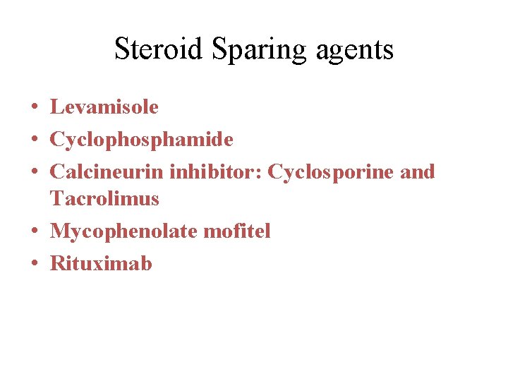
Steroid Sparing agents • Levamisole • Cyclophosphamide • Calcineurin inhibitor: Cyclosporine and Tacrolimus • Mycophenolate mofitel • Rituximab
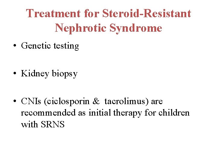
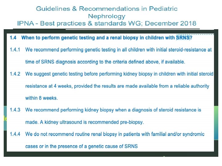
Treatment for Steroid-Resistant Nephrotic Syndrome • Genetic testing • Kidney biopsy • CNIs (ciclosporin & tacrolimus) are recommended as initial therapy for children with SRNS
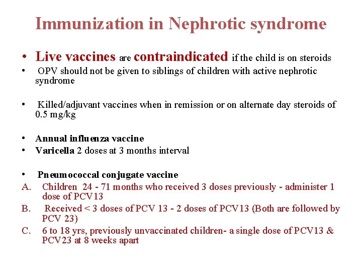
Immunization in Nephrotic syndrome • Live vaccines are contraindicated if the child is on steroids • OPV should not be given to siblings of children with active nephrotic syndrome • Killed/adjuvant vaccines when in remission or on alternate day steroids of 0. 5 mg/kg • Annual influenza vaccine • Varicella 2 doses at 3 months interval • Pneumococcal conjugate vaccine A. Children 24 - 71 months who received 3 doses previously - administer 1 dose of PCV 13 B. Received < 3 doses of PCV 13 - 2 doses of PCV 13 (Both are followed by PCV 23) C. 6 to 18 yrs, previously unvaccinated children- a single dose of PCV 13 & PCV 23 at 8 weeks apart
ACUTE KIDNEY INJURY
• Sudden deterioration in renal function results in the inability of the kidneys to maintain fluid and electrolyte homeostasis.
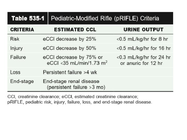
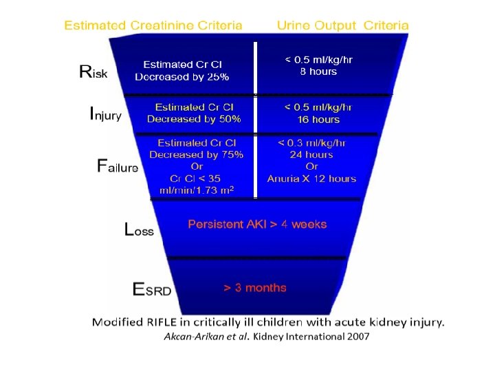
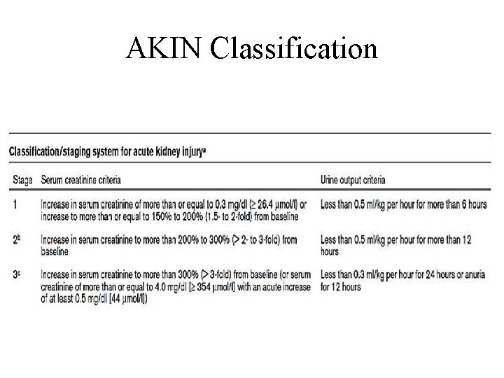
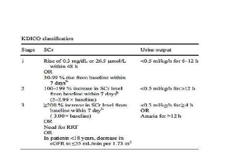
AKIN Classification
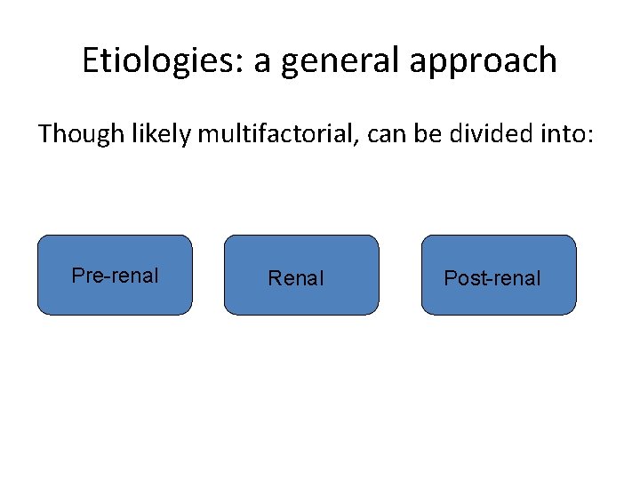
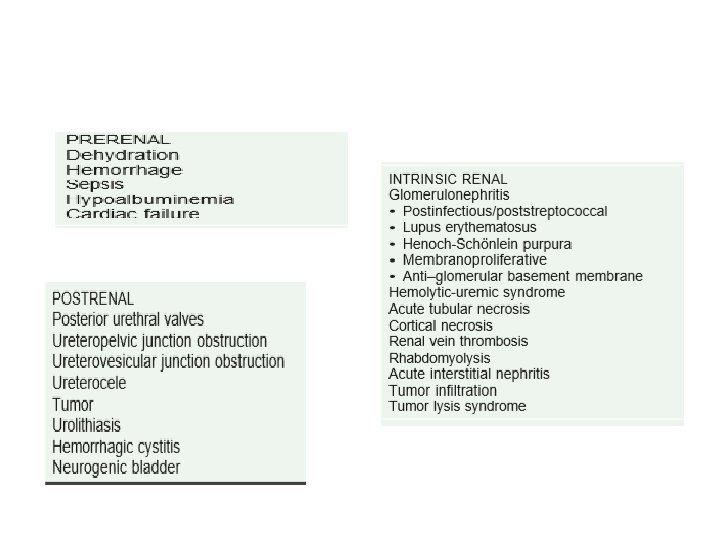
Etiologies: a general approach Though likely multifactorial, can be divided into: Pre-renal Renal Post-renal
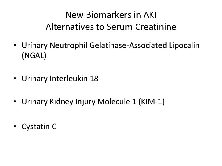
New Biomarkers in AKI Alternatives to Serum Creatinine • Urinary Neutrophil Gelatinase-Associated Lipocalin (NGAL) • Urinary Interleukin 18 • Urinary Kidney Injury Molecule 1 (KIM-1) • Cystatin C
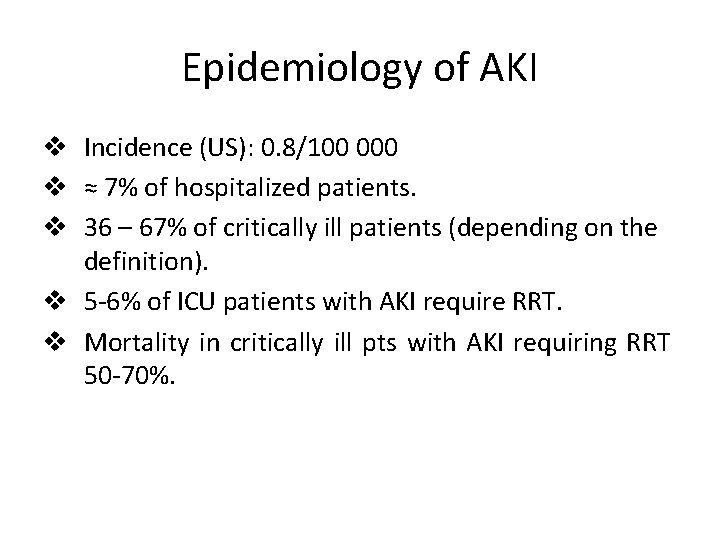
Epidemiology of AKI v Incidence (US): 0. 8/100 000 v ≈ 7% of hospitalized patients. v 36 – 67% of critically ill patients (depending on the definition). v 5 -6% of ICU patients with AKI require RRT. v Mortality in critically ill pts with AKI requiring RRT 50 -70%.
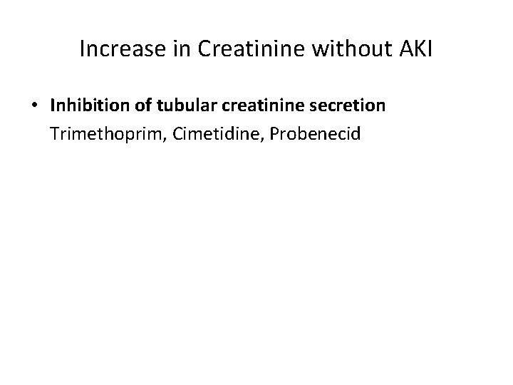
Increase in Creatinine without AKI • Inhibition of tubular creatinine secretion Trimethoprim, Cimetidine, Probenecid
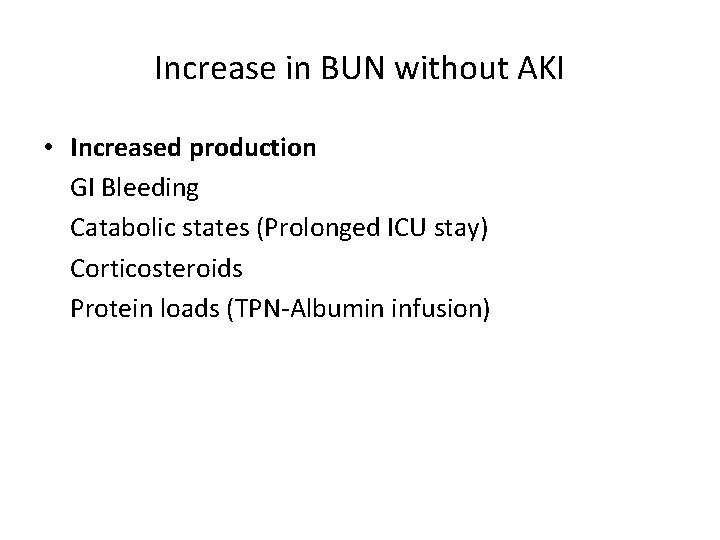
Increase in BUN without AKI • Increased production GI Bleeding Catabolic states (Prolonged ICU stay) Corticosteroids Protein loads (TPN-Albumin infusion)
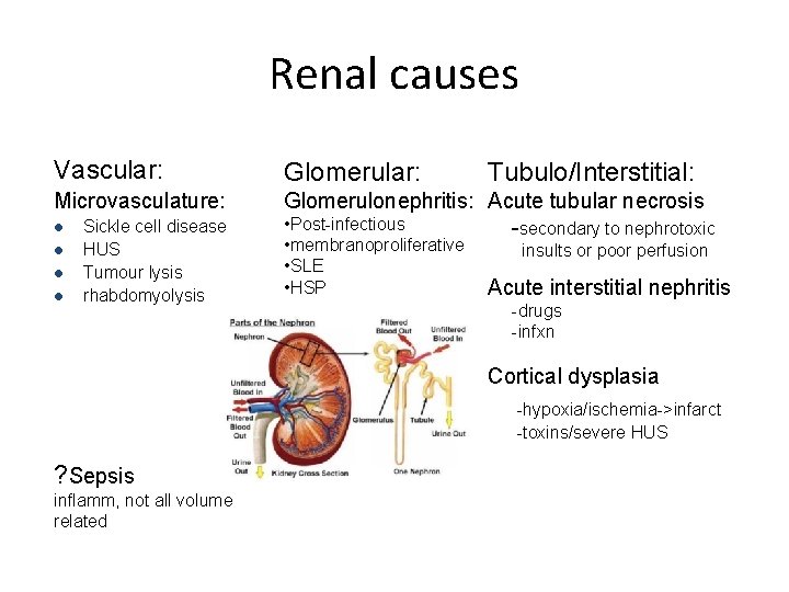
Renal causes Vascular: Glomerular: Microvasculature: Glomerulonephritis: Acute tubular necrosis • Post-infectious -secondary to nephrotoxic l l Sickle cell disease HUS Tumour lysis rhabdomyolysis • membranoproliferative • SLE • HSP Tubulo/Interstitial: insults or poor perfusion Acute interstitial nephritis -drugs -infxn Cortical dysplasia -hypoxia/ischemia->infarct -toxins/severe HUS ? Sepsis inflamm, not all volume related
Acute Tubular Necrosis Describes an end effect of tubular damage… • Secondary to perfusion insults • Secondary to toxins Change in blood flow, obstruction and passive filtrate backflow into tubular cells can cause a cycle leading to further death…
AIN Ø Drugs (71%) - 1/3 antibiotics Ø Penicillins, cephalosporins, NSAIDs, sulfonamides, cipro, rifampin, PPIs, allopurinol… and more Ø Infection (15%) Ø Strep, Legionella, leptospirosis, CMV, EBV… many Ø Tubulointersitial nephritis and uveitis (5%) Ø Autoimmune Ø Idiopathic
Nephrotoxins Vascular effect – ACEi, cyclosporine, tacrolimus Tubular effect – Proximal: aminoglycosides, amphotericin B, cisplatin, immunoglobulins, contrast – Distal: NSAIDs, ACEi, lithium, cyclophosphamide – Obstruction: sulfa, acylovir, methotrexate AIN
Post-renal causes Two kidneys - distal or bilateral proximal obstruction Single kidney - obstruction anywhere – – – – Posterior urethral valves Ureteropelvic junction obstruction Ureterovesicular junction obstruction Ureterocele Stones Tumour Hemorrhagic cystitis Neurogenic bladder
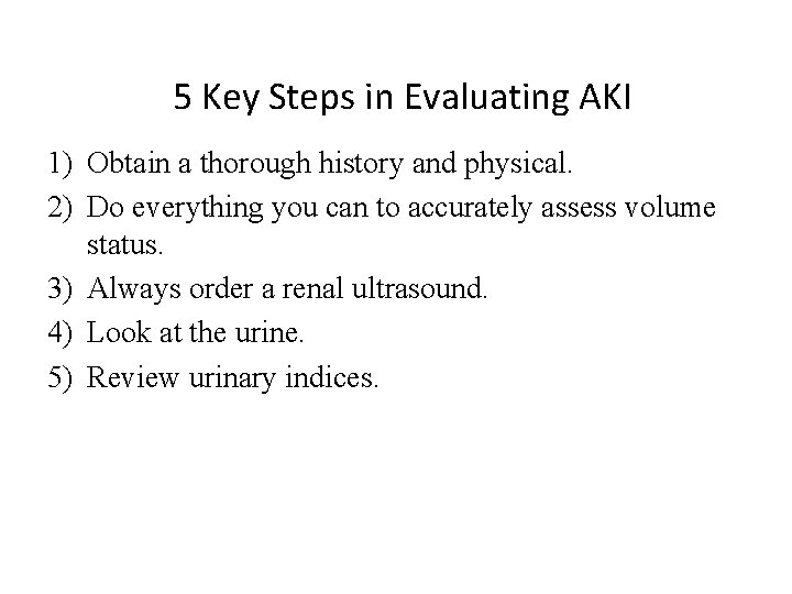
5 Key Steps in Evaluating AKI 1) Obtain a thorough history and physical. 2) Do everything you can to accurately assess volume status. 3) Always order a renal ultrasound. 4) Look at the urine. 5) Review urinary indices.
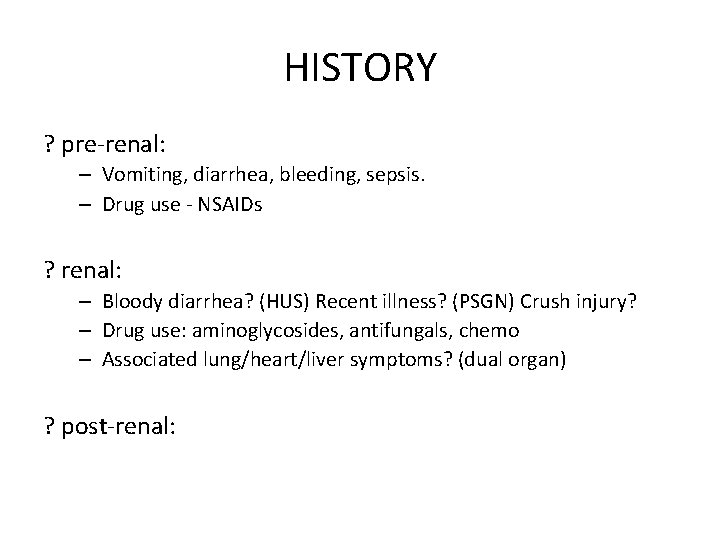
HISTORY ? pre-renal: – Vomiting, diarrhea, bleeding, sepsis. – Drug use - NSAIDs ? renal: – Bloody diarrhea? (HUS) Recent illness? (PSGN) Crush injury? – Drug use: aminoglycosides, antifungals, chemo – Associated lung/heart/liver symptoms? (dual organ) ? post-renal:
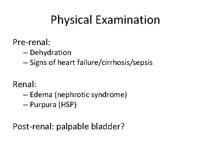
Physical Examination Pre-renal: – Dehydration – Signs of heart failure/cirrhosis/sepsis Renal: – Edema (nephrotic syndrome) – Purpura (HSP) Post-renal: palpable bladder?
What to order? BUN, Cr, lytes, fractional excretion of sodium Urinalysis
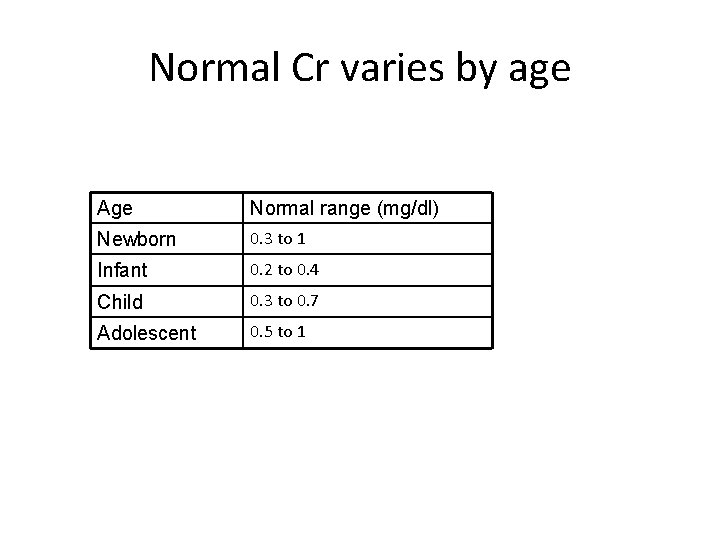
Normal Cr varies by age Age Normal range (mg/dl) Newborn 0. 3 to 1 Infant 0. 2 to 0. 4 Child 0. 3 to 0. 7 Adolescent 0. 5 to 1
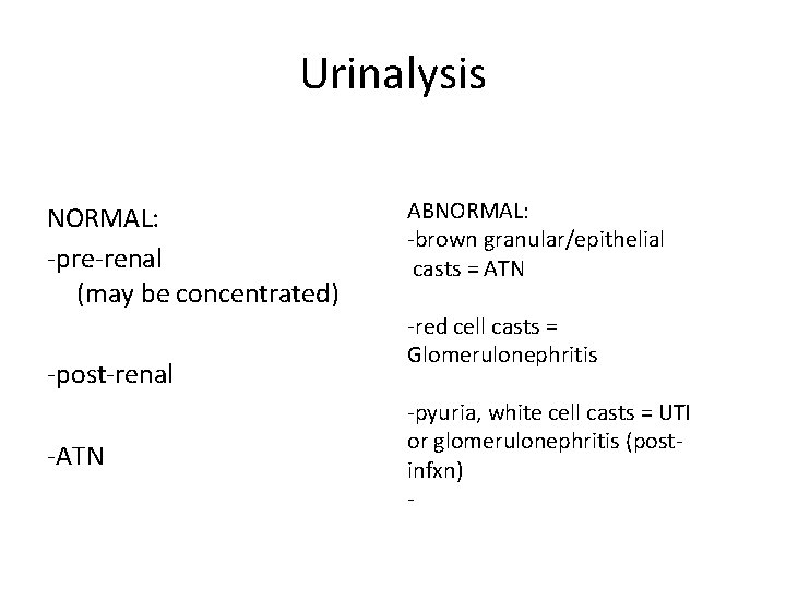
Urinalysis NORMAL: -pre-renal (may be concentrated) -post-renal -ATN ABNORMAL: -brown granular/epithelial casts = ATN -red cell casts = Glomerulonephritis -pyuria, white cell casts = UTI or glomerulonephritis (postinfxn) -
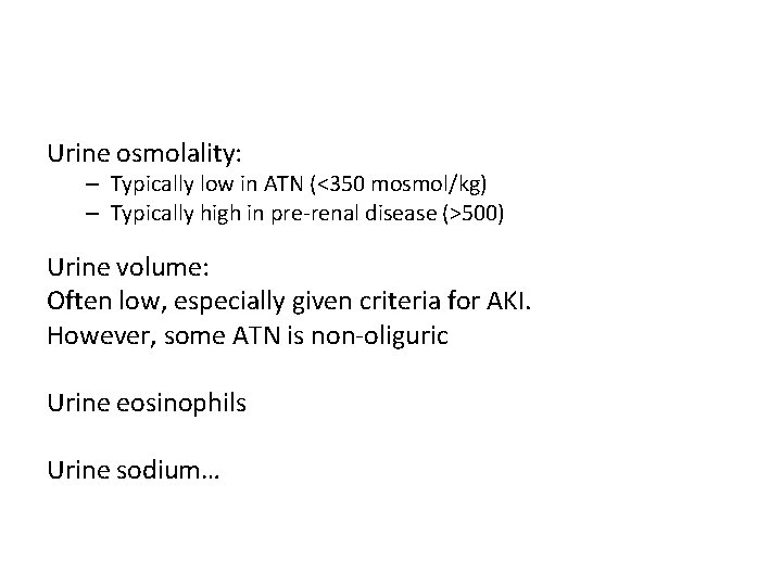
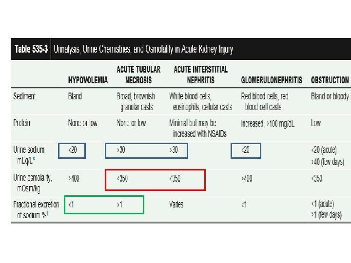
Urine osmolality: – Typically low in ATN (<350 mosmol/kg) – Typically high in pre-renal disease (>500) Urine volume: Often low, especially given criteria for AKI. However, some ATN is non-oliguric Urine eosinophils Urine sodium…
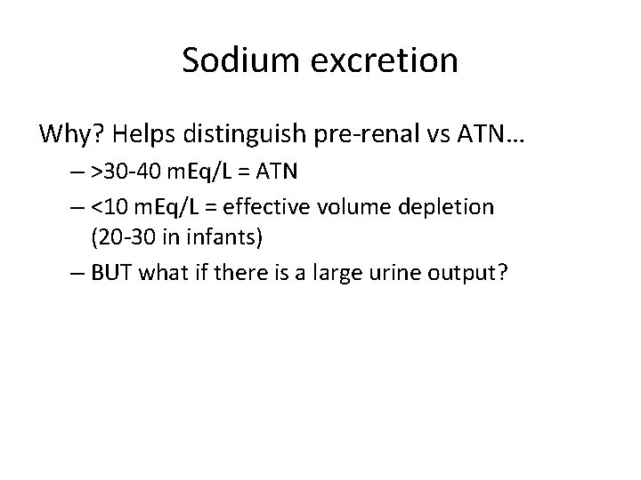
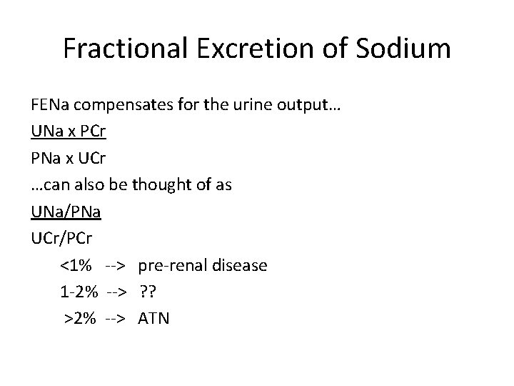
Sodium excretion Why? Helps distinguish pre-renal vs ATN… – >30 -40 m. Eq/L = ATN – <10 m. Eq/L = effective volume depletion (20 -30 in infants) – BUT what if there is a large urine output?
Fractional Excretion of Sodium FENa compensates for the urine output… UNa x PCr PNa x UCr …can also be thought of as UNa/PNa UCr/PCr <1% --> pre-renal disease 1 -2% --> ? ? >2% --> ATN
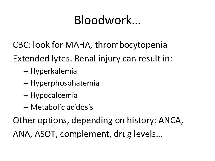
Bloodwork… CBC: look for MAHA, thrombocytopenia Extended lytes. Renal injury can result in: – Hyperkalemia – Hyperphosphatemia – Hypocalcemia – Metabolic acidosis Other options, depending on history: ANCA, ANA, ASOT, complement, drug levels…
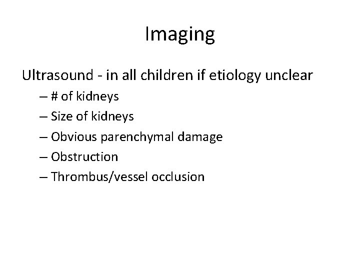
Imaging Ultrasound - in all children if etiology unclear – # of kidneys – Size of kidneys – Obvious parenchymal damage – Obstruction – Thrombus/vessel occlusion
Renal biopsy Only when diagnosis remains unknown, or there is a failure to respond to treatment
RRT Options: Continuous renal replacement therapy Peritoneal dialysis Hemodialysis
Prognosis Mortality: 60% (critically ill) 20 -25% go on to have some degree of chronic renal issues
Creatinine • None sensitive, > 50% of kidney function to be lost • Varies with age, muscle mass, gender • Tubular secretion
TREATMENT - Medical Management • Obstruction- PUV: bladder cath should be placed immediately for drainage • volume status : If there is no volume overload or cardiac failure, isotonic saline, 20 m. L/kg over 30 min. • After volume resuscitation, hypovolemic patients generally void within 2 hr; failure to do so suggests intrinsic or postrenal AKI. • Hypotension caused by sepsis: vigorous fluid resuscitation followed by norepinephrine infusion • Furosemide (2 -4 mg/kg) and mannitol (0. 5 g/kg) may be administered as a single IV dose
Hyperkalemia> 6 • Stop Exogenous sources of potassium (dietary, intravenous fluids, total parenteral nutrition) • Sodium polystyrene sulfonate resin (Kayexalate), 1 g/kg, should be given orally or by retention enema. • More severe elevations in serum potassium (>7 m. Eq/L), ECG changes • Calcium gluconate 10% solution, 1. 0 m. L/kg IV, over 3 -5 min • Sodium bicarbonate, 1 -2 m. Eq/kg IV, over 5 -10 min • Regular insulin, 0. 1 units/kg, with glucose 50% solution, 1 m. L/kg, over 1 hr • β-adrenergic agonists • Persistent hyperkalemia should be managed by dialysis.
Metabolic Acidosis • < 7. 15 to 12 • IV then oral • Correction of metabolic acidosis with intravenous bicarbonate can precipitate tetany in patients with renal failure as rapid correction of acidosis reduces the ionized calcium concentration
Management of ↑PO 4 and ↓Ca Hyperphosphatemia: – Low phosphate diet – Binders Hypocalcemia: – Calcium gluconate if Tetany. – Usually by ↓ PO 4
Others • • • Hypertension Anemia GI protection Neurological disorders Dialysis Nutritional support
Indication of Dialysis • Anuria/oliguria • Volume overload with evidence of hypertension and/or pulmonary edema refractory to diuretic therapy • Persistent hyperkalemia • Severe metabolic acidosis unresponsive to medical management • Uremia (encephalopathy, pericarditis, neuropathy) • Blood urea nitrogen >100 -150 mg/d. L (or lower if rapidly rising) • Calcium: phosphorus imbalance, with hypocalcemic tetany that cannot be controlled by other measures
Questions