Pediatric II INFECTIONS Bronchiolitis Bronchiolitis a lower respiratory

Pediatric II INFECTIONS
 that primarily affects the small airways")
Bronchiolitis Bronchiolitis, a lower respiratory tract infection (LRTI) that primarily affects the small airways (bronchioles), is a common cause of illness and hospitalization in infants and young children. 1 -Bronchiolitis is seasonal, with peak activity during winter and early spring. Bronchiolitis occurs almost exclusively during the first 2 years of life, with a peak age at 2 to 6 months. Acute bronchiolitis is characterized by bronchiolar obstruction with edema, mucus, and cellular debris.
")
Etiology � 1 -Acute bronchiolitis is predominantly a viral disease. Respiratory syncytial virus (RSV) is responsible for more than 50% of cases. � 2 -Other agents include parainfluenza, adenovirus, Mycoplasma, and occasionally other viruses.

Clinical Manifestation �The infant first develops a mild upper respiratory tract infection with sneezing and clear rhinorrhea. This may be accompanied by diminished appetite and fever. �Gradually, respiratory distress ensues, with paroxysmal wheezy cough, dyspnea, and irritability. The infant is often tachypneic, which interferes with feeding. �As a result of limited oral intake due to coughing combined with fever, infants are frequently dehydrated.

Diagnosis �The diagnosis of bronchiolitis is based primarily on history and clinical findings.

Normal Infected

Treatment �The mainstay of treatment is supportive. Therapy of bronchiolitis primarily consists of administration of supplemental oxygen and replacement of fluid deficits (hydration) as needed. �The risk of aspiration of oral feedings may be high in infants with bronchiolitis owing to tachypnea and the increased work of breathing. The infant may be fed through a nasogastric tube.

� 3 -A number of agents have been proposed as adjunctive therapies for bronchiolitis: �A-Bronchodilators produce modest short-term improvement in clinical features. Nebulized epinephrine may be more effective than β-agonists. �B-Corticosteroids, whether parenteral, or inhaled, are widely used despite conflicting studies.

�C-Ribavirin, is a compound with antiviral activity against RSV administered by aerosol, has been used for infants with congenital heart disease (CHD)or chronic lung disease (CLD) although its benefit is uncertain. �D-Antibiotics have no value unless there is secondary bacterial pneumonia.

�Prophylaxis �Palivizumab is a monoclonal antibody to RSV and can be used as prophylaxis initiated just before the onset of the RSV season (monthly IM injection for 5 months starting in October) confers some protection from severe RSV disease

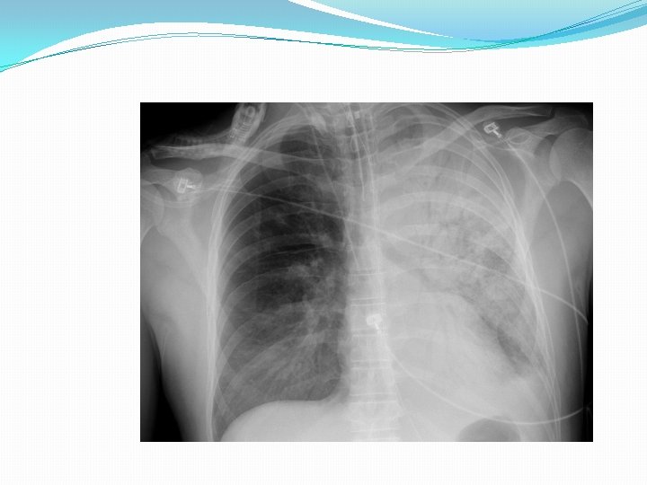
Pneumonia �Pneumonia is defined as infection of the lung parenchyma (that is of the alveoli rather than the bronchi or bronchioles) and characterized by consolidation. (Consolidation is a pathological process in which the alveoli are filled with a mixture of inflammatory exudate, bacteria and WBCs that on chest X-ray appear as an opaque shadow in the normally clear lungs)

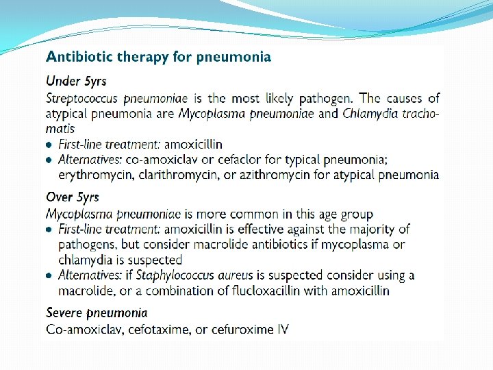
�Viruses alone account for 14– 35% of all community acquired pneumonia in childhood. M. pneumoniae and Chlamydophila pneumoniae are principal causes of atypical pneumonia. Common infecting bacterial agents by age are : � 1 -Neonates: group B streptococcus, Escherichia coli, Klebsiella, Staphylococcus aureus. � 2 -Infants: Streptoccus pneumoniae, Chlamydia. � 3 -School age: Streptococcus pneumoniae, Staphylococcus aureus, group A streptococcus, Bordetella pertussis, Mycoplasma pneumoniae

Clinical manifestation �In many cases these symptoms are preceded by minor upper respiratory tract infection symptoms. The patient may also be complaining of pleuritic chest pain or abdominal pain. The typical history will have: � Temperature > 38. 5 0 C; � Tachypnea and Shortness of breath; � Cough; [with sputum production in older children (>7 yrs)].

Diagnosis �Diagnosis of pneumonia in many cases is made based on the presence of clinical signs and symptoms. �Chest x-ray are often used to confirm the diagnosis.

Treatment �Oral antibiotics are safe and effective in the treatment of community acquired pneumonia. IV antibiotics are used in children who cannot absorb oral antibiotics or in those with severe symptoms.

�Supportive therapies Consider whether any of the following are needed: � Antipyretics for fever. � IV fluids: consider if dehydrated or not drinking. • Supplemental oxygen
, whereas encephalitis is an")
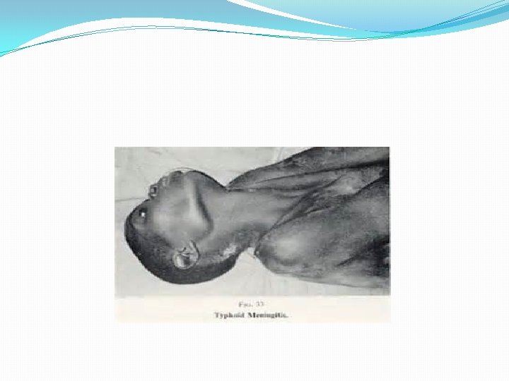
Meningitis �Meningitis is an inflammation of the membranes (the meninges), whereas encephalitis is an inflammation of the brain tissue. 75% of cases of meningitis are believed to occur in those <15 yrs of age.

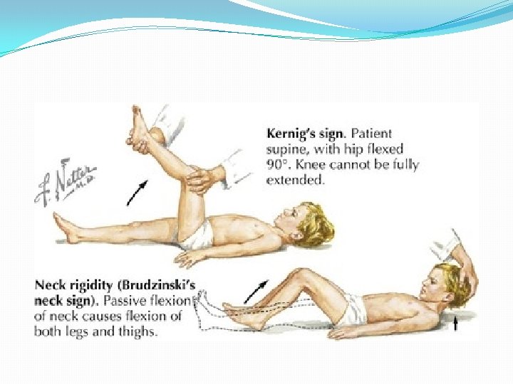
Clinical manifestation 1 -In young infants symptoms may be non-specific including fever, poor feeding, lethargy. 2 -In older children clinical features include: • General: fever, with headache. • Central: irritability, disorientation, altered mental state. • Seizures: occur in 30%. • Neck stiffness: more common in older children. • Kernig and Brudzinski signs of meningeal irritation are often positive in children older than 12 months.

Diagnosis �If bacterial meningitis is suspected, a lumbar puncture should be performed. Routine CSF examination includes a white blood cell count, differential, protein and glucose levels, and Gram stain. �

Treatment of bacterial meningitis focuses on sterilization of the CSF by antibiotics. Steroids In bacterial meningitis: • Do not use corticosteroids in children younger than 3 mths. • There is benefit from the use of dexamethasone and the dosing schedule is 0. 15 mg/kg qds for 4 days to reduce the severity of neurological sequelae, particularly deafness, after bacterial meningitis). • If dexamethasone was not given before the first dose of antibiotics, but was indicated, try to give the first dose within 4 hr of starting antibiotics

Encephalitis �Encephalitis is an inflammation of the brain tissue. Viruses are the principal causes of acute infectious encephalitis

Clinical manifestation �Acute infectious encephalitis usually is preceded by a prodrome of several days of nonspecific symptoms such as sore throat, fever, and headache followed by the characteristic symptoms of progressive lethargy, behavioral changes, and neurologic deficits. Seizures are common at presentation.

Diagnosis �The diagnosis of viral encephalitis is supported by examination of the CSF.

Treatment �With the exception of HSV, varicella-zoster virus, cytomegalovirus, and HIV, there is no specific therapy for viral encephalitis. Management is supportive. � 2 -Intravenous acyclovir is the treatment of choice for HSV and varicella-zoster virus infections. Cytomegalovirus infection is treated with ganciclovir. HIV infections may be treated with a combination of antiretroviral agents.
 is caused by the protozoon Leishmania donovani. Ø")
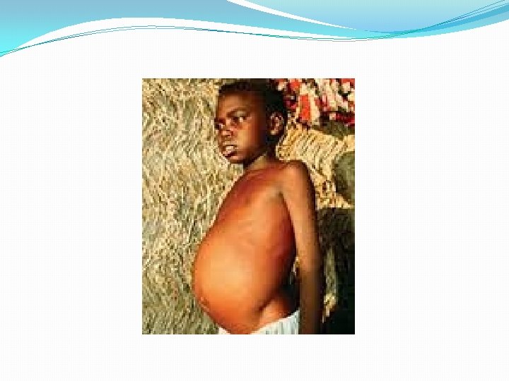
Visceral leishmaniasis Ø Visceral Leishmaniasis (VL) is caused by the protozoon Leishmania donovani. Ø Infection are introduced by the feeding female sand fly. Ø The great majority of people infected remain asymptomatic. In visceral diseases the spleen, liver, bone marrow and lymph nodes are primarily involved.

Diagnosis: Ø VL is predominantly a disease of small children and infants. Ø The first sign of infection is high fever, usually accompanied by rigor and chills. Ø Splenomegaly develops quickly in the first few weeks and becomes massive as the disease progresses. Moderate hepatomegaly occurs later. Lymphadenopathy may also seen. Ø Blackish discoloration of the skin, from which the disease derived its name, kala-azar (the Hindi word for ‘black fever’), is a feature of advanced illness and is now rarely seen. Ø Pancytopenia is a common feature. Ø Without adequate treatment most patients with clinical VL die.

Diagnosis Demonstration of amastigotes in splenic smears is the most efficient means of diagnosis, with 98% sensitivity ; however, it carries a risk of serious haemorrhage in inexperienced hands. Serodiagnosis, by ELISA or indirect immunofluorescence antibody test (IFAT). A significant proportion of the healthy population in an endemic region will be positive for these tests due to past exposure.
![Treatment The pentavalent antimony compound [sodium stibogluconate (Pentostam®)]. The daily dose is 20 mg/kg](http://slidetodoc.com/presentation_image_h2/3afe51b4d652fb9c8b2d7b468ac82fd3/image-33.jpg "Treatment The pentavalent antimony compound [sodium stibogluconate (Pentostam®)]. The daily dose is 20 mg/kg")
Treatment The pentavalent antimony compound [sodium stibogluconate (Pentostam®)]. The daily dose is 20 mg/kg body weight, given either intravenously or intramuscularly for 28 days. Side-effects are common and include arthralgias, myalgias, raised hepatic transaminases, pancreatitis and ECG changes. Amphotericin B is very useful in the treatment of antimonyunresponsive VL �
 It is an infection of the small intestine, which present")
Gastroenterology �Acute Gastroenteritis (GE) It is an infection of the small intestine, which present with a combination of diarrhea and vomiting, but sometimes present without vomiting. �Rota virus is the most common pathogen in children under 2 years, other causes include: A- Acute bacterial infections (shigellae, Salmonellae, E coli and Vibirio cholera which secrete enterotoxins). B-Parasites like E. histolytica, and Giardia lambilia

Clinical features �Rotaviruse cause watery diarrhea. Respiratory illness occur in about half of patients followed by vomiting and diarrhea. 2 -Acute bacterial infection cause invasion of GIT, so there is fever, and small volume bloody stool. Dehydration, metabolic disturbances and even death are complications of GE.

Treatment �Uncomplicated viral GE requires no specific treatment except attention to fluid and electrolyte replacement (3) Most of these episodes are self-limited. �There is no role for antiemetic or antidiarrheal in GE �Antibiotics are rarely indicated except for specific infections such as invasive salmonellosis, cholera , amebiasis or giardiasis. �The key management of GE is rehydration with correction of fluid and electrolyte imbalance.

Signs of dehydration

�Mild Dehydration: ORS are used. �Moderate dehydration: Oral rehydration is still indicated if tolerated. �I. V fluid should be reserved for those with vomiting or severe dehydration. �Zinc supplementation (10– 20 mg for 10– 14 days) has been recommended by the WHO for the treatment and prevention of diarrheal disease in children in developing countries �Continuation of oral feeding, despite diarrheal episodes, decreases the duration of illness; and improves nutritional status
- Slides: 38