Pediatric CCJ Companion Nadja Kadom Gilbert Vezina Raymond

Pediatric CCJ Companion Nadja Kadom, Gilbert Vezina, Raymond Sze

C-spine measurements Trauma Platybasia Basilar Invagination

Assess the C-Spine/skull base Trauma: • Alignment • Soft tissue swelling • Occiput-C 1 dissociation • C 1 -C 2 instability Basilar Invagination • Chamberlain • Mc. Gregor Platybasia • Standard • Modified

Trauma

Prevertebral/Retropharyngeal Soft Tissues • False thickening: flexion, end of expiration Flexed Extended

Prevertebral/Retropharyngeal Soft Tissues Normal thickness: • < 7 mm anterior to C 2 and < 5 mm anterior to C 3/C 4 • or less than half the diameter of the vertebral bodies
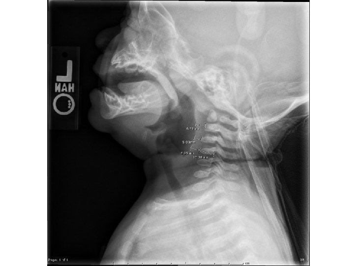

Prevertebral/Retropharyngeal Soft Tissues Loss of physiological mucosal step off ~C 4/5 is ABNORMAL! Step-off ~ C 4 -6 Lost step-off

Alignment

Soft Tissue Swelling In children: Retropharyngeal tissues should NOT exceed 1/2 to 2/3 vertebral body AP diameter

Evaluate Occiput-C 1 dissociation: Suggestion: Get C 0 -C 2 CT scan Landmarks not seen on x-ray, get limited CT
: 8. 37 (pediatric normal < 12. 5 mm) Basion-Axial Line-Interval (BAI):")
CT Basion-Dens-Interval (BDI): 8. 37 (pediatric normal < 12. 5 mm) Basion-Axial Line-Interval (BAI): 5. 56 (adult normal < 12 mm)

Other methods • Power ratio • Lee X

Power Ratio A = the anterior tubercle of the atlas. B. = the basion. C = the spinolaminar line of the atlas. O = the opisthion The value BC/AO should be less than 1. BC/AO = 30. 21/39. 59 < 1 normal
 and posterior atlanto-dens interval (PADI) ADI")
C 1 -C 2 instability Atlanto-dens interval (ADI) and posterior atlanto-dens interval (PADI) ADI = 3. 24 mm (normal < 5 mm) PADI = 21. 92 (abnormal < 13 mm)

Occiput-C 1 Pathology • Axial dislocation (dislocation in the axial plane, anterior or posterior “listhesis” of occiput versus C 1, best seen on sagittal images) • Sagittal dislocation (dislocation in the sagittal plane, increased height of space between occipital condyles and C 1 articulation, seen on coronal and sagittal images)
 1. CCI physiologically narrow normal pediatric mean is 1.")
Occipital Condyle-C 1 Interval (CCI) 1. CCI physiologically narrow normal pediatric mean is 1. 28 mm, normal range 0. 25 -2. 5 mm 2. The left and right OC 1 joints are normally highly symmetrical Right Left

Example of CCI enlargement

Example of asymmetry

Wackenheim line • Assess antlanto-occipital dissociation • Line along the posterior border of the clivus should inferiorly touch the odontoid tangentially

Examples Normal Posterior dislocation

Rotatory subluxation C 1 -C 2 • 4 types • Assess the facet joints, look for: => displaced facets on sagittal views => visualization of both articular surfaces in one axial image

Type I: simple rotatory displacement; < 3 mm with an intact transverse ligament. Type II: anterior displacement of C 1 on C 2 of 3 -5 mm (one lateral mass serving as a pivot point) + deficiency of the transverse ligament. Type III: injuries involve > 5 mm of anterior displacement. Type IV: injuries involve the posterior displacement of C 1 on C 2. Both Type III and IV are highly unstable injuries.

Basilar Invagination

Basilar Invagination

Definition • The tip of the dens projects more than 5 mm above Chamberlain's line • Or the tip of the dens is >7 mm above Mc. Gregor's line

Chamberlain’s line • line joining the hard palate to the posterior lip of the foramen magnum

Mc. Gregor’s line • the back of the hard palate to the lowest point of the occipital squama

Platybasia Standard technique: • measuring the angle formed by two lines: 1 st line: nasion to center of the pituitary fossa 2 nd line: anterior border of foramen magnum with center of the pituitary fossa (= tip of clivus to center of pituitary) Normal: • Adult: 129° +/- 6° • Pediatric: 127° +/- 5° Koenigsberg RA, Vakil N, Hong TA, Htaik T, Faerber E, Maiorano T, Dua M, Faro S, Gonzales C. Evaluation of platybasia with MR imaging. AJNR Am J Neuroradiol. 2005 Jan; 26(1): 89 -92.

Standard: Pediatric: 127° +/- 5°

Platybasia Modified technique: Uses different landmarks • measuring the angle formed by two lines: 1 st line: extending across the anterior cranial fossa to the tip to the dorsum sellae 2 nd line: connecting with a line drawn along the posterior margin of the clivus Normal: • Adult: 117° +/- 6° • Pediatric: 114. 4° +/- 5°

Modified: Pediatric: 114. 4° +/- 5°
- Slides: 32