PECTORAL REGION Happy Teachers Day 05 09 2018


 Sternal")

 Breast : nipple,")


 at the deltopectoral triangle,")

")
 n - Supraclavicular nerve (C 3 -4,")


- Slides: 29
PECTORAL REGION Happy Teacher’s Day 05. 09. 2018 Dr. K. S. Rav i
OBJECTIVES • By the end of the lecture the students should be able to : • Ø Ø Ø Ø Identify and describe the muscles of the pectoral region. Pectoralis major. Pectoralis minor. Subclavius. Serratus anterior. Action of these muscles Clavipectoral Fascia Blood & Nerve Supply • Clinical Relevance
Pectoral region 1. Bony landmark : Sternum: Jugular notch (body of T 2) Sternal angle of Louise (T 4 -5) Xiphosternal joint (T 9) Ribs & costal cartilage
Scapular: acromian, coracoid process Clavicle: Humerus: head • supraclavicular fossa • infraclavicular fossa
2 . Superficial structure - skin & derivative of skin (breast) Breast : nipple, areolar, mammary gland (F) 1. Surface Anatomy (position) : Nipple – 4 -5” from the midline, intercostal space 4 Breast – between rib 2 -7
2. Components : - subcutaneous fat, mammary gland - Lactiferous duct - Cooper’s ligament (suspensory ligament) - Retinaculum cutis fascia
The majority of the breast is in the superficial fascia, except the tail part (Tail of Spence) extends upward laterally into deep fascia at the lower border of pectoralis major. 2/3 of the gland lies on pectoralis major 1/3 of the gland lies on serratus anterior
Deltopectoral Triangle -deep fascia separating deltoid and pectoralis muscles - Platysma = superficial muscle, thin plate, extends from the mandible to the clavicle
3. Muscles of region pectoral a. Pectoralis Major b. Pectoralis Minor c. Serratus Anterior d. Subclavious
Pectoralis major Origin Anterior sternal half of the clavicle; Manubrium and Sternum upto sixth costal cartilages Cartilages of all the true ribs, Aponeurosis of the abdominal external oblique Insertion By a bilaminar tendon into the lateral lip of the bicipital groove of the humerus Innervation Medial and lateral pectoral nerves
Actions Flexion of the humerus, Adduction of the humerus and Medial rotation of the humerus. Clavicular part : flexion, adduction, and medial rotation of the humerus. Sternocostal part extension of the flexed arm as in climbing. It aids in deep inspiration.
Pectoralis minor Origin It arises from the upper margins and outer surfaces of the third, fourth, and fifth ribs, Inserted into the medial border and upper surface of the coracoid process of the scapula. Innervation Medial and lateral pectoral nerves Actions Protracts the scapula with serratus anterior Depresses the shoulder with the rhomboids and levator scapulae Important The pectoralis minor muscle is covered by the clavipectoral fascia. The medial pectoral nerve pierces the pectoralis minor. Axillary artery is divided into three parts by pectoralis minor.
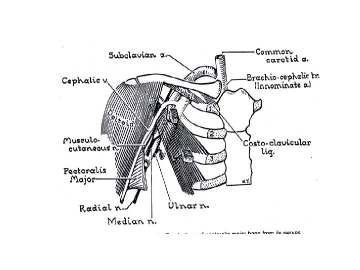
Clavipectoral fascia Encloses subclavius and Pectoralis Minor. It is pierced by : Lateral pectoral nerve. Thoraco- acromial artery Cephalic vein. Lymph nodes from pectoral region to apical group of axillary lymph nodes
Pectoral. Girdle : clavicle, scapular, ribs Clavipectoral fascia / Costocoracoid membrane - deep fascia separating the pectoralis and the subclavious
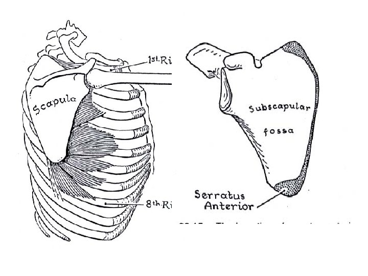
Serratus anterior Origin Arises from ribs 1 to 8, to be inserted into the medial border of the scapula. Insertion • Medial border of the scapula between the superior and inferior angles. • 1 st and 2 nddigitations to upper angle of scapula. (C 5) • 3 rd and 4 th digitations to medial border on costal surface upto the inferior angle. • Lower 4 digitations to inferior angle of scapula. Action Protraction of the scapula along with pectoralis minor. • The fibres inserted on inferior angle rotate scapula laterally and upwards in overhead abduction with trapezius. Assists in respiration. Innervation long thoracic nerve(Nerve of Bell)
-Blood supply to the pectoral region 1. Axillary artery 2. Perforating branches of the internal thoracic a.
1. Axillary artery : divided into 3 parts First part : Supreme thoracic a. Second part : 1. Thoraco-acromial trunk Acromial branch Pectoral branch Clavicular branch Deltoid branch 2. Lateral thoracic a. Third part : give branches to supply head of humerus and scapular regions
Venous drainage at the pectoral region . 1 Deep veins - axillary v. <= from the muscles
. 2 Superficial veins - cephalic v. (from upper limb) at the deltopectoral triangle, it pierces the clavipectoral fascia (or infraclavicular fossa) into the axillary v. -from mammary gland, it drains into deep veins => internal thoracic v. and lateral thoracic v.
Nerve Supply of the pectoral region . 1 - medial & lateral pectoral nerve (terminal branches from the cords of the Brachial plexus (C 58 & T 1) - nerve to subclavius (a branch from upper trunk of the Brachial plexus) - long thoracic nerve (nerve roots from C 5 -6 -7 of the Brachial plexus)
Brachial plexus (C 5 -8 & T 1)
. 2 Spinal n. / Sensory (cutaneous) n - Supraclavicular nerve (C 3 -4, medial, intermediate & lateral branches) - Intercostal nerve T 3 -7 (anterior & lateral cutaneous branches) *Dermatome at the pectoral region: C 3 -4, T 3 -7
Applied aspect • Serratus anterior is called the Boxer’s muscle since it is responsible for pushing and punching movements. • Paralysis of this muscle results in a "winged scapula" , results in protrusion of the scapula on the affected side when the patient is asked to push against the wall with both arms extended. • Winged scapula occurs in lateral thoracic nerve paralysis
Paralysis of Serratus anterior muscle.
Clinical Relevance. 1 Chest wall – heart /lung sound . 2 Clavipectoral fascia - protection of the vessels and nerves underneath -limit spreading of the abscess from upper limb to neck . 3 Fracture of clavicle -common site is at 1/3 from the lateral -Poland Anomaly -Cardiac Catheterisation- Basilic vein the
1. Which one of the following muscles performs adduction of the arm ? a. Pectoralis minor. b. Pectoralis major. c. Subclavius. d. Serratus anterior. 2. Serratus anterior is innervated by : a. Thoracodorsal nerve. b. Long thoracic nerve. c. Axillary nerve. d. Radial nerve. 3. Which one of the following muscles contributes in rotation of the scapula above the head? a. Pectoralis major. b. Pectoralis minor. c. Serratus anterior. d. Teres major. 4. Which one of the following do not pierces clavipectoral fascia? a. Lateral Pectoral Nerve. b. Lymph Nodes. c. Cephalic Vein. d. Lateral thoracic artery. 5. Nerve to subclavius is a branch from which part of brachial plexus? a. Roots. b. Divisions. c. Cords. D. Trunks.
THANK YOU