
Pectoral Region and Axilla Anatomy Team 434 Color
- Slides: 26
Pectoral Region and Axilla Anatomy Team 434 Color Index: ▪ ▪ ▪ Important Points Helping notes Explanation We do advise you to read everything If you have any complaint or suggestion please don’t hesitate to contact us on: Anatomy. Team 434@gmail. com
OBJECTIVES ● Identify and describe the muscles of the pectoral region. Pectoralis major. Pectoralis minor. Subclavius. Serratus anterior. ● Describe and demonstrate the boundaries and contents of the axilla.
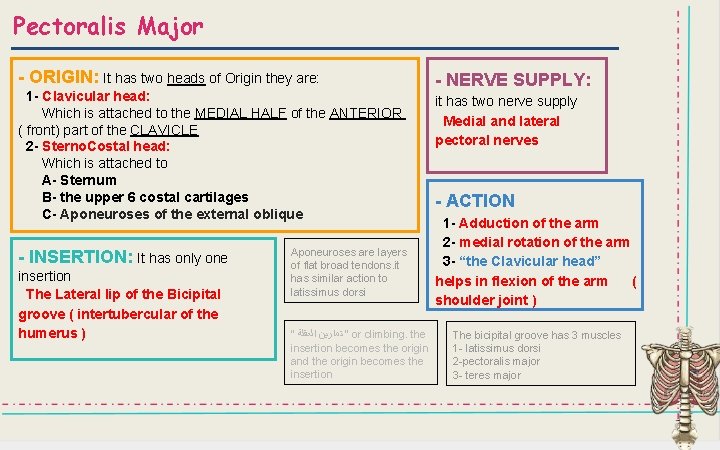
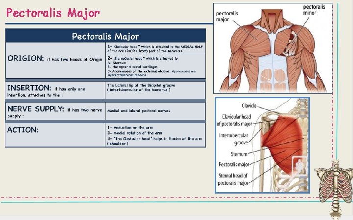
Pectoralis Major - ORIGIN: It has two heads of Origin they are: 1 - Clavicular head: Which is attached to the MEDIAL HALF of the ANTERIOR ( front) part of the CLAVICLE 2 - Sterno. Costal head: Which is attached to A- Sternum B- the upper 6 costal cartilages C- Aponeuroses of the external oblique - INSERTION: It has only one insertion The Lateral lip of the Bicipital groove ( intertubercular of the humerus ) Aponeuroses are layers of flat broad tendons. it has similar action to latissimus dorsi “ “ ﺗﻤﺎﺭﻳﻦ ﺍﻟﻌﻘﻠﺔ or climbing. the insertion becomes the origin and the origin becomes the insertion - NERVE SUPPLY: it has two nerve supply Medial and lateral pectoral nerves - ACTION 1 - Adduction of the arm 2 - medial rotation of the arm 3 - “the Clavicular head” helps in flexion of the arm ( shoulder joint ) The bicipital groove has 3 muscles 1 - latissimus dorsi 2 -pectoralis major 3 - teres major
Pectoralis Major
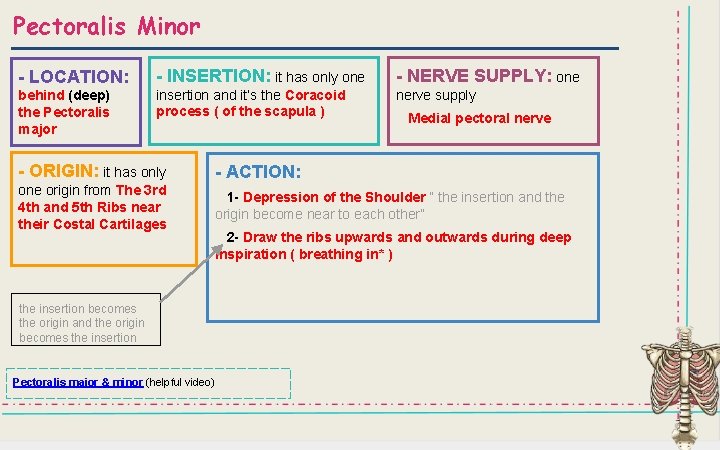
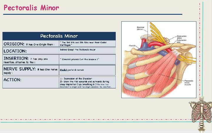
Pectoralis Minor - LOCATION: - INSERTION: it has only one - NERVE SUPPLY: one behind (deep) the Pectoralis major insertion and it’s the Coracoid process ( of the scapula ) nerve supply Medial pectoral nerve - ORIGIN: it has only - ACTION: one origin from The 3 rd 4 th and 5 th Ribs near their Costal Cartilages 1 - Depression of the Shoulder “ the insertion and the origin become near to each other” 2 - Draw the ribs upwards and outwards during deep inspiration ( breathing in* ) the insertion becomes the origin and the origin becomes the insertion Pectoralis major & minor (helpful video)
Pectoralis Minor
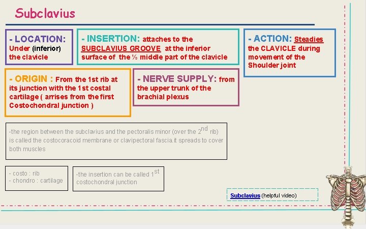
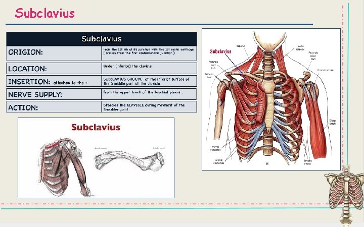
Subclavius - LOCATION: - INSERTION: attaches to the - ACTION: Steadies Under (inferior) the clavicle SUBCLAVIUS GROOVE at the inferior surface of the ⅓ middle part of the clavicle the CLAVICLE during movement of the Shoulder joint - ORIGIN : From the 1 st rib at - NERVE SUPPLY: from its junction with the 1 st costal cartilage ( arrises from the first Costochondral junction ) the upper trunk of the brachial plexus -the region between the subclavius and the pectoralis minor (over the 2 nd rib) is called the costocoracoid membrane or clavipectoral fascia. it spreads to cover both muscles - costo : rib - chondro : cartilage -the insertion can be called 1 st costochondral junction Subclavius (helpful video)
Subclavius
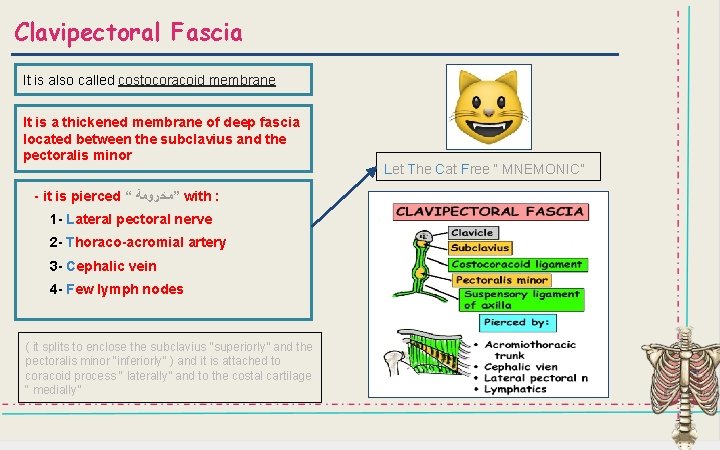
Clavipectoral Fascia It is also called costocoracoid membrane It is a thickened membrane of deep fascia located between the subclavius and the pectoralis minor - it is pierced “ ”ﻣﺨﺮﻭﻣﺔ with : 1 - Lateral pectoral nerve 2 - Thoraco-acromial artery 3 - Cephalic vein 4 - Few lymph nodes ( it splits to enclose the subclavius “superiorly” and the pectoralis minor “inferiorly” ) and it is attached to coracoid process “ laterally” and to the costal cartilage “ medially” Let The Cat Free “ MNEMONIC”
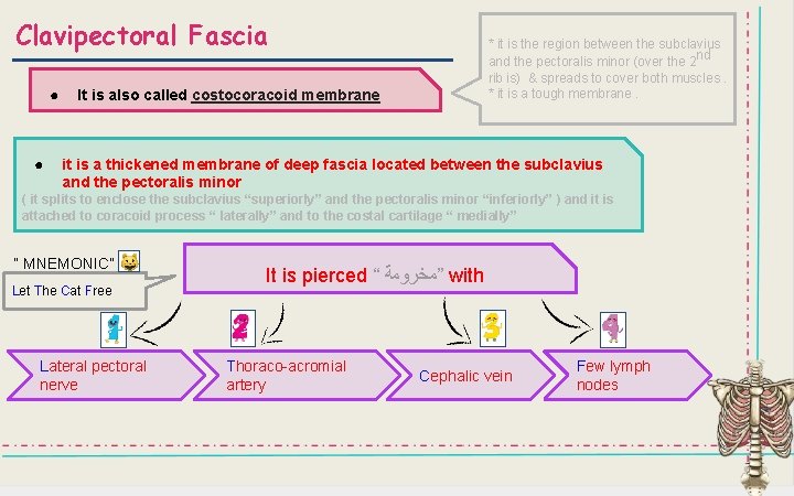
Clavipectoral Fascia ● ● * it is the region between the subclavius and the pectoralis minor (over the 2 nd rib is) & spreads to cover both muscles. * it is a tough membrane. It is also called costocoracoid membrane it is a thickened membrane of deep fascia located between the subclavius and the pectoralis minor ( it splits to enclose the subclavius “superiorly” and the pectoralis minor “inferiorly” ) and it is attached to coracoid process “ laterally” and to the costal cartilage “ medially” “ MNEMONIC” Let The Cat Free Lateral pectoral nerve It is pierced “ ”ﻣﺨﺮﻭﻣﺔ with Thoraco-acromial artery Cephalic vein Few lymph nodes
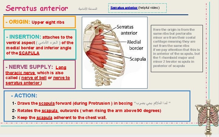
Serratus anterior ﺍﻟﻤﺴﻨﻨﺔ ﺍﻷﻤﺎﻣﻴﺔ Serratus anterior (helpful video) - ORIGIN: Upper eight ribs - INSERTION: attaches to the ventral aspect ( ) ﺍﻟﺠﺰﺀ ﺍﻷﻤﺎﻣﻲ of the medial border and inferior angle of the SCAPULA - NERVE SUPPLY: Long thoracic nerve which is also called ( nerve of bell or nerve to serratus anterior ) Here the origin is from the same ribs but pectoralis minor are from their costal cartilage meaning they are not from the same ribs if we pay attention that this is in anterior of the scapula. but the 1 rhomboid major and minor 2 levator scapula in posterior of scapula - ACTION: 1 - Draws the scapula forward (during Protrusion ) in boxing “ “ ﻟﻤﺎ ﺍﻟﻤﻼﻛﻢ ﻳﺠﻲ ﻳﻀﺮﺏ 2 - Rotates the scapula outwards ( when rising the arm above 90 degrees) 3 - Keep the scapula adherent to the chest wall.
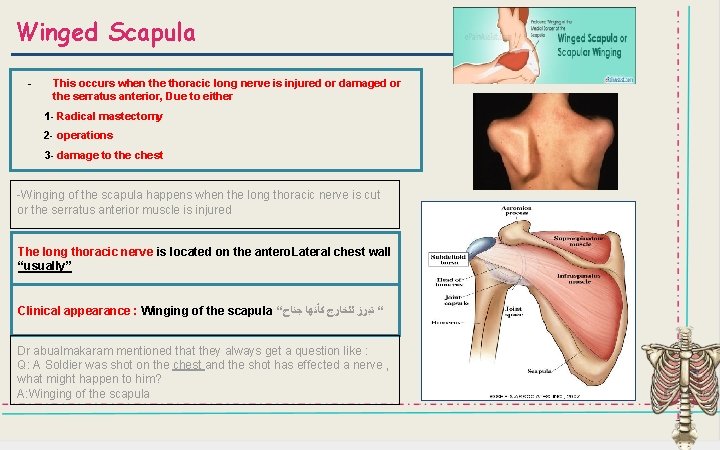
Winged Scapula - This occurs when the thoracic long nerve is injured or damaged or the serratus anterior, Due to either 1 - Radical mastectomy 2 - operations 3 - damage to the chest -Winging of the scapula happens when the long thoracic nerve is cut or the serratus anterior muscle is injured The long thoracic nerve is located on the antero. Lateral chest wall “usually” Clinical appearance : Winging of the scapula “ “ ﺗﺒﺮﺯ ﻟﻠﺨﺎﺭﺝ ﻛﺄﻨﻬﺎ ﺟﻨﺎﺡ Dr abualmakaram mentioned that they always get a question like : Q: A Soldier was shot on the chest and the shot has effected a nerve , what might happen to him? A: Winging of the scapula
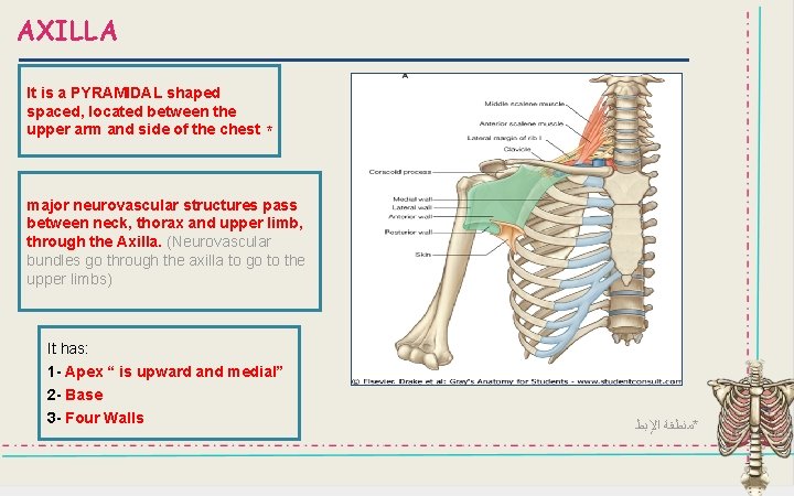
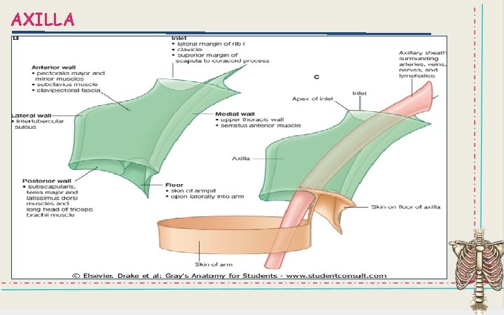
AXILLA It is a PYRAMIDAL shaped spaced, located between the upper arm and side of the chest * major neurovascular structures pass between neck, thorax and upper limb, through the Axilla. (Neurovascular bundles go through the axilla to go to the upper limbs) It has: 1 - Apex “ is upward and medial” 2 - Base 3 - Four Walls *ﻣﻨﻄﻘﺔ ﺍﻹﺑﻂ
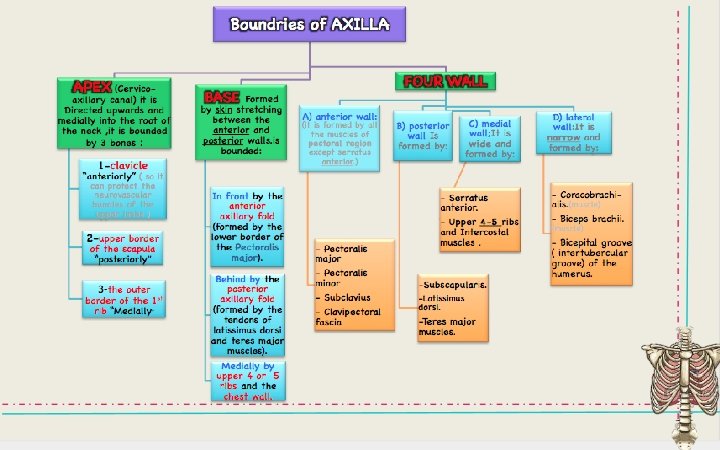
Boundaries of the Axilla 1 - Apex: ( Cervico-axillary canal): it is Directed upwards and medially into the root of the neck , it is bounded by 3 bones : 1 - clavicle “anteriorly” ( so it can protect the neurovascular bundles of the upper limbs ) 2 - upper border of the scapula “posteriorly” 3 - the outer border of the 1 st rib “Medially” 2 - Base: Formed by skin stretching between the anterior and posterior walls. it’s bounded by: In front by the anterior axillary fold (formed by the lower border of the Pectoralis major). Behind by the posterior axillary fold (formed by the tendons of latissimus dorsi and teres major muscles). Medially by upper 4 or 5 ribs and the chest wall.
Boundaries of the Axilla (cont. . ) 3 -Four walls: A) The anterior wall: it is formed by all the muscles of pectoral region except serratus anterior. - Pectoralis major - Pectoralis minor C)The medial wall: It is wide and formed by: - Serratus anterior. - Upper 4 -5 ribs and Intercostal muscles. - Subclavius - Clavipectoral fascia D)The lateral wall: It is narrow and formed by: B)The posterior wall Is formed by: - Coracobrachialis. (muscle) - Subscapularis. - Biceps brachii. (muscle) - Latissimus dorsi. - Bicipital groove ( intertubercular groove) of the humerus. - Teres major muscles.
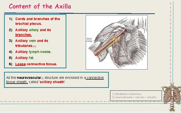
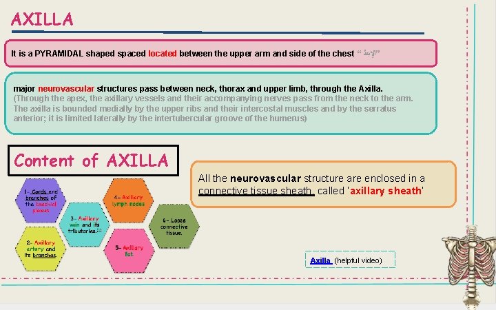
Content of the Axilla 1) Cords and branches of the brachial plexus. 2) Axillary artery and its branches. 3) Axillary vein and its tributaries. (1) 4) Axillary lymph nodes. 5) Axillary fat. 6) Loose connective tissue. All the neurovascular(2) structure are enclosed in a connective tissue sheath, called ‘axillary sheath’ 1) tributaries= branches 2) neurovascular = nerves + vessels
AXILLA It is a PYRAMIDAL shaped spaced located between the upper arm and side of the chest “ ”ﺍﻹﺑﻂ major neurovascular structures pass between neck, thorax and upper limb, through the Axilla. (Through the apex, the axillary vessels and their accompanying nerves pass from the neck to the arm. The axilla is bounded medially by the upper ribs and their intercostal muscles and by the serratus anterior; it is limited laterally by the intertubercular groove of the humerus) Content of AXILLA All the neurovascular structure are enclosed in a connective tissue sheath, called ‘axillary sheath’ Axilla (helpful video)
AXILLA
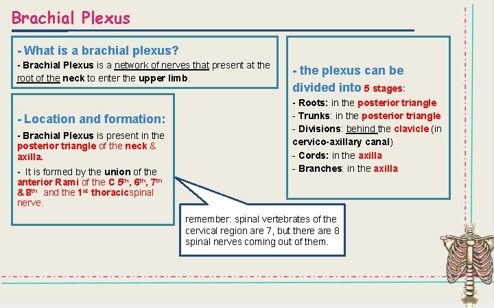
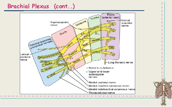
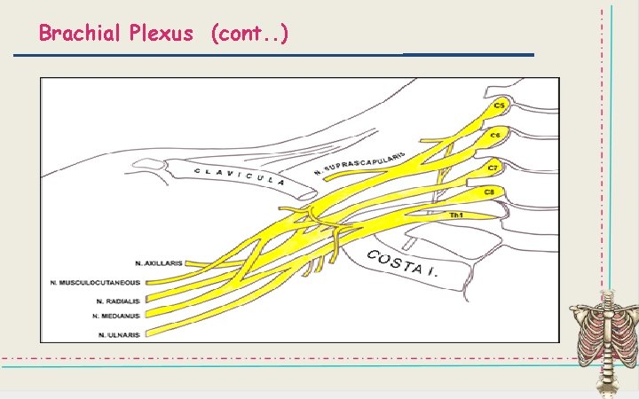
Brachial Plexus - What is a brachial plexus? - Brachial Plexus is a network of nerves that present at the root of the neck to enter the upper limb. - Location and formation: - Brachial Plexus is present in the posterior triangle of the neck & axilla. - It is formed by the union of the anterior Rami of the C 5 th, 6 th, 7 th & 8 th and the 1 st thoracicspinal nerve. - the plexus can be divided into 5 stages: - Roots: in the posterior triangle - Trunks: in the posterior triangle - Divisions: behind the clavicle (in cervico-axillary canal) - Cords: in the axilla - Branches: in the axilla remember: spinal vertebrates of the cervical region are 7, but there are 8 spinal nerves coming out of them.
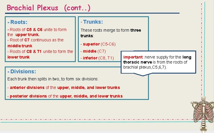
Brachial Plexus (cont. . ) - Roots: - Trunks: - Roots of C 5 & C 6 unite to form the upper trunk. - Root of C 7 continuous as the middle trunk - Roots of C 8 & T 1 unite to form the lower trunk These roots merge to form three trunks: - superior (C 5 -C 6) - middle (C 7) - inferior (C 8, T 1) - Divisions: Each trunk then splits in two, to form six divisions: - anterior divisions of the upper, middle, and lower trunks - posterior divisions of the upper, middle, and lower trunks important : nerve supply for the long thoracic nerve is from the roots of brachial plexus, C 5, 6, 7).
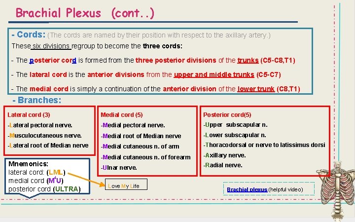
Brachial Plexus (cont. . ) - Cords: (The cords are named by their position with respect to the axillary artery. ) These six divisions regroup to become three cords: - The posterior cord is formed from the three posterior divisions of the trunks (C 5 -C 8, T 1) - The lateral cord is the anterior divisions from the upper and middle trunks (C 5 -C 7) - The medial cord is simply a continuation of the anterior division of the lower trunk (C 8, T 1) - Branches: Lateral cord (3) Medial cord (5) Posterior cord(5) -Lateral pectoral nerve. -Medial pectoral nerve. -Upper subscapular n. -Musculocutaneous nerve. -Medial root of Median nerve -Lower subscapular n. -Lateral root of Median nerve -Medial cutaneous n. of arm -Thoracodorsal or nerve to latissimus dorsi -Medial cutaneous n. of forearm -Axillary nerve. -Ulnar nerve. -Radial nerve. Mnemonics: lateral cord: (LML) 4 medial cord (M U) posterior cord (ULTRA) Love My Life Brachial plexus (helpful video)
Brachial Plexus (cont. . )
Brachial Plexus (cont. . )
MCQ 1) What is the origin of pectoralis minor? a) Coracoid process b) Clavicular head c) 3 rd, 4 th and 5 th ribs d) First rib a) Medial pectoral nerve b) Lateral pectoral nerve c) Nerve to subclavius 2) The nerve supply of serratus anterior is? d) Nerve of bell 3) The action of subclavius is? a) Steadies the clavicle b) Flexion of the shoulder c) Depression of the shoulder d) Medial rotation of the shoulder 4) The clavipectoral fascia is pierced by which artery? a) Popliteal artery b) Coracoid artery c) Thoracoacromial artery d) Lateral circumflex artery 5) The nerve supply of pectoralis major is? a) Nerve of bell b) Medial and lateral pectoral nerves c) Nerve to subclavius d) Obturator nerve Answers 6) The apex of the axilla is bounded posteriorly by? 6) The base of the axilla is bounded in the front by? 6) One of the following is a muscle of the anterior wall muscles of the axilla: a) b) c) d) 6) a) b) c) d) Clavicle Outer border of the first rib Upper border of the scapula C 4 transverse process The lower border of pectoralis major The tendons of latissimus dorsi Teres major muscles Upper 4 ribs and the chest wall Subscapularis Coracobrachialis Teres major Subclavius The roots of brachial plexus are from: Posterior rami of C 5 -T 1 spinal nerve Anterior rami of C 3 -T 1 spinal nerve Anterior rami of C 1 -T 1 spinal nerve Which of the following is true? All of the posterior divisions join to form the posterior cord All of the anterior divisions join to form the lateral cord The anterior division of the middle trunk continues to form the medial cord The anterior division of the upper and middle trunks unite to form the medial cord 1 -c 2 -d 3 -a 4 -c 5 -b 6 -c 7 -c 8 -d 9 -b 10 -a