
PDBefold SSM A webbased service for protein structure
PDBe-fold (SSM) A web-based service for protein structure comparison and structure searches Sanchayita Sen, Ph. D. PDB Depositions EBI is an Outstation of the European Molecular Biology Laboratory.
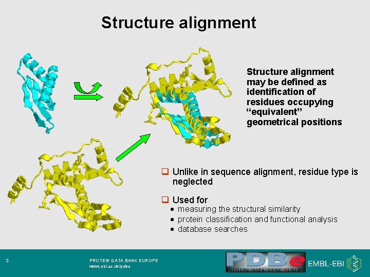
Structure alignment may be defined as identification of residues occupying “equivalent” geometrical positions q Unlike in sequence alignment, residue type is neglected q Used for · measuring the structural similarity · protein classification and functional analysis · database searches 2 PROTEIN DATA BANK EUROPE www. ebi. ac. uk/pdbe
Methods q Many methods are known: · Distance matrix alignment (DALI, Holm & Sander, EBI) · Vector alignment (VAST, Bryant et. al. NCBI) · Depth-first recursive search on SSEs (DEJAVU, Madsen & Kleywegt, Uppsala) · Combinatorial extension (CE, Shindyalov & Bourne, SDSC) · Dynamical programming on Ca (Gerstein & Levitt) · Dynamical programming on SSEs (SSA, Singh & Brutlag, Stanford University) · many other q SSM employs a 2 -step procedure: A Initial structure alignment and superposition using SSE graph matching B Ca - alignment 3 PROTEIN DATA BANK EUROPE www. ebi. ac. uk/pdbe
Three dimensional graph matching • Protein secondary structure elements (SSE)– natural and convenient objects for building three dimensional graphs. • Secondary structures provide most functionality and is conserved through evolution • Details of protein fold –expressed in terms of two SSE – helices and strands. 4 PROTEIN DATA BANK EUROPE www. ebi. ac. uk/pdbe
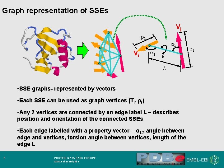
Graph representation of SSEs Vj r 2 Vi a 2 a 1 e L • SSE graphs- represented by vectors • Each SSE can be used as graph vertices (Ti, ρi) • Any 2 vertices are connected by an edge label L – describes position and orientation of the connected SSEs • Each edge labelled with a property vector – α 1/2 angle between edge and vertices, torsion angle between vertices, length of the edge L 5 PROTEIN DATA BANK EUROPE www. ebi. ac. uk/pdbe r 1
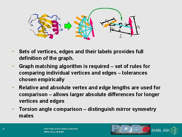
r 2 a 1 e a 2 r 1 L • Sets of vertices, edges and their labels provides full definition of the graph. • Graph matching algorithm is required – set of rules for comparing individual vertices and edges – tolerances chosen empirically • Relative and absolute vertex and edge lengths are used for comparison – allows larger absolute differences for longer vertices and edges • Torsion angle comparison – distinguish mirror symmetry mates 6 PROTEIN DATA BANK EUROPE www. ebi. ac. uk/pdbe
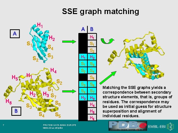
SSE graph matching H 1 A A B 7 S 3 S 2 S 3 H 4 H 1 H 2 H 6 S 1 S 4 S 2 H 5 H 1 H 2 S 1 S 2 S 1 H 3 S 7 S 6 S 4 B S 5 PROTEIN DATA BANK EUROPE www. ebi. ac. uk/pdbe H 1 H 2 S 1 S 3 S 2 S 4 S 5 S 3 S 6 S 4 S 7 H 2 H 3 H 4 H 5 H 6 Matching the SSE graphs yields a correspondence between secondary structure elements, that is, groups of residues. The correspondence may be used as initial guess for structure superposition and alignment of individual residues.
What next? • We have considered three dimensional arrangement of secondary structure element (SSE) regardless of their ordering in protein chain. • Connectivity of SSEs is significant (can be neglected in comparing mutated/engineered proteins) • In previous methods connectivity was either preserved or neglected. 8 PROTEIN DATA BANK EUROPE www. ebi. ac. uk/pdbe
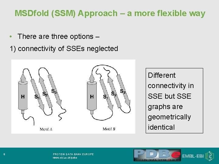
MSDfold (SSM) Approach – a more flexible way • There are three options – 1) connectivity of SSEs neglected Different connectivity in SSE but SSE graphs are geometrically identical 9 PROTEIN DATA BANK EUROPE www. ebi. ac. uk/pdbe
2) Soft connectivity – general order of SSEs along their protein chains are same in both structures BUT any number of missing/unmatched SSE between matched ones allowed 3)Strict connectivity – matched SSEs follow same order along their protein chains – separated only by equal number of matched/unmatched SSE in both structures • To obtain 3 D alignment of individual residues – represent them by their C-alpha atoms – use results of graph matching as a starting point 10 PROTEIN DATA BANK EUROPE www. ebi. ac. uk/pdbe
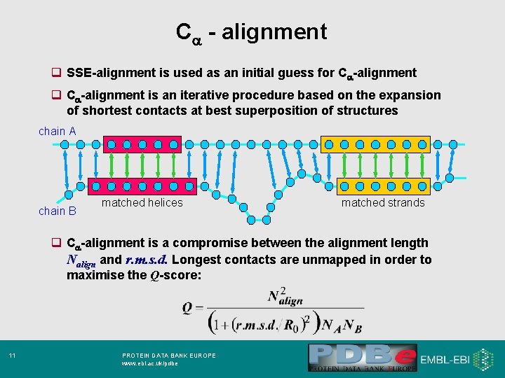
Ca - alignment q SSE-alignment is used as an initial guess for Ca-alignment q Ca-alignment is an iterative procedure based on the expansion of shortest contacts at best superposition of structures chain A chain B matched helices matched strands q Ca-alignment is a compromise between the alignment length Nalign and r. m. s. d. Longest contacts are unmapped in order to maximise the Q-score: 11 PROTEIN DATA BANK EUROPE www. ebi. ac. uk/pdbe
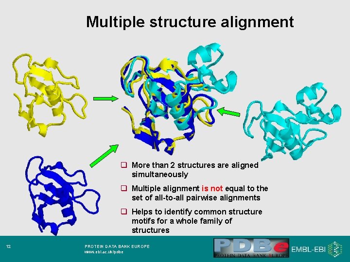
Multiple structure alignment q More than 2 structures are aligned simultaneously q Multiple alignment is not equal to the set of all-to-all pairwise alignments q Helps to identify common structure motifs for a whole family of structures 12 PROTEIN DATA BANK EUROPE www. ebi. ac. uk/pdbe
If you have to ask…. • Are there any structures in the PDB that are similar to mine? • What SCOP and/or CATH family could my structure belong to ? • Can I get some idea about the possible function of my protein based on similarity with others based on structural similarity ? • Mutiple alignment of many of my structures ? 13 31. 10. 07 Macromolecular PROTEIN DATAStructure BANK EUROPE Database www. ebi. ac. uk/pdbe USE MSDfold Upload your own PDB file for analysis !!
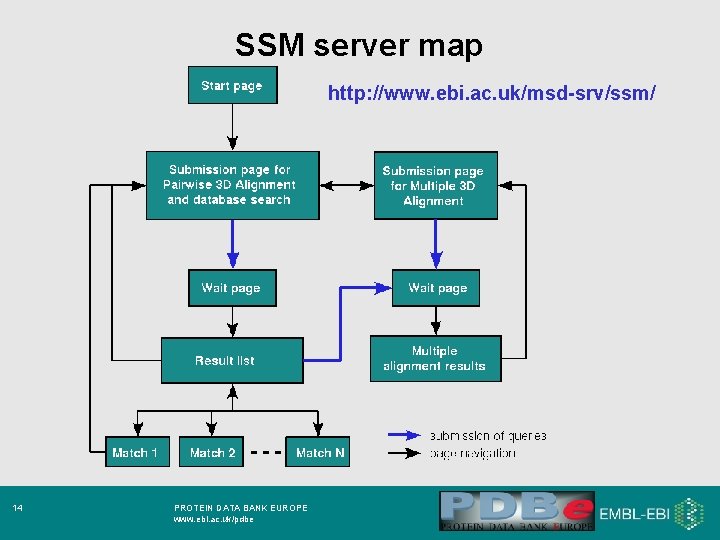
SSM server map http: //www. ebi. ac. uk/msd-srv/ssm/ 14 PROTEIN DATA BANK EUROPE www. ebi. ac. uk/pdbe
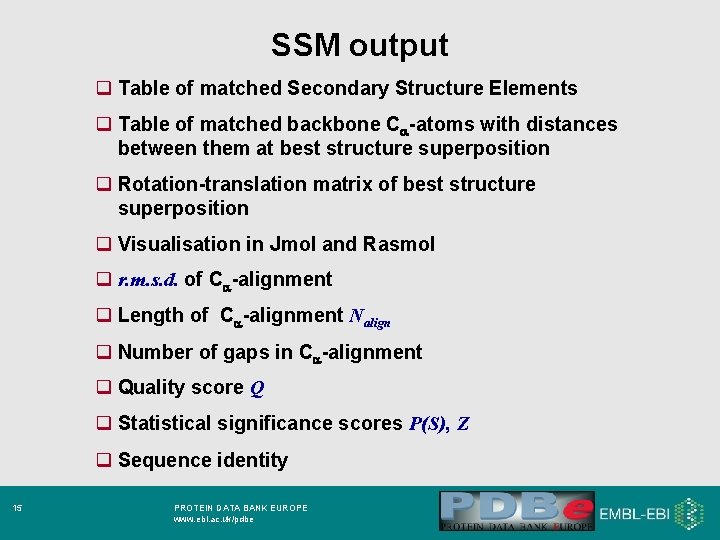
SSM output q Table of matched Secondary Structure Elements q Table of matched backbone Ca-atoms with distances between them at best structure superposition q Rotation-translation matrix of best structure superposition q Visualisation in Jmol and Rasmol q r. m. s. d. of Ca-alignment q Length of Ca-alignment Nalign q Number of gaps in Ca-alignment q Quality score Q q Statistical significance scores P(S), Z q Sequence identity 15 PROTEIN DATA BANK EUROPE www. ebi. ac. uk/pdbe
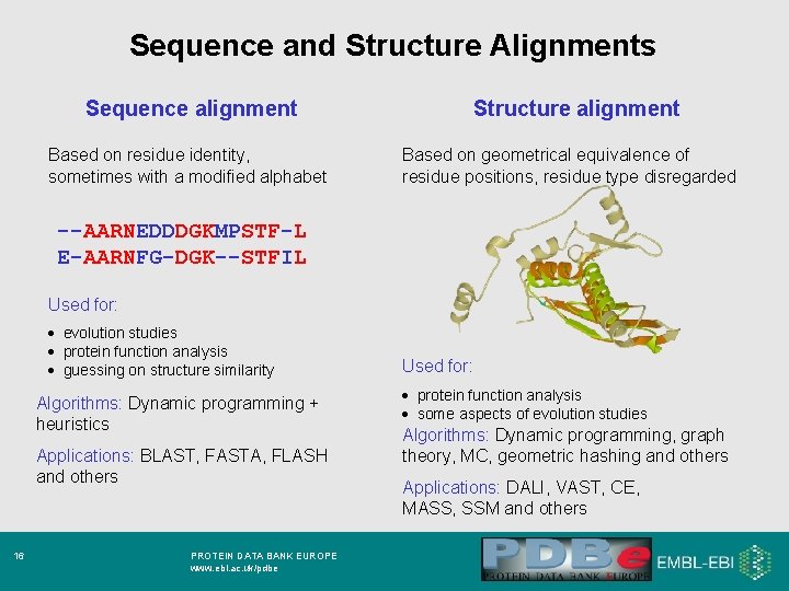
Sequence and Structure Alignments Sequence alignment Based on residue identity, sometimes with a modified alphabet Structure alignment Based on geometrical equivalence of residue positions, residue type disregarded --AARNEDDDGKMPSTF-L E-AARNFG-DGK--STFIL Used for: · evolution studies · protein function analysis · guessing on structure similarity Algorithms: Dynamic programming + heuristics Applications: BLAST, FASTA, FLASH and others 16 PROTEIN DATA BANK EUROPE www. ebi. ac. uk/pdbe Used for: · protein function analysis · some aspects of evolution studies Algorithms: Dynamic programming, graph theory, MC, geometric hashing and others Applications: DALI, VAST, CE, MASS, SSM and others
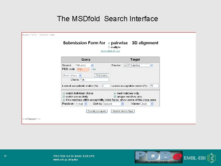
The MSDfold Search Interface 17 PROTEIN DATA BANK EUROPE www. ebi. ac. uk/pdbe
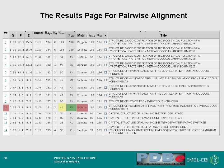
The Results Page For Pairwise Alignment 18 PROTEIN DATA BANK EUROPE www. ebi. ac. uk/pdbe
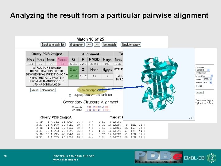
Analyzing the result from a particular pairwise alignment 19 PROTEIN DATA BANK EUROPE www. ebi. ac. uk/pdbe
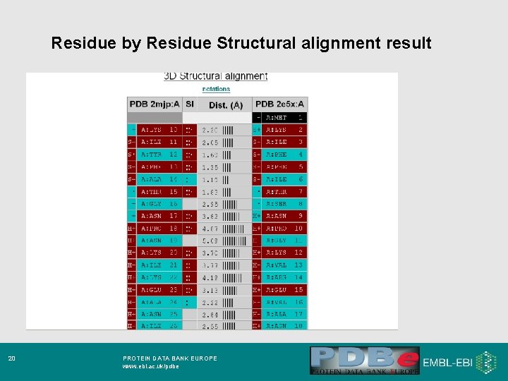
Residue by Residue Structural alignment result 20 PROTEIN DATA BANK EUROPE www. ebi. ac. uk/pdbe
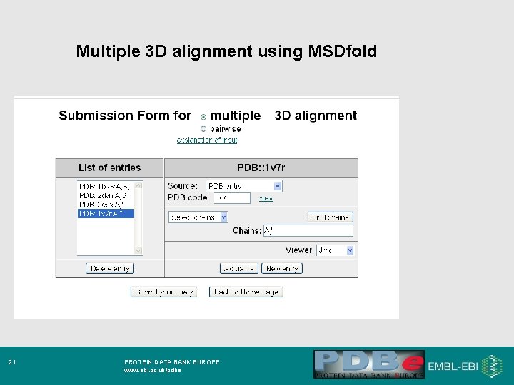
Multiple 3 D alignment using MSDfold 21 PROTEIN DATA BANK EUROPE www. ebi. ac. uk/pdbe
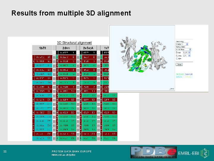
Results from multiple 3 D alignment 22 PROTEIN DATA BANK EUROPE www. ebi. ac. uk/pdbe
Conclusion · it is quite possible that residue identity plays a much less significant role in protein structure than often believed · as a consequence, the role of residue identity in protein function may be often overestimated · using sequence identity for the assessment of structural or functional features may give more false negatives than expected · physical-chemical properties of residues should be given preference over residue identity in structure and function analysis · modern methods for structure alignment are efficient; there is little sense to use sequence alignment in structure-related studies 23 PROTEIN DATA BANK EUROPE www. ebi. ac. uk/pdbe
- Slides: 23